[Preprint]. 2024 Jun 4:2024.06.04.597421.
doi: 10.1101/2024.06.04.597421.

A frameshift mutation in the murine Prkra gene causes dystonia and exhibits abnormal cerebellar development and reduced eIF2α phosphorylation

Affiliations

A frameshift mutation in the murine Prkra gene causes dystonia and exhibits abnormal cerebellar development and reduced eIF2α phosphorylation

Samuel B Burnett et al. bioRxiv. .

Update in

Abstract

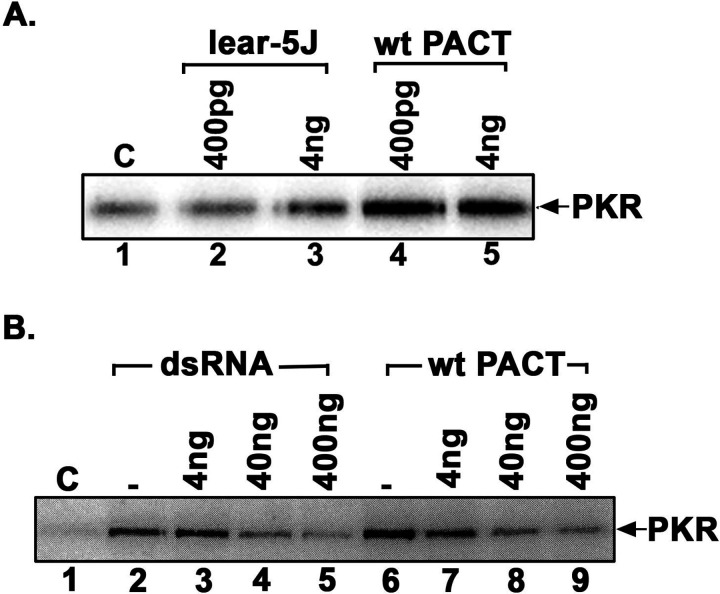
Mutations in Prkra gene, which encodes PACT/RAX cause early onset primary dystonia DYT-PRKRA, a movement disorder that disrupts coordinated muscle movements. PACT/RAX activates protein kinase R (PKR, aka EIF2AK2) by a direct interaction in response to cellular stressors to mediate phosphorylation of the α subunit of the eukaryotic translation initiation factor 2 (eIF2α). Mice homozygous for a naturally arisen, recessively inherited frameshift mutation, Prkra lear-5J exhibit progressive dystonia. In the present study, we investigate the biochemical and developmental consequences of the Prkra lear-5J mutation. Our results indicate that the truncated PACT/RAX protein retains its ability to interact with PKR, however, it inhibits PKR activation. Furthermore, mice homozygous for the mutation have abnormalities in the cerebellar development as well as a severe lack of dendritic arborization of Purkinje neurons. Additionally, reduced eIF2α phosphorylation is noted in the cerebellums and Purkinje neurons of the homozygous Prkra lear-5J mice. These results indicate that PACT/RAX mediated regulation of PKR activity and eIF2α phosphorylation plays a role in cerebellar development and contributes to the dystonia phenotype resulting from this mutation.

Keywords: DYT-PRKRA; EIF2AK2; PACT/RAX; PKR; cerebellum; dystonia.


Conflict of interest statement

Financial Disclosure/Conflict of Interest: The authors declare no financial disclosures or conflicts of interest.

Figures

Figure 1:
Figure 1:. Schematic representations of the lear-5J frameshift mutation in the Prkra gene.
(A) Functional domains of PACT/RAX and lear-5J truncated protein. Orange boxes: conserved dsRBM1 and dsRBM2 that facilitate high affinity dsRNA as well as protein-protein interactions. Green box: dsRBM3 that does not bind dsRNA but has weak binding affinity to the PACT/RAX binding motif (PBM) within the catalytic (kinase) domain (KD) of PKR. The frameshift mutation from a single nucleotide insertion results in the addition of 7 novel amino acid represented in red before the stop codon. (B) Frameshift mutation in Prkra ORF. Single adenine insertion at nucleotide position 534 (underlined) results in a truncated protein with the original 178 amino acids (AA) of PACT/RAX followed by 7 novel AA before a premature stop codon truncating the protein. This truncation occurs within the dsRBM2 functional domain.
Figure 2:
Figure 2:. The lear-5J protein binds dsRNA less efficiently but interacts with PKR similar to wt PACT/RAX.
(A) dsRNA binding assay. dsRNA binding activity of WT PACT/RAX/RAX and lear-5J truncated protein was measured by a poly(I):poly(C)-agarose binding assay with in vitro translated 35S-labeled proteins. T, total input; B, proteins bound to poly(I):poly(C)-agarose. Competition lanes 5 and 6: competition with 100- fold molar excess of single-stranded RNA (ss) or dsRNA (ds). The faint band below the parent PACT/RAX band represents products of in vitro translation from an internal methionine codon in lane 1. (B) Quantification of the dsRNA binding assay. Bands were quantified by phosphorimaging analysis, and % bound was calculated. Error bars: S.D. from three independent experiments. The p value (0.003) calculated using statistical analyses indicated significant difference between % dsRNA-binding of WT (blue bar) and lear-5J mutant (red bar) as indicated by the bracket. (C).Co-Immunoprecipitation of endogenous PKR and Flag- PACT/RAX or Flag-lear-5J overexpressed in HeLa cells. HeLa cells were transfected with Flag wt PACT/RAX or Flag-lear-5J in pCDNA3.1- expression constructs at 40% confluency and harvested 24-hour post transfection. Whole cell extracts were immunoprecipitated at 4°C overnight using 100 ng of anti-PKR antibody per IP. Samples were then analyzed via SDS-PAGE gel electrophoresis and western blot analysis probing for Flag tagged wt PACT/RAX or lear-5J (co-IP panel) using monoclonal anti-Flag-M2 (Sigma) antibody. To ascertain that an equal amount of protein was immunoprecipitated, blots were re-probed using an anti-PKR antibody (IP panel). Input blots without immunoprecipitation demonstrate equal amounts of each protein were present prior to IP.
Figure 3.
Figure 3.
(A) Effect of lear-5J protein on PKR kinase activity. PKR kinase activity assay was performed using PKR immunoprecipitated from HeLa cells, recombinant lear-5J and wt PACT/RAX proteins, and 1 μCi of [γ-32P] ATP per reaction. Either pure recombinant lear-5J (left) or wt PACT/RAX (right) protein was added as activator in amounts indicated above each lane. Phosphorylated PKR was visualized after SDS-PAGE and phosphorimager analysis. (B) The truncated lear-5J protein inhibits PKR activation. PKR immunoprecipitated from HeLa cell extracts was activated with of polyI:polyC (lanes 2–5) or 4 ng of recombinant pure wt PACT/RAX protein (lanes 6–9). Increasing amounts of recombinant pure lear-5J protein (4 ng- 400 ng) were added (lanes 2–9) as indicated to assess the effect on PKR activity. Lane 1: PKR activity without any added activator, lane 2: PKR activity in the presence of polyI:polyC, lanes 3–5: PKR activity in the presence of polyI:polyC and 4ng, 40 ng, or 400 ng of lear-5J protein, lane 6: PKR activity in the presence of 4ng of wt PACT/RAX protein, lanes 7–9: PKR activity in the presence of 4 ng of wt PACT/RAX and 4 ng, 40 ng or 400 ng of lear-5J protein. Phosphorylated proteins were analyzed by SDS-PAGE and phosphorimager analysis.
Figure 4.
Figure 4.. Tunicamycin-induced apoptosis is reduced in Prkralear-5J MEFs.
A, DNA fragmentation analysis. MEFs established from wt (BTBR T+ Itprtf/J) and Prkralear-5J mice were treated with 0.5 µg/ml tunicamycin for indicated times. Lane 1: 100 bp marker ladder, lanes 2 –4: WT (BTBR T+ Itprtf/J) MEFs; lanes 5 –7: Prkralear-5J MEFs. Lanes 2 and 5: untreated cells; lanes 3–4 and 6–7: tunicamycin-treated cells. (B) Caspase-Glo 3/7 assay. MEFs established from WT (BTBR T+ Itprtf/J) mice and Prkralear-5J mice were treated with 0.5 µg/ml tunicamycin for the indicated time points and caspase 3/7 activities were measured. Blue bars, WT (BTBR T+ Itprtf/J) MEFs; red bars, Prkralear-5J MEFs. The p values that were significant are as indicated.
Figure 5:
Figure 5:. Lear-5J truncated mutant protein is present in mouse brains but not in MEFs.
(A) Western blot analysis using brain extracts prepared from wt and Prkralear-5J brain samples. Blots were probed for PACT/RAX using a polyclonal antibody and the best of 3 representative blots is shown. The position of the full-length PACT/RAX and the truncated lear-5J protein is indicated by arrows. The blots were probed with β-actin antibody which indicates equal loading. (B) Reverse transcriptase PCR (RT-PCR) using total RNA from the brains of the indicated Prkralear-5J genotypes using ribosomal protein S15 as the positive control to ascertain presence of equal amount of total RNA in all samples. (C) Western blot analysis of cell extracts derived from MEFs of the indicated Prkralear-5J genotypes and HeLa cells overexpressing Flag-Lear-5J protein. Blots were probed for PACT/RAX using a polyclonal antibody and the best of 3 representative blots is shown. The position of the full-length PACT/RAX and the truncated lear-5J protein is indicated by arrows. (D) RT-PCR using total RNA isolated from wt and homozygous Prkralear-5J MEFs. Ribosomal protein S15 was used as a positive control to ascertain equal amount of total RNA was analyzed in each sample.
Figure 6.
Figure 6.. PACT/RAX/RAX protein is abundantly expressed in mouse cerebellum and especially in Purkinje neurons.
(A) Immunohistochemistry on day 28 sagittal section of wt C57BL/6 cerebellum using anti- PACT/RAX/RAX antibody. Brown staining indicates presence of PACT/RAX protein. (B) Immunohistochemistry of tissue described in B showing co-localization of PACT/RAX staining with a Purkinje neuron-specific marker calbindin. PACT/RAX (red), DAPI (blue), and calbindin (green).
Figure 7.
Figure 7.. The Prkralear-5J mutation affects cerebellar development and reduces arborization in Purkinje neurons.
(A-B) Hematoxylin and eosin staining on day 28 sagittal sections of wt (BTBR T+ Itprtf/J ) and Prkralear-5J cerebellum. Fully developed mouse cerebellum has ten lobules denoted here as I-X. (C-F) Immunohistochemistry of Day 28 sagittal sections of mouse cerebellar tissue stained with Purkinje neuron marker, calbindin (green), and nuclear stain DAPI (blue). Dashed boxes (left panel) indicate areas of magnification (right panel).
Figure 8.
Figure 8.. Prkralear-5J cerebellum exhibits dysregulation of eIF2α phosphorylation.
A. Immunohistochemistry of Day 28 sagittal sections of wt (BTBR T+ Itprtf/J) (left) and Prkralear-5J (right) mouse cerebellum stained with the nuclear marker DAPI (blue) and protein of interest (green). A and B: DAPI (blue) and phosphorylated eIF2α (green), C and D: DAPI (blue) and CreP (green). B. Western blot analysis of cerebellar extracts. Extracts prepared from 2 wt (BTBR T+ Itprtf/J) (lanes 1–2), 1 Prkralear-5J/+- heterozygous (lane3) and 4 Prkralear-5J/lear5-J homozygous cerebellums (lanes 4–7) were analyzed with antibodies specific for p- eIF2α, total eIF2α, CreP, and β-actin. B. Western blot analysis of wt (BTBR T+ Itprtf/J) and Prkralear-5J cerebellar extracts. Cerebellar extracts prepared from two wt (BTBR T+ Itprtf/J), one heterozygous Prkralear-5J/+, and four homozygous Prkralear-5J/lear-5J mice were analyzed by western blot analysis. Blots were probed for p-eIF2α, total eIF2α, and CreP phosphatase using specific antibodies and the best of 3 representative blots is shown. The blots were probed with β-actin antibody to ensure equal loading.

References

    1. Barber G. N. (2001) Host defense, viruses and apoptosis, Cell Death Differ 8(2): 113–26. - PubMed
    1. Beauvais G., Rodriguez-Losada N., Ying L., Zakirova Z., Watson J. L., Readhead B., Gadue P., French D. L., Ehrlich M. E. and Gonzalez-Alegre P. (2018) Exploring the Interaction Between eIF2alpha Dysregulation, Acute Endoplasmic Reticulum Stress and DYT1 Dystonia in the Mammalian Brain, Neuroscience 371: 455–468. - PubMed
    1. Bellato H. M. and Hajj G. N. (2016) Translational control by eIF2α in neurons: Beyond the stress response, Cytoskeleton (Hoboken) 73(10): 551–565. - PubMed
    1. Benkirane M., Neuveut C., Chun R. F., Smith S. M., Samuel C. E., Gatignol A. and Jeang K. T. (1997) Oncogenic potential of TAR RNA binding protein TRBP and its regulatory interaction with RNA-dependent protein kinase PKR, EMBO J 16(3): 611–24. - PMC - PubMed
    1. Bennett R. L., Blalock W. L., Abtahi D. M., Pan Y., Moyer S. A. and May W. S. (2006) RAX, the PKR activator, sensitizes cells to inflammatory cytokines, serum withdrawal, chemotherapy, and viral infection, Blood 108(3): 821–9. - PMC - PubMed

Publication types