[Preprint]. 2024 Jun 4:2024.06.04.597282.
doi: 10.1101/2024.06.04.597282.

TRIM32 inhibits Venezuelan Equine Encephalitis Virus Infection by targeting a late step in viral entry

Affiliations

TRIM32 inhibits Venezuelan Equine Encephalitis Virus Infection by targeting a late step in viral entry

Yifan Xie et al. bioRxiv. .

Update in

Abstract

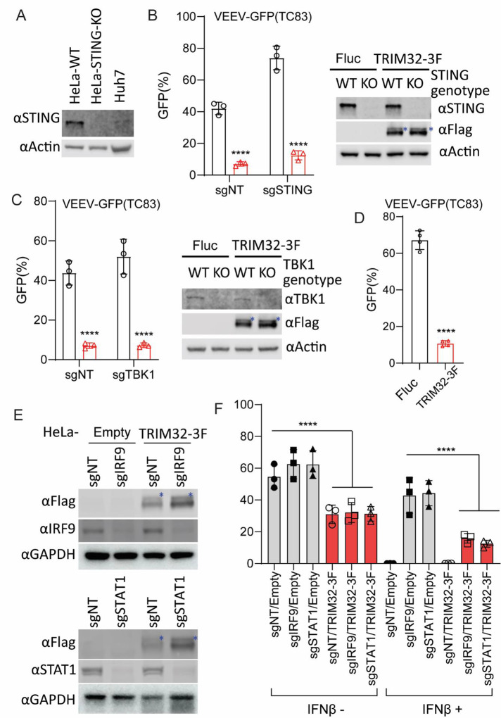
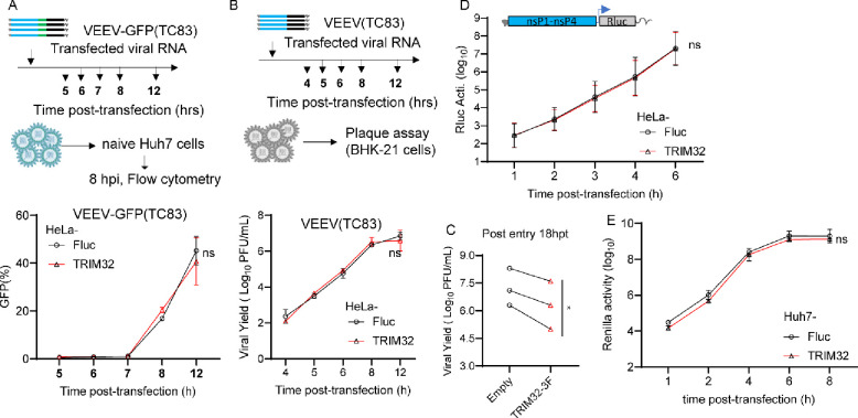
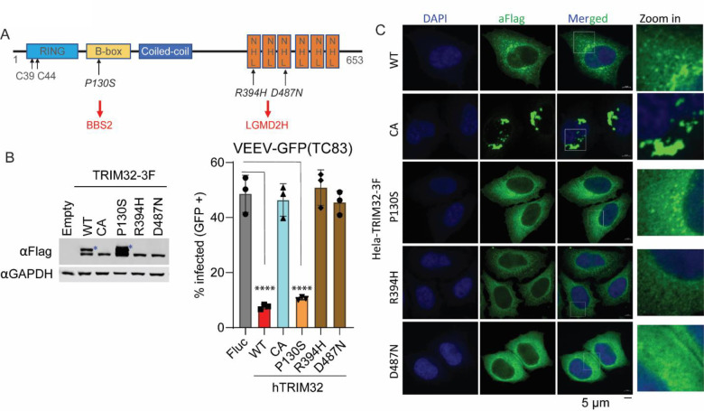
Alphaviruses are mosquito borne RNA viruses that are a reemerging public health threat. Alphaviruses have a broad host range, and can cause diverse disease outcomes like arthritis, and encephalitis. The host ubiquitin proteasome system (UPS) plays critical roles in regulating cellular processes to control the infections with various viruses, including alphaviruses. Previous studies suggest alphaviruses hijack UPS for virus infection, but the molecular mechanisms remain poorly characterized. In addition, whether certain E3 ubiquitin ligases or deubiquitinases act as alphavirus restriction factors remains poorly understood. Here, we employed a cDNA expression screen to identify E3 ubiquitin ligase TRIM32 as a novel intrinsic restriction factor against alphavirus infection, including VEEV-TC83, SINV, and ONNV. Ectopic expression of TRIM32 reduces alphavirus infection, whereas depletion of TRIM32 with CRISPR-Cas9 increases infection. We demonstrate that TRIM32 inhibits alphaviruses through a mechanism that is independent of the TRIM32-STING-IFN axis. Combining reverse genetics and biochemical assays, we found that TRIM32 interferes with genome translation after membrane fusion, prior to replication of the incoming viral genome. Furthermore, our data indicate that the monoubiquitination of TRIM32 is important for its antiviral activity. Notably, we also show two TRIM32 pathogenic mutants R394H and D487N, related to Limb-girdle muscular dystrophy (LGMD), have a loss of antiviral activity against VEEV-TC83. Collectively, these results reveal that TRIM32 acts as a novel intrinsic restriction factor suppressing alphavirus infection and provides insights into the interaction between alphaviruses and the host UPS.


Figures

Fig 1.
Fig 1.. Identification of TRIM32 as an antiviral effector against alphaviruses.
A. HeLa cells transduced with lentivirus co-expressing RFP and 118 different human RING type E3 ubiquitin ligases were infected with VEEV-TC83-GFP. Virus infectivity was determined as the percentage of GFP-positive cells within the RFP-positive gate using flow cytometry. Infection values were normalized to the empty vector control. B. HeLa cells expressing the indicated E3 or Fluc control were infected with VEEV-TC83-GFP at MOI of 0.1, and virus infectivity was measured at 24hpi by flow cytometry. C and D. HeLa-Fluc or HeLa-TRIM32 cells were infected with VEEV (C) or SINV (D) at the indicated MOI. The progeny virions in culture medium were collected at the indicated time points post-infection. and quantified by plaque assay. E. HeLa cells expressing mouse Trim32 or empty plasmid control infected with VEEV-TC83-GFP at MOI of 0.1 for 24h. Viral infectivity was quantified by flow cytometry. F. Left: C2C12 cells transduced withTrim32 targeting sgRNA or non-targeting sgRNA were infected VEEV-TC83-GFP at the indicated MOI for 8 h, and viral infectivity was determined by flow cytometry. Right: Western blot analysis of Trim32 or housekeeping Gapdh on the indicated C2C12 cells. Data represent averages of independent biological replicates and are presented as means ± SD (n=3). Statistical significance was determined by unpaired students’ t-test (*P<0.05, **P<0.01, ****P<0.0001).
Fig 2.
Fig 2.. TRIM32-mediated alphavirus inhibition is independent of the TRIM32-STING-IFN axis.
A. Western blot analysis of STING protein expression from the indicated cells. B. HeLa-sgSTING or HeLa-sgNT with or without TRIM32 expression were infected VEEV-TC83-GFP at MOI of .01 or 1 for 24h, and viral infectivity was quantified by flow cytometry. The asterisks denote the monoubiquitinated TRIM32. C. HeLa-sgTBK1 or HeLa-sgNT with or without TRIM32 expression were infected VEEV-TC83-GFP at MOI of .01 1 for 24h, and viral infectivity was quantified by flow cytometry. The asterisks denote the monoubiquitinated TRIM32. D. Huh7 cells expressing mouse TRIM32 were infected with VEEV-TC83-GFP at 0.1 MOI for 12h, virus infectivity was quantified by flow cytometry. E. Western blot analysis of IRF9 and STAT1 from the indicated cells. The asterisks denote the monoubiquitinated TRIM32. F. The indicated cells were pretreated with IFNβ at a concentration of 100U for 16h, then infected with VEEV-TC83-GFP at 0.1 MOI for 24h, and viral infectivity was determined by flow cytometry. Data represent averages of independent biological replicates and are presented as means ± SD (n=2–4). In Statistical significance was determined by unpaired students’ t-test (****P<0.0001).
Fig 3.
Fig 3.. TRIM32 does not influence post-entry stages of the VEEV infection cycle.
A. Upper: schematic of post-entry assay by transfecting VEEV-TC83-GFP infectious RNA on HeLa-empty or HeLa-TRIM32 cells. Bottom: the supernatants were harvested at the indicated time points post-transfection and used to infect naïve HeLa cells. Infectivity of progeny virus was determined by flow cytometry. B. Upper schematic of post-entry assay by transfecting VEEV-TC83 infectious RNA on HeLa-Fluc or HeLa-TRIM32 cells. Bottom: the supernatants were collected at the indicated time points post-transfection. The progeny virus in supernatants were determined by plaque assay on BHK-21 cells. C. HeLa-Fluc or HeLa-TRIM32 cells were transfected with VEEV-TC83 infectious RNA for 18 hrs, the supernatants were collected and the progeny virus in supernatants were determined by plaque assay on BHK-21 cells. D. HeLa-Empty or HeLa-TRIM32 cells were transfected with VEEV-TC83-Renilla luciferase (Rluc) replicon RNA. The cells were harvested at indicated time points, and Rluc activity was quantified. E. VEEV-TC83-Renilla luciferase (Rluc) replicon assay on Huh7-Empty or Huh7-TRIM32. Three independent biological replicates were carried out for each experiments. Statistical significance was determined by 2-way ANOVA with Sidak’s multiple comparisons test for A, B, D, E, ns, no significant. A paired t-test was used for C (*P<0.05).
Fig 4.
Fig 4.. TRIM32 does not impair viral binding, entry, and membrane fusion.
A. HeLa-Empty or HeLa-TRIM32 cells were infected with 50 MOI of VEEV-TC83 at 4°C for 1.5h, and bound virions were quantified by real-time qPCR assay. B. HeLa-Empty or HeLa-TRIM32 cells were infected with VEEV-NLuc/Cap at 4°C for 1.5h, then the cells were washed and collected to determine Nanoluc activity. C. U2OS-Empty or U2OS-TRIM32 cells were infected with VEEV- NLuc/Cap at 4°C for 1.5h, then the cells were washed and collected for to determine Nanoluc activity. D. Schematic of acid bypass assay. E. Acid bypass assay of VEEV-TC83-GFP on HeLa-Empty or HeLa-TRIM32 cells. Viral infectivity was quantified by flow cytometry. Data represent averages of independent biological replicates and are presented as means ± SD (n=3). Statistical significance was determined by unpaired students’ t-test for A, B, C, and G (****P<0.0001). For D and E, statistical significance was determined by 2-way ANOVA with Sidak’s multiple comparisons test (****P<0.0001).
Fig 5.
Fig 5.. Proximity-labeling proteomic analysis suggests TRIM32 associates with the endosome-lysosome compartment.
A. Schematic of proximity labeling proteomic approach for identification of cellular proteins and viral proteins of VEEV that interact with TRIM32. B. HeLa-TurboID.HA or HeLa-TRIM32.TurboID.HA cells were infected with VEEV-TC83-GFP at 0.1 MOI for 12h, virus infectivity was quantified by flow cytometry. C. The protein abundance of TRIM32 and viral structural and nonstructural polyproteins from the indicated groups. Triangle, HeLa-TurboID.HA infected with (blue) or without (red) infection. Circle, HeLa-TRIM32.TurboID.HA infected with (blue) or without (red) infection. D-E. Confocal images of the intracellular localization of TRIM32 and the indicated endosome or lysosome markers. Data represent averages of independent biological replicates and are presented as means ± SD (n=2–3). Statistical significance was determined by unpaired students’ t-test for A and C (****P<0.0001).
Fig 6.
Fig 6.. TRIM32 impairs the translation of incoming viral genome.
A. Schematic of the life cycle of VEEV. B. Schematic illustration of stability assay of VEEV nucleocapsids by using VEEV-NLuc/Cap infection. C. HeLa cells were infected VEEV-NLuc/Cap at 4°C for 1.5h, then the cells were washed and collected at the indicated time post temperature shift. D. HeLa-Fluc or HeLa-TRIM32 cells were infected with VEEV-NLuc/Cap at 4°C for 1.5h, then the cells were washed and collected at the indicated time post temperature shift. E. HeLa-Fluc or HeLa-TRIM32 cells were infected VEEV-NLuc/Cap at 4°C for 1.5h, then the cells were washed and collected at the indicated time post temperature shift in the presence or absence of CHX (75 ug/mL). F. HeLa-Fluc or HeLa-TRIM32 cells were infected with 1 MOI of VEEV-nsp3/NLuc at 37C for 20min, then the cells were washed and replaced with fresh media. The cells were collected at the indicated time points post infection, and the Nanoluc activity was measured. G. HeLa-Fluc or HeLa-TRIM32 cells were infected with 1 MOI of VEEV-nsp3/NLuc at 37°C for 20min, then the cells were washed and replaced with fresh media containing DMSO or 1μM ML336. The cells were collected at the indicated time points post infection, and the Nanoluc activity was measured. Data represent averages of independent biological replicates and are presented as means ± SD (n=3). Statistical significance was determined by 2-way ANOVA with Sidak’s multiple comparisons test (****P<0.0001).
Fig 7.
Fig 7.. TRIM32 LGMD2H mutants do not exhibit antiviral activity against VEEV.
A. Schematic of TRIM32 protein with domain features and important residues. B. Left: Western blot analysis of expression of wild type and mutated TRIM32 in HeLa stable cells. Right: VEEV-TC83-GFP infectivity in the indicated HeLa stable cells. The cells were infected VEEV-TC83-GFP at 0.1 MOI for 24h, viral infectivity was measured by flow cytometry. The asterisks denote the monoubiquitinated TRIM32. C. Microscope analysis the intracellular distribution of wild type and the indicated TRIM32 mutants in HeLa cells. Nuclei are stained with DAPI, blue signal. Wild type and mutated TRIM32, green signal. Scale bar, 5 μM. Data represent averages of independent biological replicates and are presented as means ± SD (n=3). Statistical significance was determined by unpaired students’ t-test for B (****P<0.0001).

Similar articles

References

    1. Lim EXY, Lee WS, Madzokere ET, Herrero LJ. Mosquitoes as suitable vectors for alphaviruses. Viruses. 2018;10: 1–17. doi:10.3390/v10020084 - DOI - PMC - PubMed
    1. Zaid A, Burt FJ, Liu X, Poo YS, Zandi K, Suhrbier A, et al. Arthritogenic alphaviruses: epidemiological and clinical perspective on emerging arboviruses. Lancet Infect Dis. 2021;21: e123–e133. doi:10.1016/S1473-3099(20)30491-6 - DOI - PubMed
    1. Sharma A, Knollmann-Ritschel B. Current understanding of the molecular basis of venezuelan equine encephalitis virus pathogenesis and vaccine development. Viruses. 2019;11. doi:10.3390/v11020164 - DOI - PMC - PubMed
    1. Charlier C, Beaudoin MC, Couderc T, Lortholary O, Lecuit M. Arboviruses and pregnancy: maternal, fetal, and neonatal effects. Lancet Child Adolesc Heal. 2017;1: 134–146. doi:10.1016/S2352-4642(17)30021-4 - DOI - PubMed
    1. Tanabe ISB, Tanabe ELL, Santos EC, Martins W V., Araújo IMTC, Cavalcante MCA, et al. Cellular and Molecular Immune Response to Chikungunya Virus Infection. Front Cell Infect Microbiol. 2018;8: 1–15. doi:10.3389/fcimb.2018.00345 - DOI - PMC - PubMed

Publication types