[Preprint]. 2024 Jun 3:2024.06.03.597171.
doi: 10.1101/2024.06.03.597171.

Protein Editing using a Concerted Transposition Reaction

Affiliations

Protein Editing using a Concerted Transposition Reaction

Yi Hua et al. bioRxiv. .

Update in

Abstract

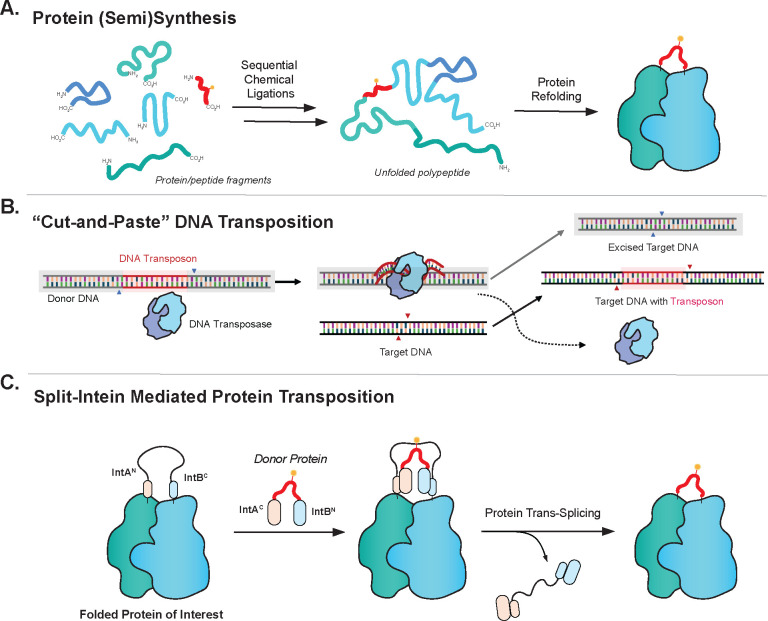
Protein engineering through the chemical or enzymatic ligation of polypeptide fragments has proven enormously powerful for studying countless biochemical processes in vitro. In general, this strategy necessitates a protein folding step following ligation of the unstructured fragments, a requirement that constrains the types of systems amenable to the approach. Here, we report an in vitro strategy that allows internal regions of target proteins to be replaced in a single operation. Conceptually, our system is analogous to a DNA transposition reaction, but employs orthogonal pairs of split inteins to swap out a designated region of a host protein with an exogenous molecular cassette. We show using isotopic labeling experiments that this 'protein transposition' reaction is concerted when the kinetics for the embedded intein pairs are suitably matched. Critically, this feature allows for efficient manipulation of protein primary structure in the context of a native fold. The utility of this method is illustrated using several protein systems including the multisubunit chromatin remodeling complex, ACF, where we also show protein transposition can occur in situ within the cell nucleus. By carrying out a molecular 'cut and paste' on a protein or protein complex under native folding conditions, our approach dramatically expands the scope of protein semisynthesis.


Conflict of interest statement

Competing interests: The authors declare that they have no competing interests.

Figures

Fig. 1.
Fig. 1.. Design of a split intein-mediated protein transposition reaction.
(a) Traditional protein semi(synthesis) using the stepwise assembly of multiple peptide fragments. Accumulated losses during the multi-step synthesis combined with an obligate final protein folding step limit the scope of this approach. (b) Schematic showing the convenience of the ‘Cut and Paste’ mechanism of DNA transposition. (c) A concerted protein transposition reaction carried out by a pair of orthogonal split inteins flanking the modification site. The approach circumvents the need to generate and manipulate protein fragments and does not require a folding step.
Fig. 2.
Fig. 2.. Efficient Protein Transposition Requires Matched Split-Intein Splicing Kinetics.
(a) Product outcomes for protein transposition depend on split-intein splicing kinetics. Mismatched splicing kinetics for the two split inteins can lead to a build-up of unwanted reaction intermediates. (b) The design of MBP-eGFP model system for optimizing the protein transposition reaction. (c) Characterization of transposition reactions using the VidaL/Cfa split intein pair (left) and Cat/mut-Cfa pair (right). MBP-eGFP recipient and transposon donor constructs (2 μM of each) were reacted for the indicated times in 100 mM phosphate, 150 mM NaCl, 1 mM EDTA, 1 mM TCEP, pH 7.2. Reactions mixtures were analyzed by SDS-PAGE with Coomassie staining (CBB). The expected transposition product is indicated. Lower bands correspond to incomplete splicing intermediates. (d) The design of an isotopic labeling experiment to differentiate between stepwise and concerted processes. A 1:1 mixture of uniformly 14N or 15N labeled MBP-eGFP recipient proteins are reacted with the transposon construct. Up to four different isotopic compositions in the transposition product are possible depending on the reaction pathway. These kinetic outcomes are denoted by colored balls. (e) Anticipated product mass spectrometry readout distinguishing the concerted versus stepwise transposition pathways. (f) Mass spectrometry characterization of transposition products in an isotopic labeling experiment for different intein pairs. Each isotopically labeled recipient (2 μM total) is reacted with an equimolar amount of HA tag transposon (2 μM) for 2 hours in 100 mM phosphate, 150 mM NaCl, 1 mM EDTA, 1 mM TCEP, pH 7.2. After the transposition, the products were isolated by RP-HPLC and the isotopic compositions were detected by ESI-MS and deconvoluted. These experiments reveal that the VidaL/Cfa split intein pair proceeds via a stepwise process whereas the Cat/mut-Cfa pair enables almost fully concerted transposition.
Fig. 3.
Fig. 3.. Protein transposition enables traceless installation of protein tags and chimeric cargo on folded proteins.
(a) Scheme illustrating the transposition reaction on an internal loop of eGFP. (b) eGFP recipient and HA tag transposon constructs (2 μM each) were reacted for the indicated times in 100 mM phosphate, 150 mM NaCl, 1 mM EDTA, 1 mM TCEP, pH 7.2. Reactions mixtures were analyzed by SDS-PAGE with Coomassie staining (CBB) and western blotting with anti-HA antibody. The expected transposition product is noted in both analyses. (c) The fluorescence emission spectra (λex = 488 nm) of the recipient eGFP protein before and after the transposition reaction. (d) Schematic of the transposition reaction within the dCas9 fold using Cat/mut-Cfa orthogonal split intein pair. In the example shown, the 6x His tag inside the recipient dCas9 gets replaced by a PEG12 abiotic polymer enabling purification of the product from the unreacted recipient. (e) Analysis of the dCas9 transposition reaction involving a PEG12-containing transposon construct. Recipient dCas9 and PEG12 transposon constructs (1.5 μM of each) were reacted for the indicated times in 50 mM Tris, 250 mM NaCl, 1 mM TCEP, 10% v/v glycerol, pH 7.5. Reactions mixtures were further purified by reverse nickel affinity chromatography. Both the reaction progress (left) and purified product (right) were analyzed by SDS-PAGE with Coomassie staining and the expected product is indicated. (f) DNA binding activity of dCas9 constructs. A wild-type dCas9 without any transposition construct, the recipient dCas9 and the purified dCas9 containing PEG12 after transposition were mixed with DNA in the presence of the complementary guide RNA (c-gRNA) or non-complementary guide RNA (nc-gRNA) and their DNA binding capability characterized by 5% native TBE gel electrophoresis with SYBR Gold staining.
Fig. 4.
Fig. 4.. Protein transposition on SMARCA5 and ACF complex enables functional evaluation of remodeling activity and binding preferences.
(a) Scheme illustrating protein transposition using the Cat/mut-Cfa orthogonal split intein pair on the SMARCA5 subunit of ACF complex; various chemical modifications can be installed via the transposon to enable different biochemical experiments. (b) The pre-made ACF complex containing the recipient SMARCA5 (500 nM) was mixed with a designated transposon construct (500 nM) in 25 mM HEPES, 60 mM KCl, 10 mM MgCl2, 1 mM TCEP, 10% v/v glycerol, 0.02% v/v IGEPAL CA630, pH 7.75. Following overnight reaction at 4 °C the products were characterized by SDS-PAGE with Coomassie staining (CBB) and western blotting with indicated antibodies. The APB transposons used in this experiment are as follows: SMARCA5 APB1 (APB), SMARCA5 APB+photoMet (APB w/photoMet), and SMARCA5 APB+pY (APB + pY) (See Fig. S22 for further details) (c) Top: Remodeling activity of modified ACF complexes on mononucleosomes (MNs) as assayed by REAA (10 nM ACF, 10 nM MNs); reintroducing the APB domain into a catalytically inactive ACF complex via protein transposition rescues its activity. Bottom: Kinetics of remodeling for the indicated ACF complexes as measured by REAA, Errors = s.e.m. (n = 3 independent experiments). Representative native gel analyses of the remodeling reactions are shown in Fig. S31. (d) Introducing a phosphotyrosine modification into SMARCA5 (pY742) markedly slows nucleosome sliding, as observed by electrophoretic mobility shift after repositioning (8.33 nM ACF, 10 nM MNs, 4 minutes). Upon λ protein phosphatase (λPP) treatment, ACF activity is fully restored. Off-center bead-on-a-string represents the un-remodeled MN whereas the on-center bead-on-a-string represents the remodeled MN. Quantification is by gel densitometry; error bars represent the s.e.m. from 3 replicates. * denotes P value < 0.05. See Fig. S32 for more details. (e) A photo-methionine residue (photoM743) installed within SMARCA5 via protein transposition enables photo-crosslinking between SMARCA5 (10 pmol) and a mononucleosome (10 pmol) upon UV-irradiation (20 min). Left: Immunostaining with an H2A antibody shows a positive crosslink between SMARCA5 and histone, which is significantly ablated upon the addition of excess LANA peptide (10 uM) — a competitive binder to the nucleosome acidic patch. The asterisk denotes nonspecific binding of histone antibody to SMARCA5. Right: densitometry analysis of the crosslinking signal in the immuno-blot normalized to total H2A signal. Errors represent the s.e.m. from 3 independent biological replicates. * denotes P value < 0.05. (f) Immunoblot analysis of in nucleo protein transposition reaction between endogenously expressed recipient SMARCA5 in HEK 293T cells containing an embedded HA tag (red signal) and exogenously added transposon construct (0.1–0.5 uM) containing a FLAG tag (green signal) over 30 min. Successful in nucleo transposition is denoted by the decrease in the HA signal with a concomitant increase in the FLAG signal.

References

    1. Thompson R. E., Muir T. W., Chemoenzymatic Semisynthesis of Proteins. Chem. Rev. 120, 3051–3126 (2020). - PMC - PubMed
    1. Gramespacher J. A., Stevens A. J., Thompson R. E., Muir T. W., Improved protein splicing using embedded split inteins. Protein Science 27, 614–619 (2018). - PMC - PubMed
    1. Conibear A. C., Watson E. E., Payne R. J., Becker C. F. W., Native chemical ligation in protein synthesis and semi-synthesis. Chem. Soc. Rev. 47, 9046–9068 (2018). - PubMed
    1. Cowieson N. P. et al., An automatable screen for the rapid identification of proteins amenable to refolding. Proteomics 6, 1750–1757 (2006). - PubMed
    1. Willis M. S. et al., Investigation of protein refolding using a fractional factorial screen: A study of reagent effects and interactions. Protein Science 14, 1818–1826 (2005). - PMC - PubMed

Publication types