[Preprint]. 2025 Jun 25:2024.06.04.597454.
doi: 10.1101/2024.06.04.597454.

Intracellular pH dynamics respond to extracellular matrix stiffening and mediate vasculogenic mimicry through β-catenin

Affiliations

Intracellular pH dynamics respond to extracellular matrix stiffening and mediate vasculogenic mimicry through β-catenin

Leah M Lund et al. bioRxiv. .

Abstract

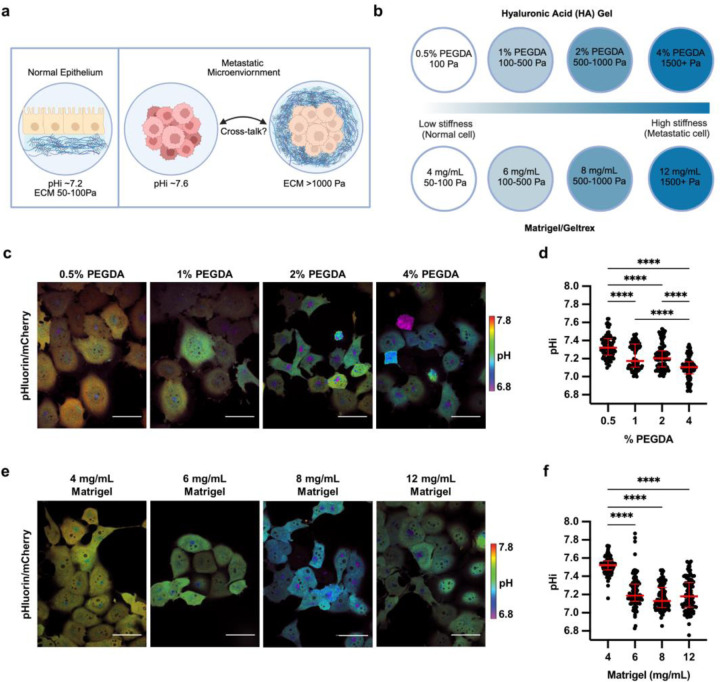
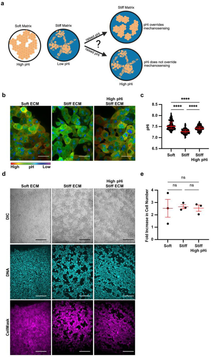
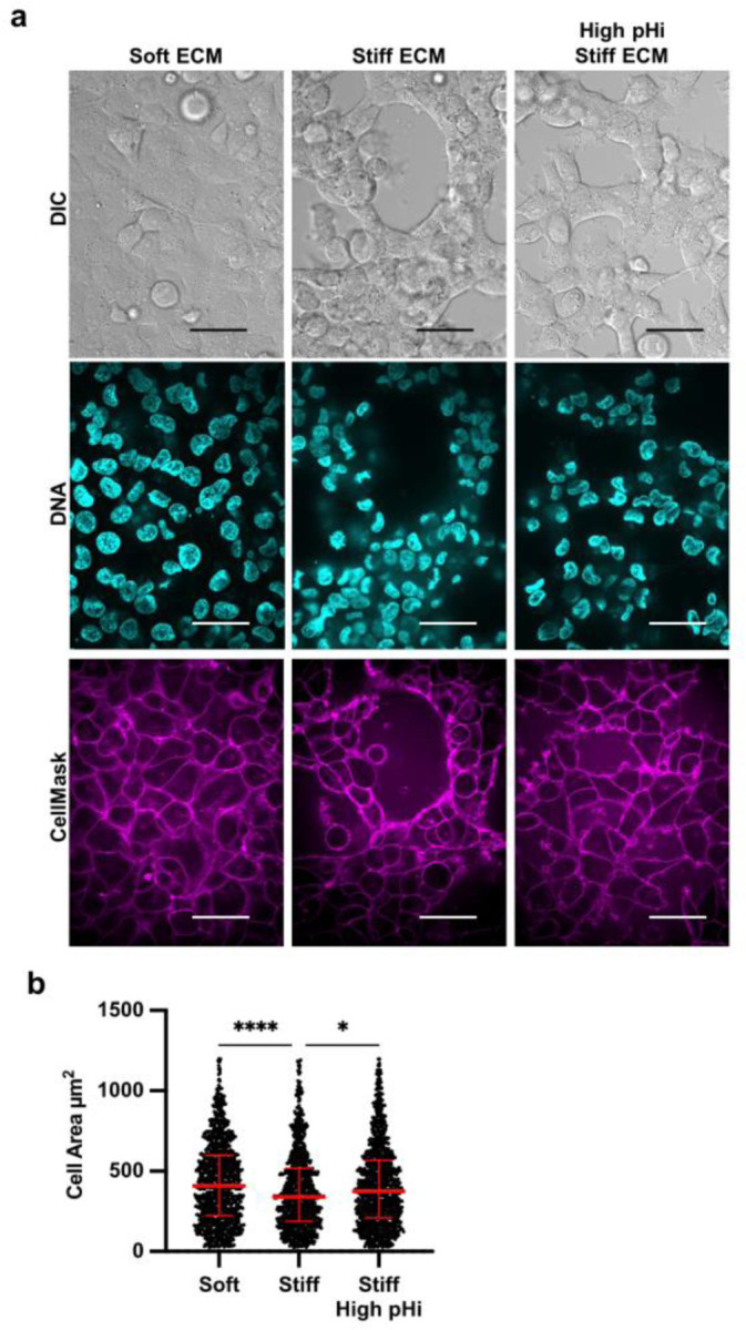
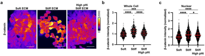
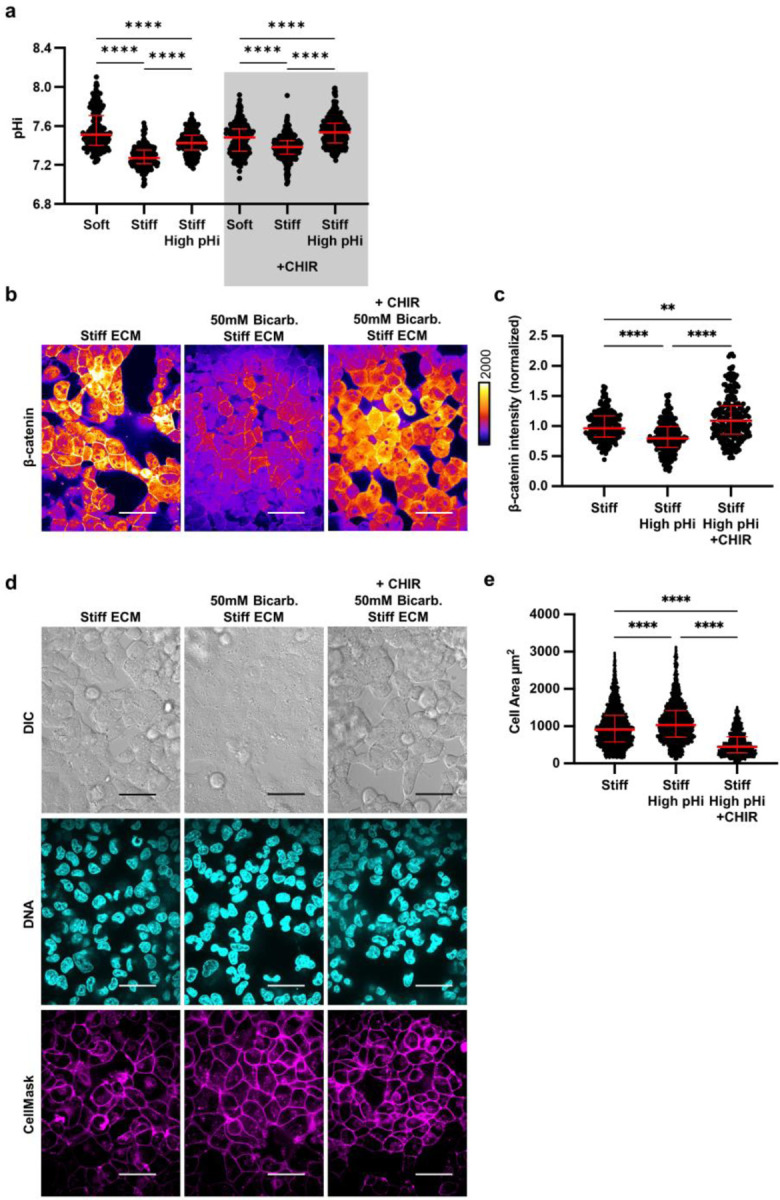
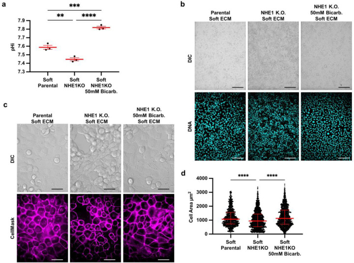
Dysregulated intracellular pH (pHi) dynamics and an altered tumor microenvironment have emerged as drivers of cancer cell phenotypes. However, the molecular integration between the physical properties of the microenvironment and dynamic intracellular signaling responses remains unclear. Here, we identify a mechanistic link between ECM stiffness and pHi dynamics in driving vasculogenic mimicry (VM), an aggressive cancer phenotype associated with poor prognosis. We performed single-cell imaging of pHi in lung and breast metastatic cell lines cultured on tunable-stiffness hydrogel systems. We used two tunable-stiffness hydrogel systems to independently model stiffness induced by increased protein secretion (Matrigel) and increased protein crosslinking (Hyaluronic acid gels). We show that increased ECM stiffness lowers single-cell pHi in both lung and breast metastatic cell lines. We also observed that stiff ECM promotes a distinct morphological phenotype called vasculogenic mimicry (VM). Importantly, we show that low pHi is a necessary mediator of VM, as raising pHi on stiff ECM reduces VM phenotypes. We also find that lowering pHi on soft ECM was sufficient to induce VM in the absence of extracellular stiffening. We characterized β-catenin as a pH-dependent molecular mediator of VM, where stiffness-driven increases in β-catenin abundance can be overridden by high pHi, which destabilizes β-catenin to reduce VM on stiff ECM. In contrast, the transcription factor FOXC2 is activated by ECM stiffness but is insensitive to pHi, and its activity alone is insufficient to maintain VM at high pHi when β-catenin is lost. We uncover a novel mechanotransduction axis in which ECM stiffness regulates intracellular pH to drive β-catenin-induced VM. We also show pHi dynamics can override mechanosensitive cell responses to the extracellular microenvironment. Thus, our work positions pHi as an integrator of mechanotransduction in cancer, suggesting a new framework for therapeutically targeting pHi in cancer and perhaps in other diseases driven by ECM remodeling.


Conflict of interest statement

Competing Interests: Authors declare no competing interests.

Figures

Figure 1:
Figure 1:. Stiffening extracellular matrix lowers pHi in metastatic human lung carcinoma (H1299).
a) Schematic of increased pHi and ECM stiffening (via increased protein secretion and increased protein crosslinking) associated with tumorigenesis. b) Schematic of synthetic ECM models with tunable-stiffness (~50 Pa-1,500 Pa). The Matrigel (or Geltrex) model mimics increased ECM protein secretion while hyaluronic acid (HA) gel system mimics increased ECM protein crosslinking. c) Representative images of H1299 cells stably expressing mCherry-pHluorin pH biosensor plated on varying HA gel stiffnesses. Images show ratiometric display of pHluorin/mCherry fluorescence. Scale bars: 50 μm. d) Quantification of single-cell pHi data collected as shown in (c). (n=3 biological replicates; n=91 0.5% PEGDA, n=90 1% PEGDA, n=102 2% PEGDA, n=89 4% PEGDA. Red lines show medians ± IQR). e) Representative images of H1299 cells stably expressing mCherry-pHluorin pH biosensor plated on varying Matrigel stiffnesses. Images show ratiometric display of pHluorin/mCherry fluorescence. Scale bars: 50 μm. f) Quantification of single-cell pHi data collected as shown in (e). (n=3 biological replicates; n=93 4mg/mL, n=92 6mg/mL, n=102 8mg/mL, n=97 12mg/mL. Red lines show medians ± IQR). For (d) and (f), significance was determined by a Kruskal-Wallis test (****P<0.0001).
Figure 2:
Figure 2:. Stiffness-dependent vasculogenic mimicry is reduced when pHi is increased on stiff ECM.
a) Schematic of vasculogenic mimicry (VM) in 2D on stiffening matrix. b) Representative images of H1299 cells stably expressing mCherry-pHluorin pH biosensor plated on soft (0.5% PEGDA) and stiff (4% PEGDA) HA gels and stiff (4% PEGDA) with raised pHi (culture media supplementation with 50mM sodium bicarbonate). Images show ratiometric display of pHluorin/mCherry fluorescence ratios. Scale bars: 50 μm. c) Quantification of single-cell pHi data collected as shown in (b) (n=3 biological replicates; n=201 soft, n=237 stiff, n=239 stiff high pHi. Red lines show medians ± IQR). d) Representative images of H1299 cells plated on soft (0.5% PEGDA) and stiff (4% PEGDA) HA gels. Images show differential interference contrast (DIC) and Hoechst stain (DNA, cyan). Scale bars: 100 μm. e) Quantification of cell proliferation across manipulation conditions. (n=3 biological replicates. Red lines show means ± SEM).
Figure 3:
Figure 3:. Vasculogenic mimicry phenotype decreases cell area on stiff ECM, which is rescued by increasing pHi in metastatic lung carcinoma.
a) Representative images of H1299 cells plated on soft (0.5% PEGDA) and stiff (4% PEGDA) HA gels and stiff (4% PEGDA) with raised pHi. Images show differential interference contrast (DIC), Hoechst 33342 (DNA, cyan) and CellMask Deep Red membrane stain (Cy5, magenta). Scale bars: 50 μm. b) Quantification of single-cell area collected as shown in (a) (n=3 biological replicates, n=1061 soft, n=954 stiff, n=1078 stiff high pHi. Red lines show medians ± IQR).
Figure 4:
Figure 4:. Increased pHi reduces β-catenin abundance and nuclear localization in stiff matrix conditions.
a) Representative images of H1299 cells plated on soft (0.5% PEGDA), stiff (4% PEGDA) and stiff with raised pHi (4% PEGDA) HA gels fixed and stained for β-catenin. β-catenin is pseudocolored according to scale. Scale bars: 50 μm. b) Quantification of whole cell β-catenin intensity collected as shown in (a). (n=3 biological replicates, n=452 soft, n=486 stiff, n=415 stiff high pHi. Red lines show medians ± IQR). c) Quantification of nuclear β-catenin intensity collected as described in (a). (n=3 biological replicates, n=1043 soft, n=975 stiff, n=1157 stiff high. Red lines show medians ± IQR). For (b) and (c), significance was determined by a Kruskal-Wallis test (*P<0.05; **P<0.01; ***P<0. 001; ****P<0.0001).
Figure 5:
Figure 5:. Stabilization of β-catenin abundance under high pHi rescues VM on a stiff matrix.
a) Quantification of pHi data from parental H1299 cells and H1299 cells treated with CHIR treatment with and without treatment with sodium bicarbonate (Bicarb.) (see methods) (n=3 biological replicates. n=191 soft, n=219 stiff, n=198 stiff high pHi, n=302 soft +CHIR, n=373 stiff +CHIR, n=408 stiff high pHi +CHIR. Red lines show means ± SEM). b) Representative images of H1299 cells plated on stiff HA gels (4% PEGDA) with and without raised pHi (Bicarb.) and with or without CHIR treatment fixed and stained for β-catenin. β-catenin is pseudocolored according to scale. Scale bars: 50 μm. c) Quantification of whole cell β-catenin intensity collected as shown in (b). (n=3 biological replicates, n=452 stiff, n=486 stiff high pHi, n=415 stiff high pHi +CHIR. Red lines show medians ± IQR). d) Representative images of H1299 cells plated on stiff HA gels (4% PEGDA) with and without raised pHi (Bicarb.) and with or without CHIR treatment. Images show differential interference contrast (DIC), Hoechst 33342 (DNA, cyan) and CellMask Deep Red membrane stain (Cy5, magenta). Scale bars: 50 μm. e) Quantification of single-cell area collected as shown in (d) (n=3 biological replicates, n=452 stiff, n=486 stiff high pHi, n=415 stiff high pHi +CHIR. Red lines show medians ± IQR).
Figure 6:
Figure 6:. Low pHi is sufficient to induce vasculogenic mimicry on a soft ECM.
a) Quantification of pHi data from parental H1299 cells and H1299 cells where NHE1 knockout via CRISPR (H1299-NHE1 K.O.). with and without treatment with sodium bicarbonate (Bicarb.) (see methods) (n=3 biological replicates. Red lines show means ± SEM). b) Representative images of H1299 cells plated on soft HA gels (0.5% PEGDA) with and without lowered pHi (H1299-NHE1 K.O.) and with or without increased pHi (H1299-NHE1 K.O. Bicarb.). Images show differential interference contrast (DIC) and Hoechst 33342 (DNA, cyan). Scale bars: 100 μm. c) Representative images of H1299 cells plated on soft HA gels (0.5% PEGDA) with and without lowered pHi (H1299-NHE1 K.O.) and with or without increased pHi (H1299-NHE1 K.O. Bicarb.). Images show differential interference contrast (DIC) and CellMask Deep Red membrane stain (Cy5, magenta). Scale bars: 50 μm. d) Quantification of single-cell area collected as shown in (a) (n=3 biological replicates, n=383 parental, n=267 NHE1 K.O., n=315 NHE1 K.O. Bicarb. Red lines show medians ± IQR).

Similar articles

References

    1. Walker C.; Mojares E.; del Río Hernández A. Role of Extracellular Matrix in Development and Cancer Progression. Int. J. Mol. Sci. 2018, 19 (10), 3028. 10.3390/ijms19103028. - DOI - PMC - PubMed
    1. Carey S. P.; Martin K. E.; Reinhart-King C. A. Three-Dimensional Collagen Matrix Induces a Mechanosensitive Invasive Epithelial Phenotype. Sci. Rep. 2017, 7 (1), 42088. 10.1038/srep42088. - DOI - PMC - PubMed
    1. Wechman S. L.; Emdad L.; Sarkar D.; Das S. K.; Fisher P. B. Vascular Mimicry: Triggers, Molecular Interactions and in Vivo Models. Adv. Cancer Res. 2020, 148, 27–67. 10.1016/bs.acr.2020.06.001. - DOI - PMC - PubMed
    1. Webb B. A.; Chimenti M.; Jacobson M. P.; Barber D. L. Dysregulated pH: A Perfect Storm for Cancer Progression. Nat. Rev. Cancer 2011, 11 (9), 671–677. 10.1038/nrc3110. - DOI - PubMed
    1. White K. A.; Grillo-Hill B. K.; Barber D. L. Cancer Cell Behaviors Mediated by Dysregulated pH Dynamics at a Glance. J. Cell Sci. 2017, 130 (4), 663–669. 10.1242/jcs.195297. - DOI - PMC - PubMed

Publication types