[Preprint]. 2024 Jun 6:2024.06.04.597210.
doi: 10.1101/2024.06.04.597210.

Evaluation in mice of cell-free produced CT584 as a Chlamydia vaccine antigen

Affiliations

Evaluation in mice of cell-free produced CT584 as a Chlamydia vaccine antigen

Steven Hoang-Phou et al. bioRxiv. .

Abstract

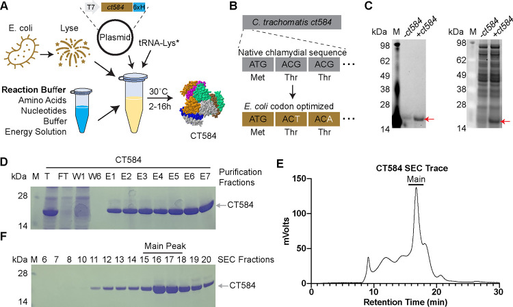
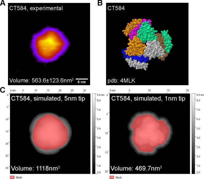
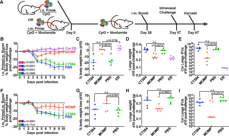
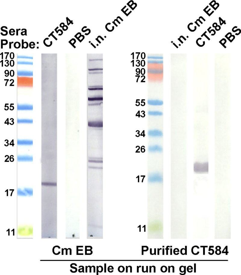
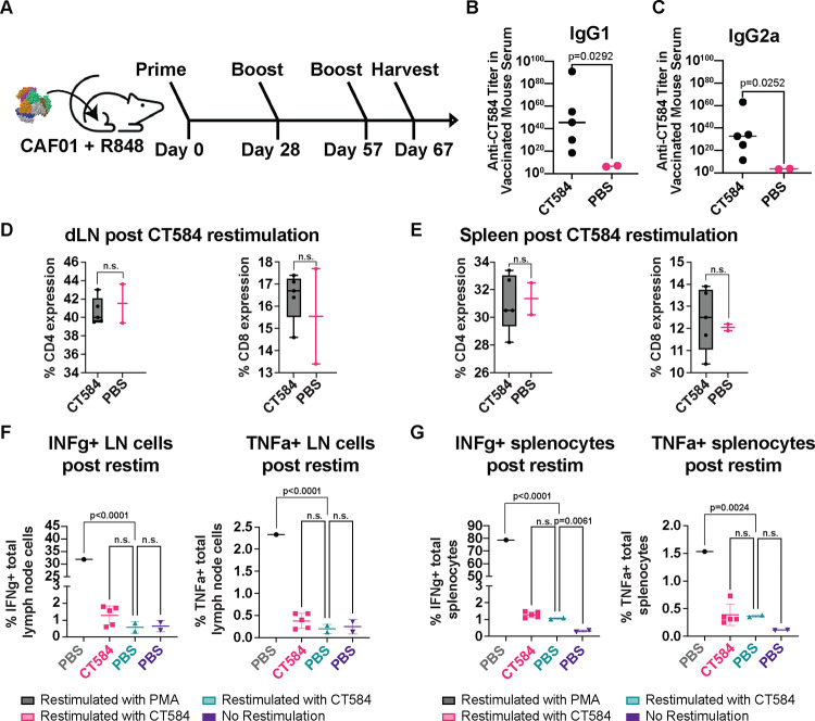
Chlamydia trachomatis is the most prevalent bacterial sexually transmitted pathogen worldwide. Since chlamydial infection is largely asymptomatic with the potential for serious complications, a preventative vaccine is likely the most viable long-term answer to this public health threat. Cell-free protein synthesis (CFPS) utilizes the cellular protein manufacturing machinery decoupled from the requirement for maintaining cellular viability, offering the potential for flexible, rapid, and de-centralized production of recombinant protein vaccine antigens. Here, we use CFPS to produce the putative chlamydial type three secretion system (T3SS) needle-tip protein, CT584, for use as a vaccine antigen in mouse models. High-speed atomic force microscopy (HS-AFM) imaging and computer simulations confirm that CFPS-produced CT584 retains a native-like structure prior to immunization. Female mice were primed with CT584 adjuvanted with CpG-1826 intranasally (i.n.) or CpG-1826 + Montanide ISA 720 intramuscularly (i.m.), followed four-weeks later by an i.m. boost before respiratory challenge with 104 inclusion forming units (IFU) of Chlamydia muridarum. Immunization with CT584 generated robust antibody responses but weak cell mediated immunity and failed to protect against i.n. challenge as demonstrated by body weight loss, increased lungs' weights and the presence of high numbers of IFUs in the lungs. While CT584 alone may not be the ideal vaccine candidate, the speed and flexibility with which CFPS can be used to produce other potential chlamydial antigens makes it an attractive technique for antigen production.

Keywords: CT584; Chlamydia; cell-free protein synthesis; immunization; mice; type-three secretion system; vaccine.


Conflict of interest statement

Conflicts of Interest The authors declare no conflict of interest.

Figures

Figure 1.
Figure 1.. CT584 can be produced and purified using cell-free protein synthesis
A) Schematic of the cell-free protein synthesis (CFPS) approach to protein production. B) Schematic of DNA codon optimization performed on chlamydial ct584. C) SDS-PAGE gel depicting fluorescent image of bodipy labeled tRNA-Lys incorporation into CT584 (left) and sypro ruby total protein stain (right). D) Coomassie blue stained SDS-PAGE gel showing affinity typical affinity purification fractions for CT584. M: Marker, T: Total, FT: Flow-through, W: Wash, E: Elutions E) Smoothed histogram of size exclusion chromatography (SEC) retention times for affinity purified CT584. F) Coomassie blue stained SDS-PAGE gel of SEC elution fractions. Fractions pooled and used for further experiments are indicated (“Main Peak”).
Figure 2.
Figure 2.. CFPS produced CT584 retains a native-like conformation
A) Atomic force microscopy (AFM) image of CT584. Scale bar: 5nm. B) Space-filling model of published CT584 crystal structure (PDB: 4MLK). C) Simulated AFM images using the CT584 crystal structure and a 5nm (left) and 1nm (right) probe size. Highlighted pink areas show coverage.
Figure 3.
Figure 3.. CT584 immunization fails to generate a protective response against an i.n. chlamydial challenge in mice
A) Schematic of immunization routes and schedule for challenge studies. B) Line graph depicting mean mouse weights +/− s.e.m. after intranasal challenge in i.n./i.m. immunized mice. P-values calculated using two-way repeated measures ANOVA. C) Scatter plot showing percent body weight loss at day 10 post-challenge in i.n./i.m. immunized mice. Line indicates median. P-values calculated using Student’s t-test. D) Scatter plot showing lung weights at day 10 post-challenge in i.n./i.m. immunized mice. Line indicates median. P-values calculated using Student’s t-test. E) Scatter plot showing number of Cm IFUs in mouse lungs at day 10 post-challenge in i.n./i.m. immunized mice. Line indicates median. P-values calculated using Mann-Whitney U test. F) Line graph depicting mean mouse weights +/− s.e.m. after intranasal challenge in i.m./i.m. immunized mice. P-values calculated using two-way repeated measures ANOVA. G) Scatter plot showing percent body weight loss at day 10 post-challenge in i.m./i.m. immunized mice. Line indicates median. P-values calculated using Student’s t-test. H) Scatter plot showing lung weights at day 10 post-challenge in i.m./i.m. immunized mice. Line indicates median. P-values calculated using Student’s t-test. I) Scatter plot showing number of Cm IFUs in mouse lungs at day 10 post-challenge in i.m./i.m. immunized mice. Line indicates median. P-values calculated using Mann-Whitney U test.
Figure 4.
Figure 4.. CT584 immune sera detects the recombinant and native protein but Cm EB immune sera does not
A) Western blots of purified recombinant CT584 probed with the serum of mice immunized with: Lane 1 – recombinant CT584; Lane 2 – PBS; Lane 3 – i.n. Cm EB. B) Western blots of Cm EB lysates probed with the serum of mice immunized with: Lane 1 - i.n. live Cm EB; Lane 2 - recombinant CT584; Lane 3 – PBS.
Figure 5.
Figure 5.. CT584 + CAF01 and R848 induces antigen-specific antibodies but not T cell-based responses
A) Schematic of i.m./i.m. immunization routes and schedule for CT584 with CAF01/R848. B) Scatter plot showing CT584 specific IgG1 antibodies in mouse immune sera. Line indicates median. P-values calculated using Welch’s t-test. C) Scatter plot showing CT584 specific IgG2a antibodies in mouse immune sera. Line indicates median. P-values calculated using Welch’s t-test. D) Box and whiskers plot showing CD4 (left) and CD8 (right) expression in T cell populations from the draining lymph nodes of immunized mice. P-values calculated by Student’s t-test. E) Box and whiskers plot showing CD4 (left) and CD8 (right) expression in T cell populations from the spleen of immunized mice. P-values calculated by Student’s t-test. F) Scatter plot showing percentage of lymph node immune cells expressing IFN-γ (left) and TNF-α (right) after ex vivo restimulation with antigen. Line indicates median. P-values calculated by one-way ANOVA. G) Scatter plot showing percentage of splenocytes expressing IFN-γ (left) and TNF-α (right) after ex vivo restimulation with antigen. Line indicates median. P-values calculated by one-way ANOVA.

Similar articles

References

    1. Newman L. et al. Global Estimates of the Prevalence and Incidence of Four Curable Sexually Transmitted Infections in 2012 Based on Systematic Review and Global Reporting. PLoS One 10, e0143304 (2015). 10.1371/journal.pone.0143304 - DOI - PMC - PubMed
    1. den Heijer C. D. J. et al. Chlamydia trachomatis and the Risk of Pelvic Inflammatory Disease, Ectopic Pregnancy, and Female Infertility: A Retrospective Cohort Study Among Primary Care Patients. Clin Infect Dis 69, 1517–1525 (2019). 10.1093/cid/ciz429 - DOI - PMC - PubMed
    1. Tang W. et al. Pregnancy and fertility-related adverse outcomes associated with Chlamydia trachomatis infection: a global systematic review and meta-analysis. Sex Transm Infect 96, 322–329 (2020). 10.1136/sextrans-2019-053999 - DOI - PMC - PubMed
    1. Du M. et al. Increasing incidence rates of sexually transmitted infections from 2010 to 2019: an analysis of temporal trends by geographical regions and age groups from the 2019 Global Burden of Disease Study. BMC Infect Dis 22, 574 (2022). 10.1186/s12879-022-07544-7 - DOI - PMC - PubMed
    1. Pinto C. N. et al. Impact of the COVID-19 Pandemic on Chlamydia and Gonorrhea Screening in the U.S. Am J Prev Med 61, 386–393 (2021). 10.1016/j.amepre.2021.03.009 - DOI - PMC - PubMed

Publication types