[Preprint]. 2024 Jun 8:2024.06.07.598004.
doi: 10.1101/2024.06.07.598004.

Fiber-based Probes for Electrophysiology, Photometry, Optical and Electrical Stimulation, Drug Delivery, and Fast-Scan Cyclic Voltammetry In Vivo

Affiliations

Fiber-based Probes for Electrophysiology, Photometry, Optical and Electrical Stimulation, Drug Delivery, and Fast-Scan Cyclic Voltammetry In Vivo

Nicolette Driscoll et al. bioRxiv. .

Abstract

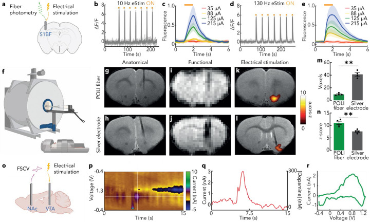
Recording and modulation of neuronal activity enables the study of brain function in health and disease. While translational neuroscience relies on electrical recording and modulation techniques, mechanistic studies in rodent models leverage genetic precision of optical methods, such as optogenetics and imaging of fluorescent indicators. In addition to electrical signal transduction, neurons produce and receive diverse chemical signals which motivate tools to probe and modulate neurochemistry. Although the past decade has delivered a wealth of technologies for electrophysiology, optogenetics, chemical sensing, and optical recording, combining these modalities within a single platform remains challenging. This work leverages materials selection and convergence fiber drawing to permit neural recording, electrical stimulation, optogenetics, fiber photometry, drug and gene delivery, and voltammetric recording of neurotransmitters within individual fibers. Composed of polymers and non-magnetic carbon-based conductors, these fibers are compatible with magnetic resonance imaging, enabling concurrent stimulation and whole-brain monitoring. Their utility is demonstrated in studies of the mesolimbic reward pathway by simultaneously interfacing with the ventral tegmental area and nucleus accumbens in mice and characterizing the neurophysiological effects of a stimulant drug. This study highlights the potential of these fibers to probe electrical, optical, and chemical signaling across multiple brain regions in both mechanistic and translational studies.

Keywords: FSCV; fiber-based interface; fiber-photometry; multifunctional neural probe; neuromodulation.


Conflict of interest statement

Conflict of Interest N.D., M.-J. A. and P.A. are co-founders and have a financial interest in NeuroBionics Inc.

Figures

Figure 1.
Figure 1.. Fabrication of the POLI fiber.
(a) schematic of the cross-section of the POLI fiber, highlighting its recording and stimulating capabilities. (b) Diagram of the convergence drawing process used to fabricate the POLI fiber. (c) Photograph of the macroscale preform highlighting the polymer combination choice and the channels used for electrodes, and microfluidics (scale bar = 5 mm). (d) Optical micrograph of the fiber cross-section highlighting the cross-sectional geometry and embedded CNT microwires (scale bar = 100 μm). (e) Photograph highlighting the flexibility and microscopic size of the POLI fiber microfluidics. (f) A fully assembled POLI fiber with electrical pins, optical ferrule and microfluidic inlet (scale bar = 1 cm). (g) Photograph of a dual-implant assembly that leverages PCB and Omnetics connector to facilitate connectorization. This implant simplifies the implantation process of fiber devices targeting the VTA and NAc (scale bar = 5 mm). (h) Photograph of a freely moving mouse with the dual-fiber implant (scale bar = 1 cm).
Figure 2.
Figure 2.. Benchtop characterization of the POLI fiber.
(a) Picture of a PMMA/THVP waveguide implanted in a fluorescein-doped agarose brain transmitting blue (470 nm) light to elicit a fluorescent response (~520 nm). Blue light is visible along the patch cord due to high input optical power, which permitted visual observation of the fluorescent response; the power of optical leakage along the fiber due to impurities or microscale defects is orders of magnitude below the power of light transmitted to the fiber tip, and as observed, did not elicit a fluorescent response along the length of the fiber (scale bar = 1 cm). (b) Evaluation of optical loss for 400 μm PC/PMMA, COC/PMMA, and PMMA/THVP polymer fibers, as well as the 200 μm PMMA/THVP optical waveguide of the POLI Fiber. Optical loss described as mean decibel loss ± s.e.m. (n = 3 samples). (c) Numerical aperture measurement of 400 μm PC/PMMA, COC/PMMA, and PMMA/THVP polymer fibers, expressed as mean ± s.e.m. (n = 3 samples, scale bar = 1 mm). (d) Bode plot of impedance magnitude and phase of the POLI fiber-embedded 20 μm-diameter CNT microelectrodes (n = 3 fibers, 18 electrodes total). (e) Cyclic voltammograms of the CNT electrodes and same-size comparison stainless-steel electrodes in phosphate-buffered saline (PBS) and their respective cathodic charge storage capacity values. (f) Voltage transient response of CNT electrodes to symmetric, biphasic, charge-balanced, current pulses of 250 μsec half phase and a 250 μsec interphase delay, and corresponding cathodic charge injection capacity. (g) Fast scan cyclic voltammetry response of a CNT electrode to a bolus of 20 μM DA solution delivered at t = 7 s recorded in a flow cell. (h) A representative voltammogram of CNT electrode response to 20 μM DA solution, showing DA oxidation peak at +0.4 V and reduction peak at 0 V. (i) DA calibration curve demonstrating a linear relationship between detected current and dopamine concentration in solution (R^2 = 0.994, n = 3). (j) Current measured at +0.4 V vs. time for n = 5 trials of 20 μM DA solution bolus delivered in the flow cell, converted to DA concentration using the calibration curve shown in (i). (k) Measured rate of fluid injection via POLI fiber microfluidic channel vs. rate set on the infusion pump. Infusions were delivered at set rates between 25, 50, and 75 nl/min and are represented as mean ± s.e.m. (n = 3). (l) Stiffness measured via dynamic mechanical analysis of POLI fibers (n = 3) compared to a similarly sized silica fiber across the frequency ranges of locomotion, respiration, and heart rate.
Figure 3.
Figure 3.. In vivo validation of POLI fiber functions.
(a) Schematic of the experiment. The POLI fiber was implanted in the whisker sensory cortices (S1BF) of Thy1-GCaMP6s mice, and used to concurrently stimulate electrically through the embedded CNT electrodes and record stimulation evoked calcium influxes using fiber photometry. (b-e) Calcium transient was measured following an electrical stimulation train of 10 Hz (b,c) and 130 Hz (d,e), with current varying between 35–215 μA. Fluorescence represented as a mean fluorescence (ΔF/F0) ± s.e.m, with (c) and (e) fluorescence normalized to the max fluorescence value. (f) High field MRI acquisition was performed with a 20 cm-bore 9.4 T Bruker small animal scanner with a custom-made 30 mm single surface coil used as a transceiver. (g-l) Anatomical scans were acquired using a T2-weighted rapid acquisition with refocused echoes (RARE) pulse sequence. Functional scans were performed using T2 *-weighted EPI sequence for detection of stimulus-induced BOLD contrast. Deep brain stimulation of 0.1mA for 2s at a frequency of 60Hz was applied preceded by 10s baseline scan and followed by 48s recovery scan, repeated over 30 cycles. (n = 8 female Sprague-Dawley rats) (m,n) Preprocessing of the functional scans was performed and scans were aligned to high-resolution anatomical images; maximum signal amplitude during an interval of 6s after stimulation onset was measured and compared to the average preceding baseline interval via Student’s t-test to evaluate statistical significance of z-scores. To quantify voxel count, a threshold of SNR< 5 was applied on slices in which the implant was observed. ** corresponds to P<0.01. (o) FSCV performed through POLI fiber was used to detect DA release in NAc in response to electrical stimulation in VTA supplied via an electrode, with a representative color plot (n = 1 Sprague-Dawley rat) (p) and voltage transient (q) shown. DA concentration was calculated from maximum at its oxidation peak in a representative voltammogram (n = 6 stimulations) (r).
Figure 4.
Figure 4.. Probing mesolimbic DA dynamics and drug-induced perturbations with POLI fiber.
(a) Mice were implanted with the dual implant device, comprised of two POLI fibers targeting the NAc and the VTA, and transfected with DA fluorescent indicator dLight1.1 into the NAc via AAV delivery through the POLI fiber fluidic channel. (b) Endogenous electrophysiology activity recorded in VTA, showing multiunit neural activity with phasic and tonic firing. Blue dots indicate spikes detected for sorting. (c) Fiber photometry recording of endogenous DA dynamics in NAc concomitant with recordings in (b). Shaded areas in (b) and (c) indicate phasic firing interspersed with tonic firing in unshaded areas (see Supporting Figure S5). Large DA transients observed in NAc photometry recordings are time-locked to bursts of tonic firing of the neurons in VTA. (d) Principal components analysis and clustering of neural spike waveforms recorded in VTA, and (e) mean spike waveforms for the two neuronal units. (f) Mice were implanted with the same dual implant POLI fiber device and transfected with a red excitatory opsin ChrimsonR in the VTA via AAV9 delivery through the POLI fiber channel to enable simultaneous optogenetic stimulation and electrophysiology recording. (g,h) Optogenetic stimulation-evoked activity of VTA neurons with (g) 5 Hz stimulation and (h) 20 Hz stimulation. (i,j) Fluorescent images show expression of (i) dLight1.1 in the NAc and (j) ChrimsonR in the VTA (HC PLAPO CS2 10x/0.40 Dry objective, WLL: 85% Power, Speed: 400Hz, 405: Intensity (2.00), Gain (12.54); 499: Intensity (2.00), Gain (22.30); 554: Intensity (2.00), Gain (10.86), scale bar = 1mm). In the same cohort of animals used in (a-e), electrical stimulation was applied either to the VTA or to the NAc while recording DA dynamics in NAc via fiber photometry. (l) Electrical stimulation of DA axon terminals in the NAc produced robust DA transients recorded via photometry. Stimulation epochs were recorded before and after intraperitoneal (IP) administration of cocaine (20 mg/kg, n = 5 epochs for each animal). Data are presented as mean ± s.e.m. (m) Electrical stimulation of cell bodies in the VTA produced robust DA transients in the NAc, though lower in amplitude compared to NAc stimulation. Stimulation epochs were recorded before and after cocaine administration (20 mg/kg, IP, n = 5 epochs for each animal). Data are presented as mean ± s.e.m. (n,o) Following administration of cocaine, stimulation-evoked dLight1.1 transients exhibited a significantly slower decay compared to the pre-cocaine transients due to inhibition of DA reuptake.

Similar articles

References

    1. Liu A., Vöröslakos M., Kronberg G., Henin S., Krause M. R., Huang Y., Opitz A., Mehta A., Pack C. C., Krekelberg B., Berényi A., Parra L. C., Melloni L., Devinsky O., Buzsáki G., Nat Commun 2018, 9, 5092. - PMC - PubMed
    1. Deisseroth K., Nat Methods 2011, 8, 26. - PMC - PubMed
    1. Deisseroth K., Feng G., Majewska A. K., Miesenböck G., Ting A., Schnitzer M. J., J. Neurosci. 2006, 26, 10380. - PMC - PubMed
    1. Wiegert J. S., Mahn M., Prigge M., Printz Y., Yizhar O., Neuron 2017, 95, 504. - PMC - PubMed
    1. Govorunova E. G., Sineshchekov O. A., Li H., Spudich J. L., Annual Review of Biochemistry 2017, 86, 845. - PMC - PubMed

Publication types