[Preprint]. 2024 Jun 3:2024.05.31.596892.
doi: 10.1101/2024.05.31.596892.

A predominately pulmonary activation of complement in a mouse model of severe COVID-19

Affiliations

A predominately pulmonary activation of complement in a mouse model of severe COVID-19

Peter J Szachowicz et al. bioRxiv. .

Abstract

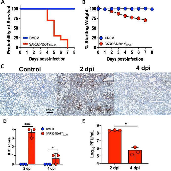
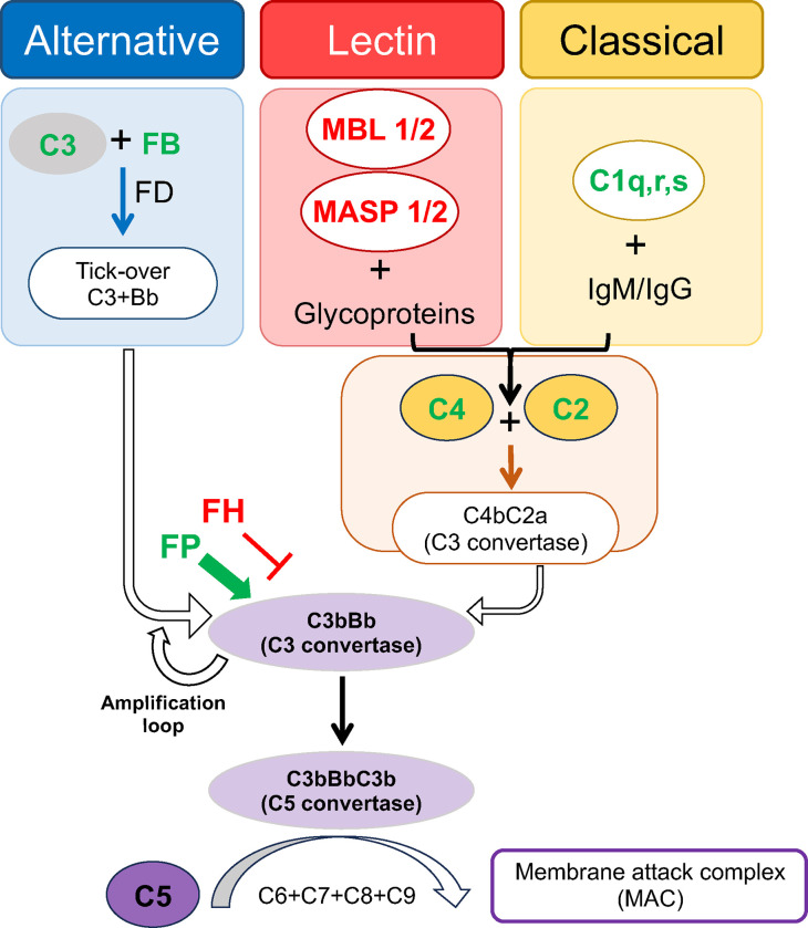
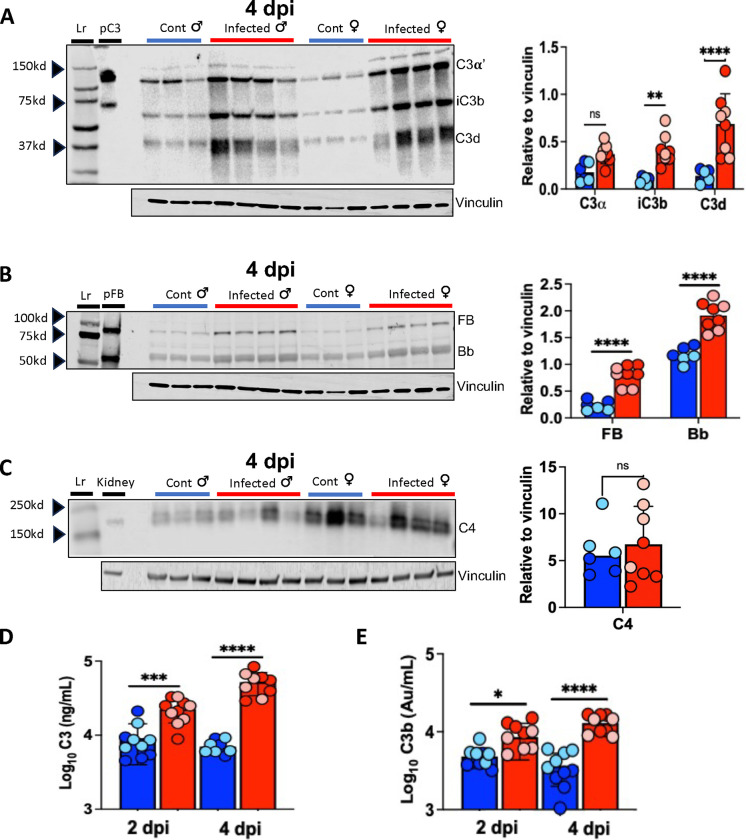
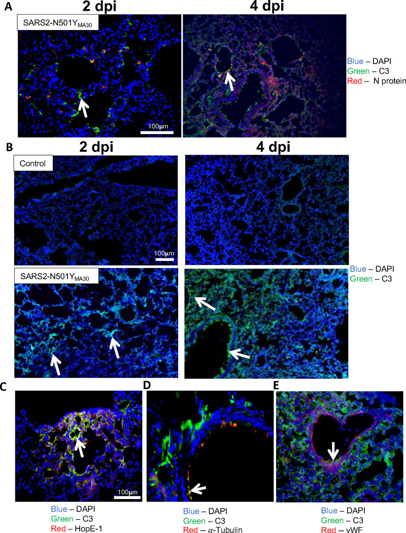
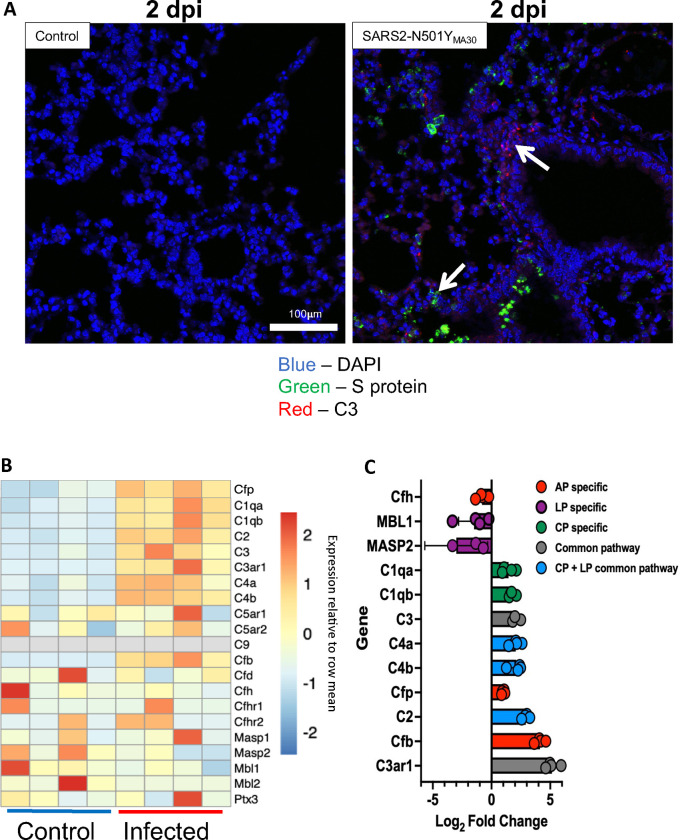
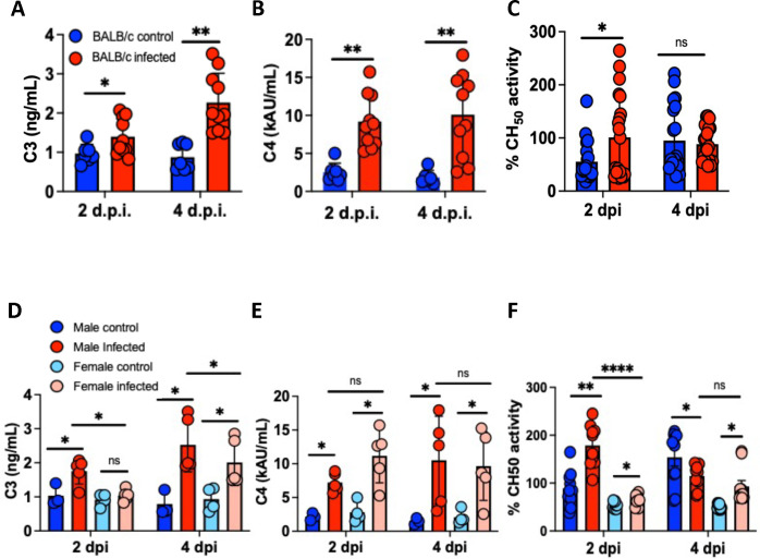
Evidence from in vitro studies and observational human disease data suggest the complement system plays a significant role in SARS-CoV-2 pathogenesis, although how complement dysregulation develops in patients with severe COVID-19 is unknown. Here, using a mouse-adapted SARS-CoV-2 virus (SARS2-N501YMA30) and a mouse model of severe COVID-19, we identify significant serologic and pulmonary complement activation following infection. We observed C3 activation in airway and alveolar epithelia, and in pulmonary vascular endothelia. Our evidence suggests that while the alternative pathway is the primary route of complement activation, components of both the alternative and classical pathways are produced locally by respiratory epithelial cells following infection, and increased in primary cultures of human airway epithelia in response to cytokine exposure. This locally generated complement response appears to precede and subsequently drive lung injury and inflammation. Results from this mouse model recapitulate findings in humans, which suggest sex-specific variance in complement activation, with predilection for increased C3 activity in males, a finding that may correlate with more severe disease. Our findings indicate that complement activation is a defining feature of severe COVID-19 in mice and lay the foundation for further investigation into the role of complement in COVID-19.

Keywords: COVID-19; SARS-CoV-2; complement; inflammation; pulmonary.


Conflict of interest statement

The authors have declared that no conflict of interest exists.

Figures

Figure 1.
Figure 1.
BALB/c mice infected with SARS2-N501YMA30 develop severe disease with peak respiratory viral burden 2 dpi. (A) Probability of survival following a 5,000 PFU inoculum of SARS2-N501YMA30 (red) compared to control group (blue) with significance achieved by 5 dpi, p<0.05 (n=10 mice per group). (B) Mean +/− SEM weight loss post-infection assessed by percent decrease from initial weight at each time point with significant difference achieved by 3 dpi, p<0.05 (n=10 mice per group). (C) Immunohistochemical (IHC) staining for SARS2-N501YMA30 nucleocapsid protein (N-protein) (brown = N protein, n=3 mice per group). (D) IHC scoring for severity by percent area of lung stained positive for viral antigen and (E) viral titer obtained from plaque assay on lung homogenates (n=3 mice per group). Red = SARS2-N501YMA30 and blue = DMEM (significance determined by log-rank test and unpaired t-test, where * indicates p<0.05 and *** indicates p<0.0001). DMEM = Dulbecco’s Modified Eagle’s Medium.
Figure 2.
Figure 2.
Schematic of complement cascade and components upregulated by SARS-CoV-2 infection. Green text indicates proteins with increased mRNA transcripts in mice following SARS2-N501YMA30 infection, red text indicates proteins with reduced mRNA transcript abundance in mice following SARS2-N501YMA30 infection. As depicted, the alternative pathway is continuously and spontaneously activated at low levels (tick-over) to generate C3bBb (C3 convertase), though rapid self-enhancement occurs by feedback amplification loop once activation is initiated. The lectin and classical pathways have different triggering substrates (glycoproteins and immunoglobulins, respectively), but both converge on the generation of C4 and C2, which join to create the C3 convertase C4bC2a. Although the lectin and classical pathways are initiated via separate mechanisms, a majority of their activity ultimately converges on C3bBb (C3 convertase), which cleaves C3 into C3a (anaphylatoxin) and C3b (opsonin). All three pathways coalesce with C5 convertase cleavage of C5, and the generation of C5a (anaphylatoxin) and C5b (joins with C6-C9 to form C5b-9, membrane attack complex). In addition, there are various cofactor proteins that help regulate cascade activity, such as properdin (FP) and complement factor H (FH), which can augment or inhibit further activation, respectively. FB = Factor B. MBL = mannose binding lectin. MASP = MBL-associated serine protease. FD = Factor D.
Figure 3.
Figure 3.
Increased C3 activation in lungs of mice infected with SARS2-N501YMA30. Western blot of lung homogenates with corresponding densitometry (right) demonstrates increases in iC3b and C3d (A) and FB and Bb (B), but not C4 (C) on 4 dpi. Purified human C3 (pC3), purified Factor B and Bb (pFB), and uninfected mouse kidney homogenates were used as controls for C3, FB, and C4, respectively. (D and E) Lung homogenate ELISA for C3 and C3b demonstrates increases with SARS2-N501YMA30 infection compared to control lung at 2 and 4 dpi. Blue bars indicate samples from control mice, red bars indicate samples from infected mice, light blue and light red circles indicate females, dark blue and dark red circles indicate males. n = 10 mice per group. Significance as determined by one-way ANOVA and unpaired t-test, * p<0.05,** p<0.001, *** p<0.0001, **** p<0.00001. Lr = ladder.
Figure 4.
Figure 4.
Complement C3 is colocalized with cells in various pulmonary sub compartments. (A) Colocalization of C3 (green) and SARS2-N501YMA30 nucleocapsid protein (red) on 2 and 4 dpi. n= 5, blue = DAPI, red = nucleocapsid protein, green = C3, white arrows indicate areas with C3 and nucleocapsid protein colocalization. (B) Staining for complement C3 on lung tissue from control (top row) and following infection with SARS2-N501YMA30 (bottom row) on 2 and 4 dpi. White arrows indicate areas of C3 staining. n = 5, green = C3, blue = DAPI. (C) Colocalization of C3 with alveolar type I cells, (D) ciliated airway cells, and (E) endothelial cells on 4 dpi. White arrows indicate regions of colocalization with C3. dpi = days post-infection, vWF = von Willebrand Factor.
Figure 5.
Figure 5.
Messenger RNA expression analysis suggests local production of complement proteins in lung tissue following SARS2-N501YMA30 infection. (A) RNAscope for S protein RNA and C3 RNA from lung section on 2 dpi following 5,000 PFU of SARS2-N501YMA30. White arrows indicate C3 RNA and SARS2-N501YMA30 S protein RNA, red = C3, green = S protein, and blue = DAPI. (B) Heat map depicting the results of bulk RNA sequencing for genes associated with complement pathways on lung tissue collected 5 dpi follow inoculation with 1,000 PFU SARS2-N501YMA30. (C) Mean log2 fold change in transcript abundance post-infection. Complement pathway indicated by bar color: red = alternative pathway specific genes, green = classical pathway specific genes, purple = lectin pathway specific genes, blue = shared lectin and classical pathway genes, grey = common pathway genes. n = 4. dpi = days post-infection.
Figure 6.
Figure 6.
Characterization of systemic complement activity in mice infected with SARS2-N501YMA30. (A and B) Serum C3 and C4 ELISA from control (blue) and infected (red) mice on 2 and 4 dpi. (C) Serum CH50 activity assay from control (blue) and SARS2-N501YMA30 mice (red). Comparison of sex difference by serum ELISA for C3 (D) and C4 (E) on 2 and 4 dpi, with data from sex specific CH50 activity assay shown in (F). dpi = days post-infection. Significance determined by one-way ANOVA or unpaired t-test, * p<0.05, ** p<0.001, **** p<0.00001. ns = nonsignificant. C3 and C4 measured from n = 7. DMEM and n = 10 SARS2-N501YMA30 mice per group. CH50 assay from n = 10 mice per group, samples run in duplicate.
Figure 7.
Figure 7.
Lung histopathology and inflammatory response is consistent with complement mediated disease. (A) Representative images of H&E staining of lung slides from histopathologic exam, with control and infected mice on 2 and 4 dpi which demonstrates cellular infiltrates (2 dpi) (black arrows) and edema (4 dpi) (black asterisks). n = 3 mice per time point. (B) Serum and lung cytokine profiling for 1–5 dpi, title above graph in each column indicates whether sample is from serum or lung homogenate. n = 3 mice per group at each time point. DMEM (Dulbecco’s Modified Eagle’s Medium), dpi = days post-infection. Significance determined by unpaired t-test, * p < 0.05.

Similar articles

References

    1. Yuki K, Fujiogi M, and Koutsogiannaki S. COVID-19 pathophysiology: A review. Clin Immunol. 2020;215:108427. - PMC - PubMed
    1. Baden LR, El Sahly HM, Essink B, Kotloff K, Frey S, Novak R, et al. Efficacy and Safety of the mRNA-1273 SARS-CoV-2 Vaccine. N Engl J Med. 2021;384(5):403–16. - PMC - PubMed
    1. Thomas SJ, Moreira ED Jr., Kitchin N, Absalon J, Gurtman A, Lockhart S, et al. Safety and Efficacy of the BNT162b2 mRNA Covid-19 Vaccine through 6 Months. N Engl J Med. 2021;385(19):1761–73. - PMC - PubMed
    1. Hammond J, Leister-Tebbe H, Gardner A, Abreu P, Bao W, Wisemandle W, et al. Oral Nirmatrelvir for High-Risk, Nonhospitalized Adults with Covid-19. N Engl J Med. 2022;386(15):1397–408. - PMC - PubMed
    1. Group RC, Horby P, Lim WS, Emberson JR, Mafham M, Bell JL, et al. Dexamethasone in Hospitalized Patients with Covid-19. N Engl J Med. 2021;384(8):693–704. - PMC - PubMed

Publication types