[Preprint]. 2024 Jun 9:2024.06.08.598080.
doi: 10.1101/2024.06.08.598080.

ProEnd: A Comprehensive Database for Identifying HbYX Motif-Containing Proteins Across the Tree of Life

Affiliations

ProEnd: A Comprehensive Database for Identifying HbYX Motif-Containing Proteins Across the Tree of Life

David Salcedo-Tacuma et al. bioRxiv. .

Update in

Abstract

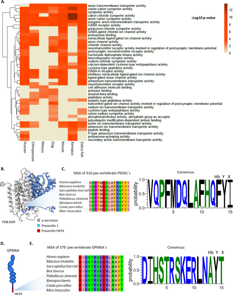
The proteasome plays a crucial role in cellular homeostasis by degrading misfolded, damaged, or unnecessary proteins. Understanding the regulatory mechanisms of proteasome activity is vital, particularly the interaction with activators containing the hydrophobic-tyrosine-any amino acid (HbYX) motif. Here, we present ProEnd, a comprehensive database designed to identify and catalog HbYX motif-containing proteins across the tree of life. Using a simple bioinformatics pipeline, we analyzed approximately 73 million proteins from 22,000 reference proteomes in the UniProt/SwissProt database. Our findings reveal the widespread presence of HbYX motifs in diverse organisms, highlighting their evolutionary conservation and functional significance. Notably, we observed an interesting prevalence of these motifs in viral proteomes, suggesting strategic interactions with the host proteasome. As validation two novel HbYX proteins found in this database were tested and found to directly interact with the proteasome. ProEnd's extensive dataset and user-friendly interface enable researchers to explore the potential proteasomal regulator landscape, generating new hypotheses to advance proteasome biology. This resource is set to facilitate the discovery of novel therapeutic targets, enhancing our approach to treating diseases such as neurodegenerative disorders and cancer. Link: http://proend.org/.


Figures

Figure 1.
Figure 1.. Prevalence of HbYX motif across organisms identified by ProEnd
A. Workflow for the design of the ProEnd database and the pipeline used to retrieve proteomes from UniProt/SwissProt. AWK was utilized to develop the regular expressions. B. Distribution of HbYX Proteins Across Life Domains and Viruses. Barplot indicates the number of HbYX proteins within various life domains, represented as percentages indicating the average occurrence of HbYX proteins per domain. On the left side, organisms possessing a 20S proteasome—specifically Eukaryotes, Actinobacteria, and Archaea—are grouped as highlighted by the line below. On the right, organisms lacking a 20S proteasome, including viruses and other bacteria, are displayed. C. Number of known proteasome activators found in the ProEnd database. Structural representation of a 20S proteasome with α-rings in blue and β-rings in gray. The green box illustrates the eukaryotic 19S regulatory particle complexed with the 20S proteasome (PDB: 6MSB), highlighting the conserved ATPases in orange. Additional regulatory proteins are outlined to enable comparison with the archaeal proteasome regulator. The orange box displays the structural complex of the archaeal 20S proteasome and PAN (PDB: 6HEC), with the PAN cap shown in salmon. In total 3,266 known activators form the 19S were identified in Eukarya and 60 PAN-like proteins were identified in Archaea. D. Percentage of HbYX proteins independently validated as proteasome interactors in other studies for Homo sapiens and Thermoplasma acidophilum, an archaeal model organism.
Figure 2.
Figure 2.. Conserved enrichment of HbYX proteins associated with neuronal pathways among jawed vertebrates indicates conserved functions for the HbYX motif
A. Heatmap representing molecular function enrichment among model jawed vertebrates. The heatmap is colored by the significance of enrichment (‘-log10 p-value’). Higher values indicate increased significance associated with the pathway. Rows indicate the enriched processes, and columns represent the model animals. Enrichment was determined using ClusterProfiler. Zero values indicate no enrichment found for this process among the HbYX proteins. B. Presenilin interaction with γ-secretase (PDB: 6IDF). This model represents the HbYX interaction of PSEN1 with γ-secretase, suggesting a potential binding role of the HbYX motif. γ-secretase is shown in gray, PSEN1 in blue, and the HbYX motif in red. C. Conservation of the HbYX motif in jawed vertebrates. A total of 316 sequences of PSEN1 were recovered from different vertebrates, and a multiple sequence alignment (MSA) was constructed using Jalview. On the right, the hidden Markov model (HMM) logo for the consensus sequence of the 316 PSEN1 sequences shows the high conservation of the HbYX motif (FYI). D. Representation of the neuronal membrane glycoprotein GPM6A and its HbYX motif. E. Conservation of the HbYX motif in jawed vertebrates A total of 379 GPM6A sequences from jawed vertebrates were retrieved, and an MSA was constructed using Jalview, highlighting the conserved features of this protein. The consensus HMM logo was then plotted, showing a highly conserved HbYX motif (AYT).
Figure 3.
Figure 3.. Neurotensin as a conserved HbYX-containing protein with the ability to bind the proteasome
A. AlphaFold prediction of the neurotensin precursor (AAF-P30990-F1) showing the Neurotensin peptide in blue and the HbYX motif in red. For the HbYX motif, a multiple sequence alignment (MSA) was constructed using neurotensin sequences from 130 vertebrates to show conservation of the HbYX motif. The consensus for the HbYX motif was constructed using an HMM logo, revealing a highly conserved YYY motif, with occasional occurrences of valine and alanine in the hydrophobic position. B. Validation pull-down assay performed with the Neurotensin HbYX peptide fused to biotin. Briefly, biotinylated Neurotensin was incubated with avidin beads. After washing off unbound HbYX, cell lysates from HEK293 cells were added for binding. Following incubation, unbound proteins were washed off, and detection of the proteasome was performed through western blotting for the alpha subunits. C. Western blot for the pull-down assay described in B, probing for alpha subunits with an expected molecular weight of 28 kDa. Three different replicates of HEK293 cell pull-downs were tested, showing the presence of alpha subunit proteasome bands. This indicates that the neurotensin HbYX peptide can pull down proteasomes, demonstrating that neurotensin is a proteasome interactor protein. The upper band in replicate 1 indicates the presence of the 26S proteasome.
Figure 4.
Figure 4.. Archaeal HbYX ATPase MJ1494 is a PAN-like protein that binds and activates the archaeal proteasome T20S
A. Rate of substrate degradation of fluorogenic LLVY after pulldown with T20S on MJ1494, pulldown with proteasome activator PA26, or T20S alone. Stimulation of degradation was measured by the increase in LLVY hydrolysis (rfu). B. AlphaFold multimer prediction LDDT indicating overall confidence in the predicted structure. Scores above 0.51 are generally accepted. Scores above 0.70 are considered to be of good quality, suggesting that most regions of the protein are predicted with high accuracy. C. Predicted Aligned Error (PAE) provides insights into the accuracy of the predicted inter-residue distances. The diagonal blocks with lower PAE values (indicated by blue regions) suggest that the intra-monomeric contacts are predicted with high confidence. This aligns with the high pLDDT score (0.78), indicating reliable local structure predictions within each monomer. The off-diagonal blocks show higher PAE values (indicated by red regions), reflecting moderate confidence in the inter-monomeric contacts, indicating some uncertainty in the predicted interactions between monomers. The repeating pattern of the blocks in the PAE plot is consistent with the hexameric nature of the protein, suggesting that each monomer’s predicted structure is similar and forms repetitive interactions within the hexamer. D. Bottom and side views of the structural representation predicted by AlphaFold multimer visualized with ChimeraX. The complex is shown in gray with the HbYX motif highlighted in orange for visualization. The HbYX motif appears to be buried inside the ATPase ring, a common feature shared with PAN when not engaged with the 20S proteasome. E. Screenshot of the main page of ProEnd. The database is available at http://proend.org/

Similar articles

References

    1. Glickman M. H. & Ciechanover A. The ubiquitin-proteasome proteolytic pathway: Destruction for the sake of construction. Physiol. Rev. 82, 373–428 (2002). - PubMed
    1. Hoyt M. A. & Coffino P. Ubiquitin-proteasome system. Cell. Mol. Life Sci. C. 2004 6113 61, 1596–1600 (2004). - PMC - PubMed
    1. Deshmukh F. K., Yaffe D., Olshina M. A., Ben-Nissan G. & Sharon M. The Contribution of the 20S Proteasome to Proteostasis. Biomol. 2019, Vol. 9, Page 190 9, 190 (2019). - PMC - PubMed
    1. Spataro V., Norbury C. & Harris A. L. The ubiquitin-proteasome pathway in cancer. Br. J. Cancer 1998 773 77, 448–455 (1998). - PMC - PubMed
    1. Bennett E. J., Bence N. F., Jayakumar R. & Kopito R. R. Global impairment of the ubiquitin-proteasome system by nuclear or cytoplasmic protein aggregates precedes inclusion body formation. Mol. Cell 17, 351–365 (2005). - PubMed

Publication types