

Intravitreal conbercept injection with panretinal photocoagulation for high-risk proliferative diabetic retinopathy with vitreous hemorrhage
- PMID: 38895681
- PMCID: PMC11144759
- DOI: 10.18240/ijo.2024.06.11
Intravitreal conbercept injection with panretinal photocoagulation for high-risk proliferative diabetic retinopathy with vitreous hemorrhage
Abstract
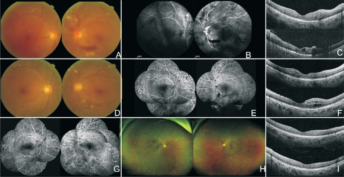
Aim: To assess the clinical efficacy and safety of combining panretinal photocoagulation (PRP) with intravitreal conbercept (IVC) injections for patients with high-risk proliferative diabetic retinopathy (HR-PDR) complicated by mild or moderate vitreous hemorrhage (VH), with or without diabetic macular edema (DME).
Methods: Patients diagnosed with VH with/without DME secondary to HR-PDR and received PRP combined with IVC injections were recruited in this retrospective study. Upon establishing the patient's diagnosis, an initial IVC was performed, followed by prompt administration of PRP. In cases who significant bleeding persisted and impeded the laser operation, IVC was sustained before supplementing with PRP. Following the completion of PRP, patients were meticulously monitored for a minimum of six months. Laser therapy and IVC injections were judiciously adjusted based on fundus fluorescein angiography (FFA) results. Therapeutic effect and the incidence of adverse events were observed.
Results: Out of 42 patients (74 eyes), 29 were male and 13 were female, with a mean age of 59.17±12.74y (33-84y). The diabetic history was between 1wk and 26y, and the interval between the onset of visual symptoms and diagnosis of HR-PDR was 1wk-1y. The affected eye received 2.59±1.87 (1-10) IVC injections and underwent 5.5±1.02 (4-8) sessions of PRP. Of these, 68 eyes received PRP following 1 IVC injection, 5 eyes after 2 IVC injections, and 1 eye after 3 IVC injections. Complete absorption of VH was observed in all 74 eyes 5-50wk after initial treatment, with resolution of DME in 51 eyes 3-48wk after initial treatment. A newly developed epiretinal membrane was noted in one eye. Visual acuity significantly improved in 25 eyes. No complications such as glaucoma, retinal detachment, or endophthalmitis were reported.
Conclusion: The study suggests that the combination of PRP with IVC injections is an effective and safe modality for treating diabetic VH in patients with HR-PDR.
Keywords: conbercept; high-risk proliferative diabetic retinopathy; panretinal photocoagulation; vitreous hemorrhage.
International Journal of Ophthalmology Press.
Conflict of interest statement
Conflicts of Interest: Xu Y, None; Ye Q, None; Shen W, None.
Figures
Similar articles
-
Panretinal photocoagulation after or prior to intravitreal conbercept injection for diabetic macular edema: a retrospective study.BMC Ophthalmol. 2021 Apr 1;21(1):160. doi: 10.1186/s12886-021-01920-8. BMC Ophthalmol. 2021. PMID: 33789617 Free PMC article.
-
Panretinal photocoagulation versus panretinal photocoagulation plus intravitreal bevacizumab for high-risk proliferative diabetic retinopathy.Int J Ophthalmol. 2016 Dec 18;9(12):1772-1778. doi: 10.18240/ijo.2016.12.12. eCollection 2016. Int J Ophthalmol. 2016. PMID: 28003978 Free PMC article.
-
Intravitreal bevacizumab (Avastin) and panretinal photocoagulation in the treatment of high-risk proliferative diabetic retinopathy.J Ocul Pharmacol Ther. 2013 Jul-Aug;29(6):550-5. doi: 10.1089/jop.2012.0202. Epub 2013 Mar 15. J Ocul Pharmacol Ther. 2013. PMID: 23495932 Free PMC article. Clinical Trial.
-
Regression of Neovascularization after Panretinal Photocoagulation Combined with Anti-VEGF Injection for Proliferative Diabetic Retinopathy-A Review.Diagnostics (Basel). 2023 Dec 22;14(1):31. doi: 10.3390/diagnostics14010031. Diagnostics (Basel). 2023. PMID: 38201340 Free PMC article. Review.
-
Anti-Vascular Endothelial Growth Factor Injections: The New Standard of Care in Proliferative Diabetic Retinopathy?Dev Ophthalmol. 2017;60:131-142. doi: 10.1159/000459699. Epub 2017 Apr 20. Dev Ophthalmol. 2017. PMID: 28427072 Review.
Cited by
-
Renal dysfunction associated with clinical response to intravitreal conbercept therapy for diabetic macular edema.Int J Ophthalmol. 2025 Mar 18;18(3):454-461. doi: 10.18240/ijo.2025.03.12. eCollection 2025. Int J Ophthalmol. 2025. PMID: 40103962 Free PMC article.
References
-
- Cheung N, Mitchell P, Wong TY. Diabetic retinopathy. Lancet. 2010;376(9735):124–136. - PubMed
-
- Flaxman SR, Bourne RRA, Resnikoff S, Ackland P, Braithwaite T, Cicinelli MV, Das A, Jonas JB, Keeffe J, Kempen JH, Leasher J, Limburg H, Naidoo K, Pesudovs K, Silvester A, Stevens GA, Tahhan N, Wong TY, Taylor HR, Vision Loss Expert Group of the Global Burden of Disease Study Global causes of blindness and distance vision impairment 1990-2020: a systematic review and meta-analysis. Lancet Glob Health. 2017;5(12):e1221–e1234. - PubMed
-
- Pandit S, Ho AC, Yonekawa Y. Recent advances in the management of proliferative diabetic retinopathy. Curr Opin Ophthalmol. 2023;34(3):232–236. - PubMed
LinkOut - more resources
Full Text Sources
Research Materials