. 2023 Jun:105:205-266.
doi: 10.3114/sim.2023.105.03. Epub 2023 Jun 12.

Revising Clonostachys and allied genera in Bionectriaceae

Affiliations

Revising Clonostachys and allied genera in Bionectriaceae

L Zhao et al. Stud Mycol. 2023 Jun.

Abstract

Clonostachys (Bionectriaceae, Hypocreales) species are common soil-borne fungi, endophytes, epiphytes, and saprotrophs. Sexual morphs of Clonostachys spp. were placed in the genus Bionectria, which was further segregated into the six subgenera Astromata, Bionectria, Epiphloea, Myronectria, Uniparietina, and Zebrinella. However, with the end of dual nomenclature, Clonostachys became the single depository for sexual and asexual morph-typified species. Species of Clonostachys are typically characterised by penicillate, sporodochial, and, in many cases, dimorphic conidiophores (primary and secondary conidiophores). Primary conidiophores are mononematous, either verticillium-like or narrowly penicillate. The secondary conidiophores generally form imbricate conidial chains that can collapse to slimy masses, particularly on sporodochia. In the present study, we investigated the species diversity within a collection of 420 strains of Clonostachys from the culture collection of, and personal collections at, the Westerdijk Fungal Biodiversity Institute in Utrecht, the Netherlands. Strains were analysed based on their morphological characters and molecular phylogeny. The latter used DNA sequence data of the nuclear ribosomal internal transcribed spacer regions and intervening 5.8S nrDNA (ITS) and partial 28S large subunit (LSU) nrDNA and partial protein encoding genes including the RNA polymerase II second largest subunit (RPB2), translation elongation factor 1-alpha (TEF1) and β-tubulin (TUB2). Based on these results, the subgenera Astromata, Bionectria, Myronectria and Zebrinella are supported within Clonostachys. Furthermore, the genus Sesquicillium is resurrected to accommodate the former subgenera Epiphloea and Uniparietina. The close relationship of Clonostachys and Sesquicillium is strongly supported as both are inferred phylogenetically as sister-genera. New taxa include 24 new species and 10 new combinations. Recognition of Sesquicillium distinguishes species typically forming a reduced perithecial stroma superficially on plant tissue from species in Clonostachys often forming well-developed, through bark erumpent stromata. The patterns of observed perithecial wall anatomies, perithecial wall and stroma interfaces, and asexual morph diversifications described in a previously compiled monograph are used for interpreting ancestral state reconstructions. It is inferred that the common ancestor of Clonostachys and Sesquicillium may have formed perithecia superficially on leaves, possessed a perithecial wall consisting of a single region, and formed intercalary phialides in penicilli of conidiophores. Character interpretation may also allow hypothesising that diversification of morphs occurred then in the two genera independently and that the frequently stroma-linked Clonostachys morphs evolved together with the occupation of woody host niches and mycoparasitism. Taxonomic novelties: New species: Clonostachys aurantiaca L. Zhao & Crous, Clonostachys australiana L. Zhao & Crous, Clonostachys bambusae L. Zhao & Crous, Clonostachys buxicola L. Zhao & Crous, Clonostachys cylindrica L. Zhao & Crous, Clonostachys ellipsoidea L. Zhao & Crous, Clonostachys flava L. Zhao, Crous & Schroers, Clonostachys fujianensis L. Zhao & Crous, Clonostachys fusca L. Zhao, Crous & Schroers, Clonostachys garysamuelsii L. Zhao & Crous, Clonostachys hongkongensis L. Zhao & Crous, Clonostachys longiphialidica L. Zhao, Crous & Schroers, Clonostachys obovatispora, L. Zhao & Crous, Clonostachys palmae L. Zhao, Crous & Schroers, Clonostachys parasporodochialis L. Zhao & Crous, Clonostachys penicillata L. Zhao, Crous & Schroers, Clonostachys reniformis L. Zhao & Crous, Clonostachys vacuolata L. Zhao, Crous & Schroers, Clonostachys venezuelae L. Zhao, Crous & Schroers, Mycocitrus synnematus L. Zhao & Crous, Nectriopsis didymii L. Zhao & Crous, Sesquicillium intermediophialidicum L. Zhao & Crous, Sesquicillium neerlandicum L. Zhao & Crous, Sesquicillium symmetricum L. Zhao & Crous. New combinations: Mycocitrus coccicola (J.A. Stev.) L. Zhao & Crous, Mycocitrus coxeniae (Y.P. Tan et al.) L. Zhao & Crous, Sesquicillium essexcoheniae (Y.P. Tan et al.) L. Zhao & Crous, Sesquicillium lasiacidis (Samuels) L. Zhao, Crous & Schroers, Sesquicillium phyllophilum (Schroers) L. Zhao, Crous & Schroers, Sesquicillium rossmaniae (Schroers) L. Zhao, Crous & Schroers, Sesquicillium saulense (Lechat & J. Fourn.) L. Zhao & Crous, Sesquicillium sesquicillii (Samuels) L. Zhao, Crous & Schroers, Sesquicillium spinulosisporum (Lechat & J. Fourn.) L. Zhao & Crous, Sesquicillium tornatum (Höhn.) Schroers. New synonyms: Clonostachys aranearum W.H. Chen et al., Clonostachys chuyangsinensis H. Yu & Y. Wang, Clonostachys eriocamporesiana R.H. Perera & K.D. Hyde, Clonostachys granuligera (Starbäck) Forin & Vizzini, Clonostachys indica Prasher & R. Chauhan, Clonostachys spinulosa R.H. Perera et al., Clonostachys squamuligera (Sacc.) Forin & Vizzini, Clonostachys wenpingii (J. Luo & W.Y. Zhuang) Z.Q. Zeng & W.Y. Zhuang. Epitypes (basionyms): Fusidium buxi J.C. Schmidt ex Link, Verticillium candelabrum Bonord. Citation: Zhao L, Groenewald JZ, Hernández-Restrepo M, Schroers H-J, Crous PW (2023). Revising Clonostachys and allied genera in Bionectriaceae. Studies in Mycology 105: 205-266. doi: 10.3114/sim.2023.105.03.

Keywords: Biocontrol; Bionectriaceae; multi-locus; mycoparasitism; new taxa; phylogeny; soil-borne; taxonomy.


Conflict of interest statement

The authors declare that there is no conflict of interest.

Figures

Fig. 1.
Fig. 1.
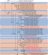
Phylogenetic tree inferred from a Maximum Likelihood (RAxML) analysis based on aligned and concatenated ITS, LSU, RPB2, TEF1 and TUB2 sequences of 269 strains representing Bionectriaceae and outgroups. Numbers at branches indicate support values (RAxML-BS / IQ-TREE-BS / BI-PP) above 50 % / 90 % / 0.9. New species are printed in red font, new combinations in blue font and coloured boxes highlight genera. Roman numerals indicate subgenera as coded in the legend. “T” indicates ex-type strains. The tree is rooted to Flammocladiella aceris CBS 138906, F. decora CBS 142776, F. anomiae CLL 16017, Tilachlidium brachiatum CBS 363.97, and T. brachiatum CBS 505.67 (Hypocreales, Flammocladiellaceae & Tilachlidiaceae). Scale bar represents expected number of changes per site.
Fig. 1.
Fig. 1.
Phylogenetic tree inferred from a Maximum Likelihood (RAxML) analysis based on aligned and concatenated ITS, LSU, RPB2, TEF1 and TUB2 sequences of 269 strains representing Bionectriaceae and outgroups. Numbers at branches indicate support values (RAxML-BS / IQ-TREE-BS / BI-PP) above 50 % / 90 % / 0.9. New species are printed in red font, new combinations in blue font and coloured boxes highlight genera. Roman numerals indicate subgenera as coded in the legend. “T” indicates ex-type strains. The tree is rooted to Flammocladiella aceris CBS 138906, F. decora CBS 142776, F. anomiae CLL 16017, Tilachlidium brachiatum CBS 363.97, and T. brachiatum CBS 505.67 (Hypocreales, Flammocladiellaceae & Tilachlidiaceae). Scale bar represents expected number of changes per site.
Fig. 2.
Fig. 2.
Phylogenetic tree inferred from a Maximum Likelihood (RAxML-ML) analysis based on aligned and concatenated ITS, LSU, RPB2, TEF1 and TUB2 sequences of 394 strains representing Clonostachys and outgroups. Numbers at branches indicate support values (RAxML-BS / IQ-TREE-BS / BI-PP) above 50 % / 90 % / 0.9. New species are printed in red font and coloured boxes highlight species clades / lineages. “T” indicates ex-type strains. A detailed view of the collapsed clade at the bottom of the phylogenetic tree can be found in Fig. S9. The tree is rooted to Acremonium alternatum CBS 407.66 and A. stroudii CBS 138820 (Hypocreales, Bionectriaceae). Scale bar represents expected number of changes per site.
Fig. 2.
Fig. 2.
Phylogenetic tree inferred from a Maximum Likelihood (RAxML-ML) analysis based on aligned and concatenated ITS, LSU, RPB2, TEF1 and TUB2 sequences of 394 strains representing Clonostachys and outgroups. Numbers at branches indicate support values (RAxML-BS / IQ-TREE-BS / BI-PP) above 50 % / 90 % / 0.9. New species are printed in red font and coloured boxes highlight species clades / lineages. “T” indicates ex-type strains. A detailed view of the collapsed clade at the bottom of the phylogenetic tree can be found in Fig. S9. The tree is rooted to Acremonium alternatum CBS 407.66 and A. stroudii CBS 138820 (Hypocreales, Bionectriaceae). Scale bar represents expected number of changes per site.
Fig. 3.
Fig. 3.
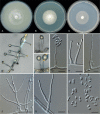
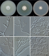
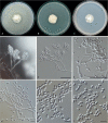
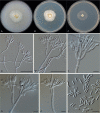
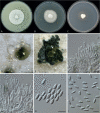
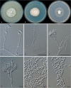
Nectriopsis didymii (ex-type CBS 852.70A). A–C. Colonies on OA, PDA and SNA after 7 d at 25 °C. D–F. Conidiophores. G, H. Conidia. Black arrows indicate intercalary phialides. Scale bars: D = 50 μm; E–H = 10 μm.
Fig. 4.
Fig. 4.
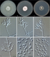
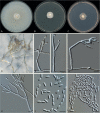
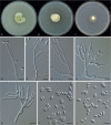
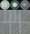
Nectriopsis microspora (ex-type CBS 933.69). A–C. Colonies on OA, PDA, SNA after 7 d at 25 °C. D, E. Conidiophores. F. Conidia. Black arrows indicate intercalary phialides. Scale bars = 10 μm.
Fig. 5.
Fig. 5.
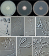
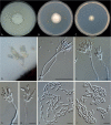
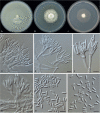
Mycocitrus odorus (CBS 120610). A–C. Colonies on OA, PDA and SNA after 7 d at 25 °C. D–K. Conidiophores. L. Conidia. Scale bars = 10 μm.
Fig. 6.
Fig. 6.
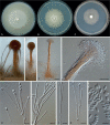
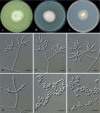
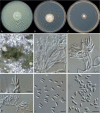
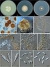
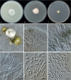
Mycocitrus synnematus (ex-type CBS 126677). A–C. Colonies on OA, PDA and SNA after 7 d at 25 °C. D–F. Synnemata. G. Detail of the apical portion of a synnema. H–K. Conidiogenous cells. L. Conidia. Scale bars: F = 100 μm; G = 50 μm; H–L = 10 μm.
Fig. 7.
Fig. 7.
Stephanonectria chromolaenae (CBS 475.91). A–C. Colonies on OA, PDA and SNA after 7 d at 25 °C. D–I. Conidiophores. J. Conidia. Scale bars: E, F = 50 μm; G–J = 10 μm.
Fig. 8.
Fig. 8.
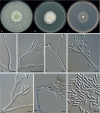
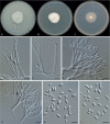
Sesquicillium candelabrum (ex-type CBS 119045). A–C. Colonies on OA, PDA and SNA after 7 d at 25 °C. D–H. Conidiophores. I. Conidia. Scale bars = 10 μm.
Fig. 9.
Fig. 9.
Sesquicillium essexcoheniae (CBS 918.97). A–C. Colonies on OA, PDA and SNA after 7 d at 25 °C. D–G. Conidiophores. H, I. Conidia. Scale bars = 10 μm.
Fig. 10.
Fig. 10.
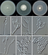
Sesquicillium intermediophialidicum (ex-type CBS 685.96). A–C. Colonies on OA, PDA and SNA after 7 d at 25 °C. D–H. Conidiophores. I, J. Conidia. Scale bars: E = 50 μm; F–J = 10 μm.
Fig. 11.
Fig. 11.
Sesquicillium lasiacidis (CBS 504.67). A–C. Colonies on OA, PDA and SNA after 7 d at 25 °C. D–G. Conidiophores. H, I. Conidia. Scale bars = 10 μm.
Fig. 12.
Fig. 12.
Sesquicillium neerlandicum (ex-type CBS 148203). A–C. Colonies on OA, PDA and SNA after 7 d at 25 °C. D–G. Conidiophores. H, I. Conidia. Scale bars: E, F = 50 μm; G–I = 10 μm.
Fig. 13.
Fig. 13.
Sesquicillium symmetricum (ex-type CBS 124.79). A–C. Colonies on OA, PDA and SNA after 7 d at 25 °C. D–H. Conidiophores. I, J. Conidia in linear chains. Scale bars = 10 μm.
Fig. 14.
Fig. 14.
Clonostachys aurantiaca (ex-type CBS 124757). A–C. Colonies on OA, PDA and SNA after 7 d at 25 °C. D, E. Perithecia. F. Section through perithecium. G. Ostiole. H. Lateral perithecial wall showing two regions. I. Perithecial base and stroma or stroma below a perithecium. J. Immature asci with slightly visible ring. K. Mature asci. L, M. Discharged ascospores in optical section (L) and surface view (M). Scale bars: F = 50 μm; G–M = 10 μm.
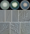
Fig. 15.
Fig. 15.
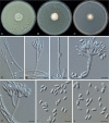
Clonostachys australiana (ex-type CBS 102421). A–C. Colonies on OA, PDA and SNA after 7 d at 25 °C. D–F. Primary conidiophores. G, H. Secondary conidiophores. I, J. Conidia. Black arrows indicate intercalary phialides. Scale bars = 10 μm.
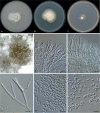
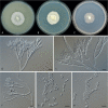
Fig. 16.
Fig. 16.
Clonostachys bambusae (ex-type CBS 139411). A–C. Colonies on OA, PDA and SNA after 7 d at 25 °C. D–H. Conidiophores. I. Conidia. Black arrows indicate intercalary phialides. Scale bars: D, E = 50 μm; F–I = 10 μm.
Fig. 17.
Fig. 17.
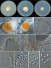
Clonostachys buxicola (ex-type CBS 102419). A–C. Colonies on OA, PDA and SNA after 7 d at 25 °C. D. Sporodochia producing green conidial masses on OA. E, F. Sporodochia. G. Phialides. H. Conidiophores. I, J. Conidia. Scale bars = 10 μm.
Fig. 18.
Fig. 18.
Clonostachys cylindrica (ex-type CBS 101113). A–C. Colonies on OA, PDA and SNA after 7 d at 25 °C. D–F. Primary conidiophores. G–I. Secondary conidiophores. J. Conidia. Scale bars: E = 50 μm; D, F–J = 10 μm.
Fig. 19.
Fig. 19.
Clonostachys ellipsoidea (ex-type CBS 175.76). A–C. Colonies on OA, PDA and SNA after 7 d at 25 °C. D–H. Conidiophores. I, J. Conidia. Scale bars = 10 μm.
Fig. 20.
Fig. 20.
Clonostachys eriocamporesii (CBS 647.91). A–C. Colonies on OA, PDA and SNA after 7 d at 25 °C. D. Sporodochia after 1 wk. E. Sporodochia after 2 wk. F, G. Sporodochia. H, I. Conidia. Scale bars = 10 μm.
Fig. 21.
Fig. 21.
Clonostachys flava (ex-type CBS 915.97). A–C. Colonies on OA, PDA and SNA after 7 d at 25 °C. D–F. Primary conidiophores. G, H. Secondary conidiophores. I, J. Conidia. Scale bars = 10 μm.
Fig. 22.
Fig. 22.
Clonostachys fusca (ex-type CBS 207.93). A–C. Colonies on OA, PDA and SNA after 7 d at 25 °C. D, E. Perithecia. F. Section through perithecium. G. Ostiole. H. Perithecial wart. I. Lateral perithecial wall showing two regions. J, K. Asci. L, M. Discharged ascospores in optical section (L) and surface view (M). Scale bars: F = 50 μm; G–M = 10 μm.
Fig. 23.
Fig. 23.
Clonostachys garysamuelsii (ex-type CBS 123964). A–C. Colonies on OA, PDA and SNA after 7 d at 25 °C. D, E. Primary conidiophores. F, G. Secondary conidiophores. H, I. Conidia. Scale bars = 10 μm.
Fig. 24.
Fig. 24.
Clonostachys hongkongensis (ex-type CBS 115291). A–C. Colonies on OA, PDA and SNA after 7 d at 25 °C. D, E. Primary conidiophores. F, G. Secondary conidiophores. H, I. Conidia. Scale bars: E, F = 50 μm; D, G–I = 10 μm.
Fig. 25
Fig. 25
Clonostachys kunmingensis (CBS 101920). A–C. Colonies on OA, PDA and SNA after 7 d at 25 °C. D–F. Primary conidiophores. G–I. Secondary conidiophores. J, K. Conidia. Scale bars: G = 50 μm; D–F, H–K = 10 μm.
Fig. 26.
Fig. 26.
Clonostachys longiphialidica (ex-type CBS 112.87). A–C. Colonies on OA, PDA and SNA after 7 d at 25 °C. D–G. Conidiophores. H, I. Conidia. Scale bars = 10 μm.
Fig. 27.
Fig. 27.
Clonostachys obovatispora (ex-type CBS 118752). A–C. Colonies on OA, PDA and SNA after 7 d at 25 °C. D, E. Sporodochia. F, G. Phialides. H, I. Conidia. Scale bars = 10 μm.
Fig. 28
Fig. 28
Clonostachys palmae (ex-type CBS 119.87). A–C. Colonies on OA, PDA and SNA after 7 d at 25 °C. D–F. Primary conidiophores. G, H. Secondary conidiophores. I, J. Conidia. Scale bars = 10 μm.
Fig. 29.
Fig. 29.
Clonostachys parasporodochialis (ex-type CBS 192.93). A–C. Colonies on OA, PDA and SNA after 7 d at 25 °C. D–F. Sporodochia. G. Phialides. H, I. Conidia. Scale bars = 10 μm.
Fig. 30.
Fig. 30.
Clonostachys penicillata (ex-type CBS 729.87). A–C. Colonies on OA, PDA and SNA after 7 d at 25 °C. D–F. Conidiophores. G, H. Conidia. Black arrows indicate intercalary phialides. Scale bars = 10 μm.
Fig. 31.
Fig. 31.
Clonostachys reniformis (ex-type CBS 695.86). A–C. Colonies on OA, PDA and SNA after 7 d at 25 °C. D, E. Primary conidiophores. F, G. Secondary conidiophores. H, I. Conidia. Scale bars: D, F = 50 μm; E, G–I = 10 μm.
Fig. 32.
Fig. 32.
Clonostachys vacuolata (ex-type CBS 191.93). A–C. Colonies on OA, PDA and SNA after 7 d at 25 °C. D–G. Conidiophores. H, I. Conidia. Scale bars = 10 μm.

Similar articles

  • Bionectriaceae: a poorly known family of hypocrealean fungi with major commercial potential.
    Zhao L, Groenewald JZ, Hou LW, Summerbell RC, Crous PW. Zhao L, et al. Stud Mycol. 2025 Jun;111:115-198. doi: 10.3114/sim.2025.111.04. Epub 2025 Apr 17. Stud Mycol. 2025. PMID: 40371418 Free PMC article.
  • Redisposition of acremonium-like fungi in Hypocreales.
    Hou LW, Giraldo A, Groenewald JZ, Rämä T, Summerbell RC, Huang GZ, Cai L, Crous PW. Hou LW, et al. Stud Mycol. 2023 Jun;105:23-203. doi: 10.3114/sim.2023.105.02. Epub 2023 Jun 2. Stud Mycol. 2023. PMID: 38895703 Free PMC article.
  • Generic concepts in Nectriaceae.
    Lombard L, van der Merwe NA, Groenewald JZ, Crous PW. Lombard L, et al. Stud Mycol. 2015 Mar;80:189-245. doi: 10.1016/j.simyco.2014.12.002. Epub 2015 Jan 29. Stud Mycol. 2015. PMID: 26955195 Free PMC article.
  • Fusarium: more than a node or a foot-shaped basal cell.
    Crous PW, Lombard L, Sandoval-Denis M, Seifert KA, Schroers HJ, Chaverri P, Gené J, Guarro J, Hirooka Y, Bensch K, Kema GHJ, Lamprecht SC, Cai L, Rossman AY, Stadler M, Summerbell RC, Taylor JW, Ploch S, Visagie CM, Yilmaz N, Frisvad JC, Abdel-Azeem AM, Abdollahzadeh J, Abdolrasouli A, Akulov A, Alberts JF, Araújo JPM, Ariyawansa HA, Bakhshi M, Bendiksby M, Ben Hadj Amor A, Bezerra JDP, Boekhout T, Câmara MPS, Carbia M, Cardinali G, Castañeda-Ruiz RF, Celis A, Chaturvedi V, Collemare J, Croll D, Damm U, Decock CA, de Vries RP, Ezekiel CN, Fan XL, Fernández NB, Gaya E, González CD, Gramaje D, Groenewald JZ, Grube M, Guevara-Suarez M, Gupta VK, Guarnaccia V, Haddaji A, Hagen F, Haelewaters D, Hansen K, Hashimoto A, Hernández-Restrepo M, Houbraken J, Hubka V, Hyde KD, Iturriaga T, Jeewon R, Johnston PR, Jurjević Ž, Karalti I, Korsten L, Kuramae EE, Kušan I, Labuda R, Lawrence DP, Lee HB, Lechat C, Li HY, Litovka YA, Maharachchikumbura SSN, Marin-Felix Y, Matio Kemkuignou B, Matočec N, McTaggart AR, Mlčoch P, Mugnai L, Nakashima C, Nilsson RH, Noumeur SR, Pavlov IN, Peralta MP, Phillips AJL, Pitt JI, Polizzi G, Quaedvlieg W, Rajeshkumar KC, Restrepo S, Rhaiem A, Robert J, Robert V, Ro… See abstract for full author list ➔ Crous PW, et al. Stud Mycol. 2021 Aug 17;98:100116. doi: 10.1016/j.simyco.2021.100116. eCollection 2021 Apr. Stud Mycol. 2021. PMID: 34466168 Free PMC article.
  • Taxonomy, phylogeny and identification of Chaetomiaceae with emphasis on thermophilic species.
    Wang XW, Han PJ, Bai FY, Luo A, Bensch K, Meijer M, B K, Han DY, Sun BD, Crous PW, Houbraken J. Wang XW, et al. Stud Mycol. 2022 Jul;101:121-243. doi: 10.3114/sim.2022.101.03. Epub 2022 Apr 1. Stud Mycol. 2022. PMID: 36059895 Free PMC article.

Cited by

References

    1. Bainier G. (1905). Mycothèque de l’École de Pharmacie. II. Acrostalagmus roseus Bainier. Bulletin Trimestriel de la Société Mycologique de France 21: 225–228.
    1. Bainier G. (1907). Gliocladium roseum sp. nov. et Cephalosporium acremonium (Corda). Bulletin Trimestriel de la Société Mycologique de France 23: 111–114.
    1. Barnett HL, Lilly VG. (1962). A destructive mycoparasite, Gliocladium roseum. Mycologia 54: 72–77.
    1. Berkeley MJ, Broome CE. (1873). Enumeration of the fungi of Ceylon. Part II. Journal of the Linnean Society. Botany 14: 29–140.
    1. Berkeley MJ, Broome CE. (1876). Notices of British fungi. Annals and Magazine of Natural History. Series 4. 17: 129–145.

LinkOut - more resources