

Can artificial intelligence-driven cephalometric analysis replace manual tracing? A systematic review and meta-analysis
- PMID: 38895901
- PMCID: PMC11185929
- DOI: 10.1093/ejo/cjae029
Can artificial intelligence-driven cephalometric analysis replace manual tracing? A systematic review and meta-analysis
Abstract
Objectives: This systematic review and meta-analysis aimed to investigate the accuracy and efficiency of artificial intelligence (AI)-driven automated landmark detection for cephalometric analysis on two-dimensional (2D) lateral cephalograms and three-dimensional (3D) cone-beam computed tomographic (CBCT) images.
Search methods: An electronic search was conducted in the following databases: PubMed, Web of Science, Embase, and grey literature with search timeline extending up to January 2024.
Selection criteria: Studies that employed AI for 2D or 3D cephalometric landmark detection were included.
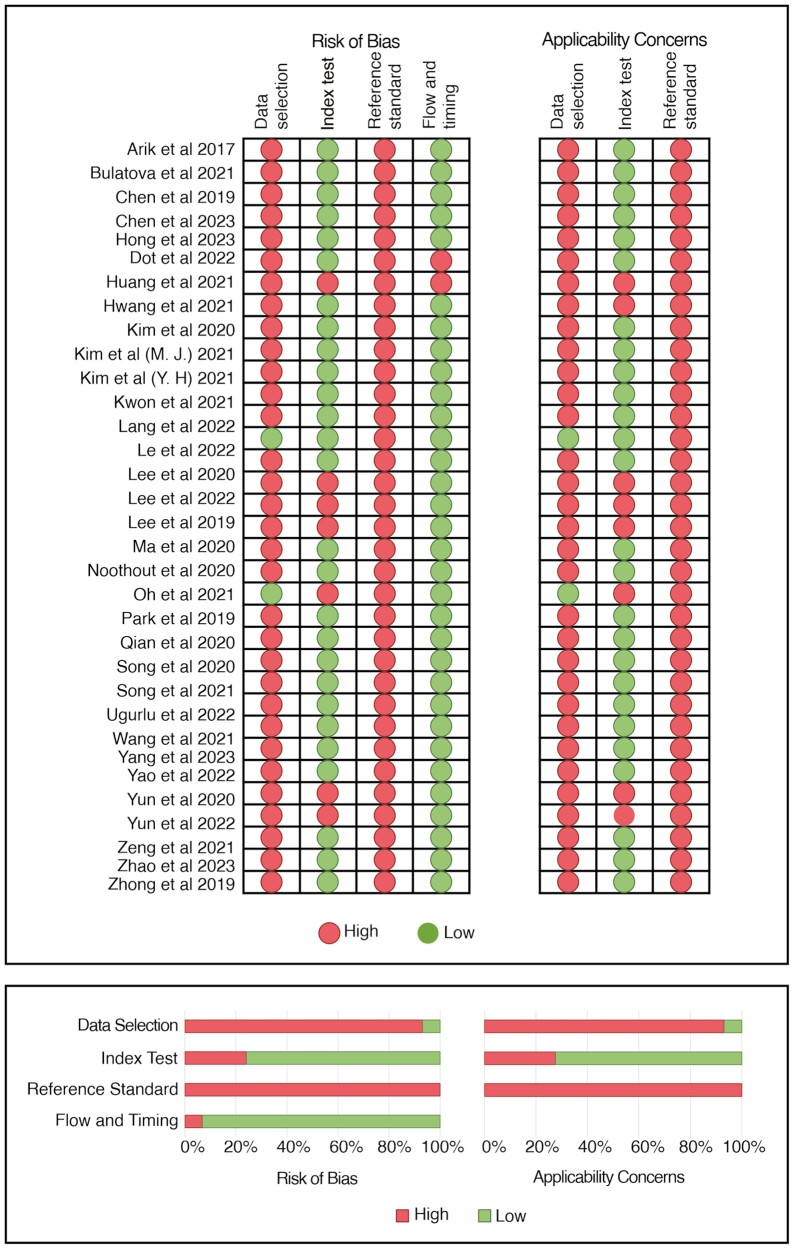
Data collection and analysis: The selection of studies, data extraction, and quality assessment of the included studies were performed independently by two reviewers. The risk of bias was assessed using the Quality Assessment of Diagnostic Accuracy Studies-2 tool. A meta-analysis was conducted to evaluate the accuracy of the 2D landmarks identification based on both mean radial error and standard error.
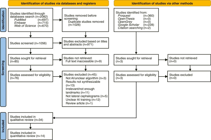
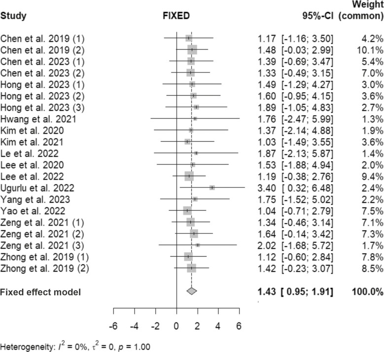
Results: Following the removal of duplicates, title and abstract screening, and full-text reading, 34 publications were selected. Amongst these, 27 studies evaluated the accuracy of AI-driven automated landmarking on 2D lateral cephalograms, while 7 studies involved 3D-CBCT images. A meta-analysis, based on the success detection rate of landmark placement on 2D images, revealed that the error was below the clinically acceptable threshold of 2 mm (1.39 mm; 95% confidence interval: 0.85-1.92 mm). For 3D images, meta-analysis could not be conducted due to significant heterogeneity amongst the study designs. However, qualitative synthesis indicated that the mean error of landmark detection on 3D images ranged from 1.0 to 5.8 mm. Both automated 2D and 3D landmarking proved to be time-efficient, taking less than 1 min. Most studies exhibited a high risk of bias in data selection (n = 27) and reference standard (n = 29).
Conclusion: The performance of AI-driven cephalometric landmark detection on both 2D cephalograms and 3D-CBCT images showed potential in terms of accuracy and time efficiency. However, the generalizability and robustness of these AI systems could benefit from further improvement.
Registration: PROSPERO: CRD42022328800.
Keywords: anatomic landmarks; artificial intelligence; cephalometry; orthodontics.
© The Author(s) 2024. Published by Oxford University Press on behalf of the European Orthodontic Society.
Conflict of interest statement
The authors declare no conflict of interest.
Figures
References
-
- Chen R, Ma Y, Chen N, et al.. Cephalometric landmark detection by attentive feature pyramid fusion and regression-voting. In: Medical Image Computing and Computer Assisted Intervention – MICCAI 2019. Cham: Springer International Publishing; 2019; 873–81. 10.1007/978-3-030-32248-9_97 - DOI
-
- Lagravere M, Low C, Flores-Mir C, et al.. Intraexaminer and interexaminer reliabilities of landmark identification on digitized lateral cephalograms and formatted 3-dimensional cone-beam computerized tomography images. American Journal of Orthodontics and Dentofacial Orthopedics 2010;137:598–604. 10.1016/j.ajodo.2008.07.018 - DOI - PubMed
