. 2024 Jul;33(7):e5080.
doi: 10.1002/pro.5080.

Interplay between protease and reverse transcriptase dimerization in a model HIV-1 polyprotein

Affiliations

Interplay between protease and reverse transcriptase dimerization in a model HIV-1 polyprotein

Brisa Caroline Alves Chagas et al. Protein Sci. 2024 Jul.

Abstract

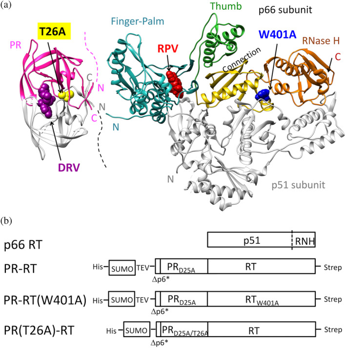
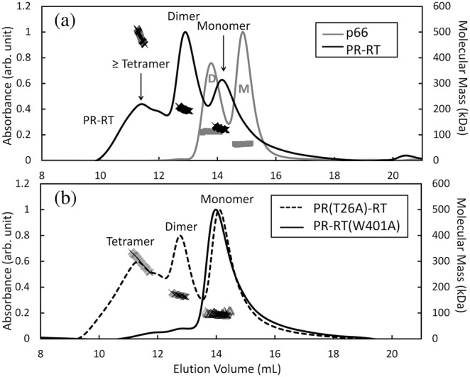
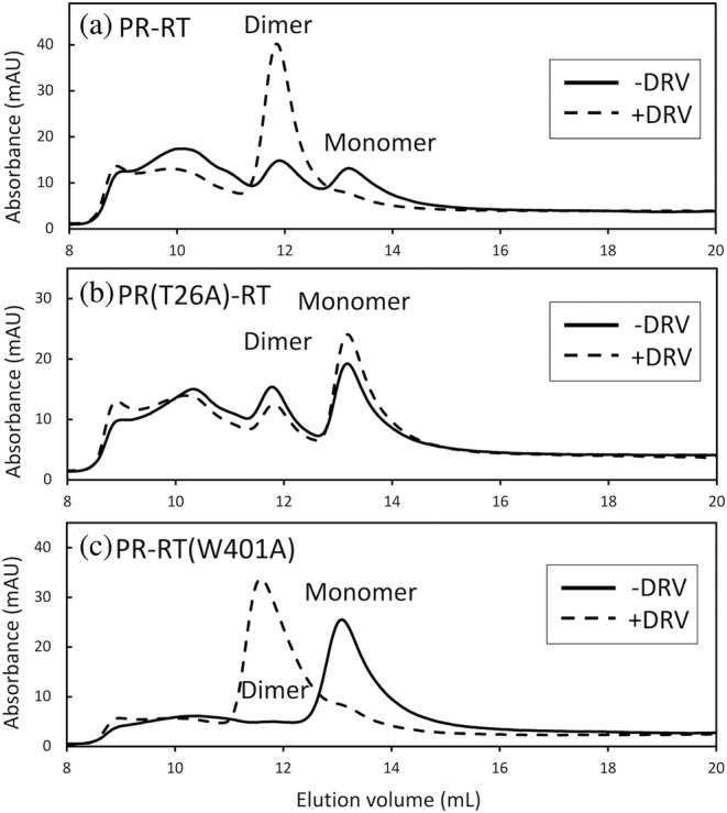
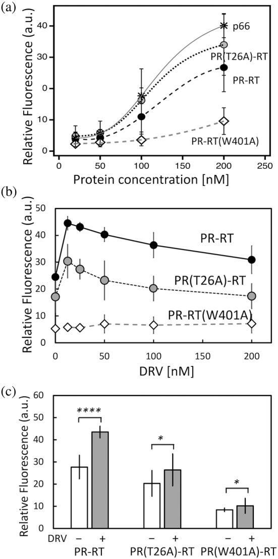
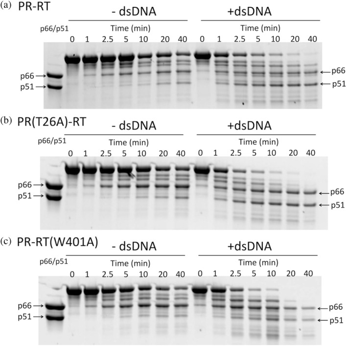
The Gag-Pol polyprotein in human immunodeficiency virus type I (HIV-1) encodes enzymes that are essential for virus replication: protease (PR), reverse transcriptase (RT), and integrase (IN). The mature forms of PR, RT and IN are homodimer, heterodimer and tetramer, respectively. The precise mechanism underlying the formation of dimer or tetramer is not yet understood. Here, to gain insight into the dimerization of PR and RT in the precursor, we prepared a model precursor, PR-RT, incorporating an inactivating mutation at the PR active site, D25A, and including two residues in the p6* region, fused to a SUMO-tag, at the N-terminus of the PR region. We also prepared two mutants of PR-RT containing a dimer dissociation mutation either in the PR region, PR(T26A)-RT, or in the RT region, PR-RT(W401A). Size exclusion chromatography showed both monomer and dimer fractions in PR-RT and PR(T26A)-RT, but only monomer in PR-RT(W401A). SEC experiments of PR-RT in the presence of protease inhibitor, darunavir, significantly enhanced the dimerization. Additionally, SEC results suggest an estimated PR-RT dimer dissociation constant that is higher than that of the mature RT heterodimer, p66/p51, but slightly lower than the premature RT homodimer, p66/p66. Reverse transcriptase assays and RT maturation assays were performed as tools to assess the effects of the PR dimer-interface on these functions. Our results consistently indicate that the RT dimer-interface plays a crucial role in the dimerization in PR-RT, whereas the PR dimer-interface has a lesser role.

Keywords: Gag‐Pol polyprotein; HIV‐1; darunavir; dimerization; inhibitor; protease; reverse transcriptase.


Conflict of interest statement

The authors declare no conflict of interest.

Figures

FIGURE 1
FIGURE 1
(a) Known structures of the mature PR homodimer and RT p66/p51, and (b) schematic representation of p66 RT and model precursors prepared in this study. (a) PR‐RT mutation sites, T26A in PR and W401A in RT, and the locations of the PR inhibitor darunavir (DRV) and of the RT inhibitor rilpivirine (RPV), each known to enhance dimerization of the mature enzymes, respectively, are indicated. Structures were generated using PDB 1T3R and 4G1Q (Kuroda et al., ; Surleraux et al., 2005). Only one subunit of PR and the p66 subunit of RT are colored: PR, pink; Finger‐Palm subdomain, cyan; Thumb subdomain, green; Connection subdomain, yellow; RNH domain, orange. How PR and RT fold in PR‐RT is unknown. The N‐ and C‐termini of PR and RT are indicated by N and C, respectively, using the same colors as those of the backbones. (b) Δp6* indicates two residues preceding PR, Asn‐Phe, and these proteins are fused to a hexa‐histidine tag and SUMO domain. Details of the amino acid sequences are described in the Section 5.
FIGURE 2
FIGURE 2
SDS‐PAGE of (a) PR‐RT, (b) PR(T26A)‐RT, (c) PR‐RT(W401A), each following the final heparin column elution, and (d) each protein following incubation at the indicated temperatures to confirm the absence of significant E. coli protease contamination. In the experiments in (d), proteins (8 μM, calculated as monomer) were incubated in the presence of 2 μM dsDNA for 0, 3, and 14 h at 37°C. If serine protease contamination was present, the dsDNA would enhance protease digestion at the RT site (see the Section 5 for RT maturation).
FIGURE 3
FIGURE 3
Normalized SEC‐MALS profiles to identify monomer, dimer and oligomer states of (a) p66 (gray line) and PR‐RT (black line), and those of (b) PR(T26A)‐RT (dashed line) and PR‐RT(W401A) (black line). Symbols in the graph indicate the observed molecular mass (the right vertical axis): p66 (gray squares), PR‐RT (black cross marks), PR(T26A)‐RT (thin cross marks), and PR‐RT(W401A) (gray triangles). Proteins at 8 μM concentration (calculated as a monomer) were injected into a Superdex Increase 200 10/300 GL column, with a pre‐filter, equilibrated with a 25 mM HEPES buffer containing 2% glycerol, 250 mM NaCl, and 1 mM DTT at pH 7.5.
FIGURE 4
FIGURE 4
SEC profiles of (a) PR‐RT, (b) PR(T26A)‐RT, and (c) PR‐RT(W401A) in the presence (dashed line) and absence (solid line) of DRV. Proteins at 8 μM protein concentration (calculated as a monomer) were injected into a Superdex Increase 200 10/300 GL column, equilibrated with a 25 mM HEPES buffer containing 10% glycerol, 250 mM NaCl and 1 mM DTT at pH 7.5. Note, due to differences in the instruments, including the presence or absence of a prefilter, and in the glycerol concentration, the elution positions differ slightly from those observed in SEC‐MALS.
FIGURE 5
FIGURE 5
Reverse transcription assay of PR‐RT, PR(T26A)‐RT, and PR‐RT(W401A): (a) protein concentration dependence, (b) DRV concentration dependence and (c) quantification at 50 nM DRV. In (a), the indicated protein concentrations are dimer concentration, since dimer is the functional form of RT. p66 was included as a control. Data from independent experiments that were used to generate the summary data in panel (a) are shown in Figure S5a–d. Data are shown as the average of the four experiments with the standard deviation as uncertainty. In (c), statistical significance of differences between ±DRV groups was evaluated from p values of the student's t‐tests: *0.01 < p < 0.05; ****p < 0.0001.
FIGURE 6
FIGURE 6
Processing of PR‐RT, PR(T26A)‐RT, and PR‐RT(W401A) by active HIV‐1 PR in the presence or absence of dsDNA: (a) PR‐RT, (b) PR(T26A)‐RT, and (c) PR‐RT(W401A). Processing of PR‐RT proteins at 4 μM protein concentration (calculated as a dimer) was performed by adding 1 μM PR (calculated as a dimer) with/without 4 μM dsDNA at pH 5.0. The entirety of these gels is shown in Figure S9. Data reproducibility was confirmed (Figure S10). A version with molecular markers is shown in Figure S11.

Similar articles

References

    1. Agniswamy J, Sayer JM, Weber IT, Louis JM. Terminal interface conformations modulate dimer stability prior to amino terminal autoprocessing of HIV‐1 protease. Biochemistry. 2012;51:1041–1050. - PMC - PubMed
    1. Bavand MR, Wagner R, Richmond TJ. HIV‐1 reverse transcriptase: polymerization properties of the p51 homodimer compared to the p66/p51 heterodimer. Biochemistry. 1993;32:10543–10552. - PubMed
    1. Bhavyasri KSM, Sumakanth M. Simultaneous method development, validation and stress studies of darunavir and ritonavir in bulk and combined dosage form using UV spectroscopy. Scholars Academic Journal of Pharmacy. 2020;9:244–252.
    1. Chattopadhyay D, Evans DB, Deibel MR, Vosters AF, Eckenrode FM, Einspahr HM, et al. Purification and characterization of heterodimeric human immunodeficiency virus type 1 (HIV‐1) reverse transcriptase produced by in vitro processing of p66 with recombinant HIV‐1 protease. Journal of Biological Chemistry. 1992;267:14227–14232. - PubMed
    1. Cherepanov P, Maertens G, Proost P, Devreese B, van Beeumen J, Engelborghs Y, et al. HIV‐1 integrase forms stable tetramers and associates with LEDGF/p75 protein in human cells. Journal of Biological Chemistry. 2003;278:372–381. - PubMed

Substances