. 2024 Jun 19;8(7):e0450.
doi: 10.1097/HC9.0000000000000450. eCollection 2024 Jul 1.

A novel experimental model of MetALD in male mice recapitulates key features of severe alcohol-associated hepatitis

Affiliations

A novel experimental model of MetALD in male mice recapitulates key features of severe alcohol-associated hepatitis

Mrigya Babuta et al. Hepatol Commun. .

Abstract

Background: The recent increase in the incidence of alcohol-associated hepatitis (AH) coincides with the obesity epidemic in the United States. However, current mouse models do not fully replicate the combined insults of obesity, metabolic dysfunction-associated steatohepatitis, and alcohol. The aim of this study was to develop a new mouse model that recapitulates the robust inflammatory and fibrotic phenotype characteristic of human MetALD.

Methods: Eight- to 10-week-old male C57BL/6 mice were fed chow or high fat-cholesterol-sugar diet (metabolic dysfunction-associated steatohepatitis diet) and in each group, some received alcohol in drinking water (ad libitum) and weekly alcohol binges (EtOH) for 3 months. The liver was assessed for features of AH.

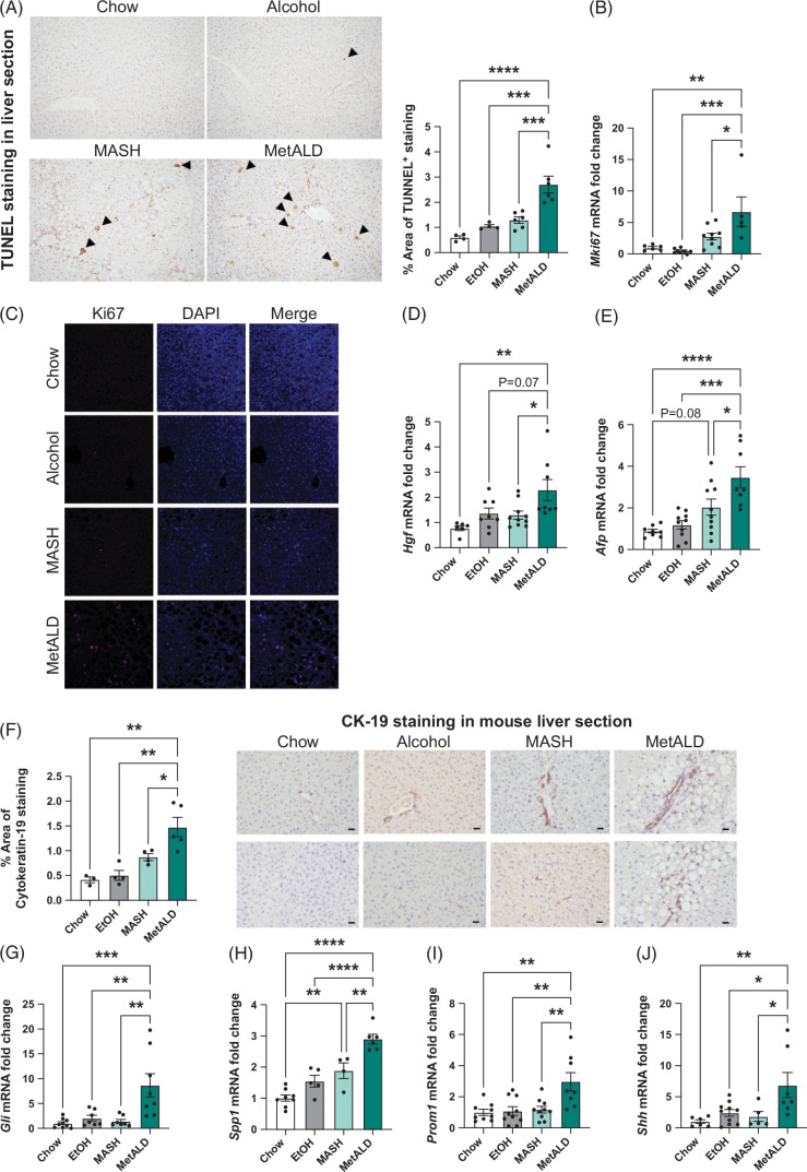
Results: MetALD mice displayed increased liver damage indicated by highly elevated ALT and bilirubin levels compared to all other groups. Liver steatosis was significantly greater in the MetALD mice compared to all other experimental groups. The inflammatory phenotype of MetALD was also recapitulated, including increased IL-6 and IL-1β protein levels as well as increased CD68+ macrophages and Ly6G+ neutrophils in the liver. Sirius red staining and expression of collagen 1, alpha-smooth muscle actin indicated advanced fibrosis in the livers of MetALD mice. In addition, indicators of epithelial-to-mesenchymal transition markers were increased in MetALD mice compared to all other groups. Furthermore, we found increased ductular reaction, dysregulated hedgehog signaling, and decreased liver synthetic functions, consistent with severe AH.

Conclusions: Alcohol administration in mice combined with metabolic dysfunction-associated steatohepatitis diet recapitulates key characteristics of human AH including liver damage, steatosis, robust systemic inflammation, and liver immune cell infiltration. This model results in advanced liver fibrosis, ductular reaction, decreased synthetic function, and hepatocyte dedifferentiation, suggesting a robust model of MetALD in mice.


Conflict of interest statement

Gyongyi Szabo consults and owns stock in Zomagen Bioscience/Ventyx Biosciences. She advises and owns stock in Glympse Bio, Inc. (acquired by Sunbird Bio) and Satellite Biosciences. She consults for Durect Corporation, CYTA Therapeutics, Merck, Pandion Therapeutics, Inc., Pfizer, Terra Firma, LabCorp, Takeda, NIAAA Board of Scientific Councilors, and Yale University School of Medicine. She advises Surrozen, Intercept, Evive, Baylor Texas Medical Center, University of Pennsylvania, and Nature Reviews gastroenterology and hepatology. She received grants from NIAAA and NIA. She received royalties from Springer Nature Group and UpToDate Inc. The remaining authors have no conflicts to report.

Figures

None
Graphical abstract
FIGURE 1
FIGURE 1
Alcohol worsens liver injury in MetALD mice. (A) Feeding schematics for liver injury by alcohol and MASH diet. C57BL/6 WT were fed either on a chow diet or a MASH diet. Some of the mice from the chow and MASH diet received alcohol in drinking water and 1 binge/wk for 12 weeks. (B) Kaplan-Meier graph representing the percent survival of mice fed on chow or MASH diet in the presence and absence of alcohol for 12 weeks. (C) Body weight gain by chow, alcohol, MASH, and MetALD mice. (D) Alcohol consumption in alcohol and MetALD mice. (E) The blood alcohol level was measured from serum. (F–I) Whole-cell liver lysates were used to detect CYP2E1 (F), ADH1a (G), ALDH1 (H), and ALDH2 (I) by ELISA. (J) ALT levels were measured from serum at different times during feeding. (K) Bilirubin level was measured from serum in alcohol, MASH, and MetALD mice. (L) Formalin-fixed liver sections were stained with hematoxylin and eosin and representative images are shown. Scale bar = 100 µm and inset scale bar is 50 µm. Data are presented as mean ± SEM. *p < 0.05, **p < 0.005, ***p < 0.0005. n = 4–8 mice in the MetALD and alcohol groups and n = 3–6 in the chow and MASH diet groups. Abbreviations: ADH, alcohol dehydrogenase; ALDH, aldehyde dehydrogenase; ALT, alanine aminotransferase; MASH, metabolic dysfunction–associated steatohepatitis; WT, wild type.
FIGURE 2
FIGURE 2
Alcohol and MASH diet promote steatosis in MetALD mice. (A) Triglyceride levels were measured from the livers. Free fatty acid levels (B) and total cholesterol levels (C) were measured in serum by ELISA. (D) Representative images of formalin-fixed liver sections stained with oil-red-O stain are shown. Scale bar = 50 µm. Liver RNA was isolated, and qRT-PCR was performed to determine mRNA levels of Dgat1 and Dgat2 (E, F), Plin2 (G), Fgf21 (H), Fabp4 (I), Cpt1a and Cpt1b (J, K), Cd36 (L), Lpl (M), Fas (N), Srebf1 (O), and Cebpa (P). 18S mRNA was used to normalize Cq values. Data are presented as mean ± SEM. *p < 0.05, **p < 0.005, ***p < 0.0005, ****p < 0.00005. ns is not significant. n = 4–8 mice/group. Abbreviations: MASH, metabolic dysfunction–associated steatohepatitis; qRT-PCR, Quantitative reverse transcription polymerase chain reaction.
FIGURE 3
FIGURE 3
MetALD mice exhibit increased inflammation and macrophage polarization. Whole-cell liver lysates were used to detect IL-1β (A), IL-6 (B), and MCP-1 (C) by ELISA. Paraffin-embedded liver sections were stained for CD68, and representative images are shown (D, left panel). CD68+ cells were quantified using ImageJ software and mean intensity was presented as graph (D, right panel). Scale bar=50 µm (n=5/group, with an average of 3–4 images/slide). Macrophage activation markers, Cd163 (E), Cd86 (F), and Il10 (G), in the liver were evaluated by qRT-PCR assay. 18S mRNA was used to normalize the Cq values. Data are presented as mean ± SEM. *p < 0.05, **p < 0.005, ***p < 0.0005, ****p < 0.00005. ns is not significant. n=4–8 mice/group. Abbreviations: MASH, metabolic dysfunction–associated steatohepatitis; MCP-1, monocyte chemoattractant protein-1; qRT-PCR, quantitative reverse transcription polymerase chain reaction.
FIGURE 4
FIGURE 4
Alcohol and MASH diet promote neutrophil infiltration in MetALD mice. Liver neutrophil chemokines, Cxcl1 (A), Cxcl2 (B), and Cxcl5 (C), chemokine receptor, Cxcr1 (D), and Itgam (E), were evaluated by qRT-PCR assay. 18S mRNA was used to normalize the Cq values. n=6–8 mice/ group. (F), Paraffin-embedded liver sections were stained for Ly6G, and representative images are shown. Scale bar= 50 µm. Data are presented as mean ± SEM. *p<0.05, **p<0.005. ns is not significant. Abbreviations: MASH, metabolic dysfunction–associated steatohepatitis; qRT-PCR, quantitative reverse transcription polymerase chain reaction.
FIGURE 5
FIGURE 5
Increased fibrosis and upregulation of EMT markers in MetALD mice. (A) Histological scoring of fibrosis. (B) Formalin-fixed liver sections were stained with Sirius red stain and representative images are shown. ImageJ software was used to quantify Sirius red stained (A, right panel) (n=4–6/group, with an average of 3–4 images/slide). (C) qRT-PCR was performed to determine liver mRNA levels of Col1a1, Tgfb1, and Timp1. Acta2/α-SMA levels were evaluated by qRT-PCR (D), and by western blot (E). GAPDH was used as equal loading and the densitometry analysis is shown as a bar graph (E, right panel) (n = 6). (F) Liver mRNA was used to evaluate the level of epithelial markers such as Ocln and cldn1. Liver mRNA and whole-cell lysate were used to evaluate the level of mesenchymal marker Vim by qPCR (G) and by western blot (H). mRNA of transcription factors involved in EMT such as Zeb1, Snai2, and Snai1 (I) was evaluated by qRT-PCR. 18S mRNA was used to normalize the Cq values. n = 6–8 mice/group. Data are presented as mean ± SEM. *p < 0.05, **p < 0.005, ***p < 0.0005, ****p < 0.00005. ns is not significant. n=4–8 mice/group. Abbreviations: α-SMA, alpha-smooth muscle actin; EMT, epithelial-to-mesenchymal transition; MASH, metabolic dysfunction–associated steatohepatitis; qRT-PCR, quantitative reverse transcription polymerase chain reaction.
FIGURE 6
FIGURE 6
Hepatocyte proliferation, ductular reaction, and upregulation of hedgehog signaling in MetALD mice. Paraffin-embedded liver sections were stained for TUNEL (A), Ki67 (C), and CK-19 (F), and representative images are shown. TUNEL+ (A, right panel) cells were quantified using ImageJ software, and mean intensity was presented as a graph (n=4–6/group, with an average of 3–4 images/slide). qRT-PCR was performed to determine liver mRNA levels of Mki67 (B), Hgf (D), Afp (E), Gli1 (G), Spp1 (H), Prom1 (I), and Shh (J). 18S mRNA was used to normalize the Cq values. n = 3–8 mice/group. Data are presented as mean ± SEM. *p < 0.05, **p < 0.005, ***p < 0.0005, ****p < 0.00005. ns is not significant. Abbreviations: DAPI, 4',6-diamidino-2-phenylindole; MASH, metabolic dysfunction–associated steatohepatitis; qRT-PCR, quantitative reverse transcription polymerase chain reaction; TUNEL, terminal deoxynucleotidyl transferase dUTP nick end labeling.
FIGURE 7
FIGURE 7
Liver synthetic function is impaired in MetALD mice. (A) vWF levels were measured from serum by ELISA. qRT-PCR was performed to determine liver mRNA levels of Vwf (B) and Hnf4a (E). 18S mRNA was used to normalize the Cq values. Circulating levels of albumin (C) and thrombin (D) were measured in serum by ELISA. (F) Model summarizing the pathways upregulated in MetALD mice. n=4–8 mice/group. Data are presented as mean ± SEM. *p < 0.05, **p < 0.005, ***p < 0.0005, ****p < 0.00005. Abbreviations: AFP, alpha-fetoprotein; ALT, alanine aminotransferase; EMT, epithelial-to-mesenchymal transition; HGF, hepatocyte growth factor; MASH, metabolic dysfunction–associated steatohepatitis; qRT-PCR, quantitative reverse transcription polymerase chain reaction; vWF, von Willebrand factor.

References

    1. Basra S. Definition, epidemiology and magnitude of alcoholic hepatitis. World J Hepatol. 2011;3:108–113. - PMC - PubMed
    1. Osna NA, Donohue TM, Jr, Kharbanda KK. Alcoholic liver disease: Pathogenesis and current management. Alcohol Res. 2017;38:147–161. - PMC - PubMed
    1. Jinjuvadia R, Liangpunsakul S. Evolving alcoholic hepatitis treatment C. Trends in alcoholic hepatitis-related hospitalizations, financial burden, and mortality in the United States. J Clin Gastroenterol. 2015;49:506–511. - PMC - PubMed
    1. Malnick S, Maor Y. The interplay between alcoholic liver disease, obesity, and the metabolic syndrome. Visc Med. 2020;36:198–205. - PMC - PubMed
    1. Abassa KK, Wu XY, Xiao XP, Zhou HX, Guo YW, Wu B. Effect of alcohol on clinical complications of hepatitis virus-induced liver cirrhosis: A consecutive ten-year study. BMC Gastroenterol. 2022;22:130. - PMC - PubMed

Publication types