. 2024 Aug:140:103700.
doi: 10.1016/j.dnarep.2024.103700. Epub 2024 Jun 4.

Oncometabolite 2-hydroxyglutarate suppresses basal protein levels of DNA polymerase beta that enhances alkylating agent and PARG inhibition induced cytotoxicity

Affiliations

Oncometabolite 2-hydroxyglutarate suppresses basal protein levels of DNA polymerase beta that enhances alkylating agent and PARG inhibition induced cytotoxicity

Kate M Saville et al. DNA Repair (Amst). 2024 Aug.

Abstract

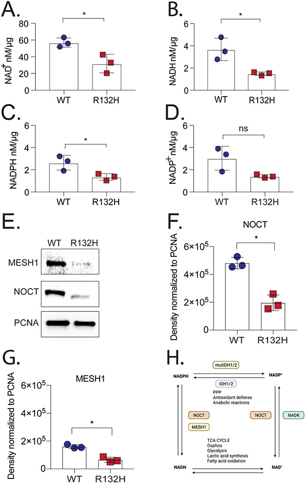
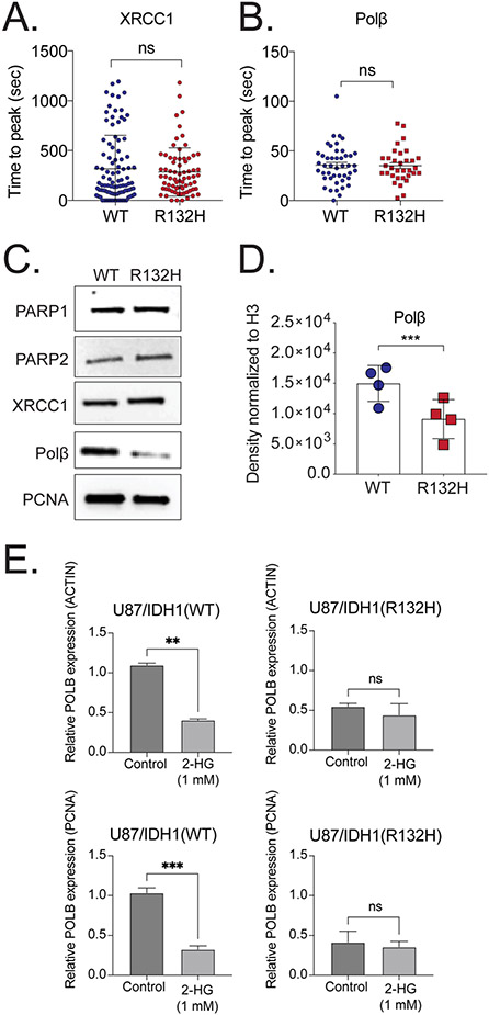
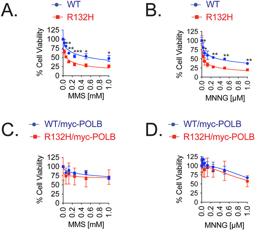
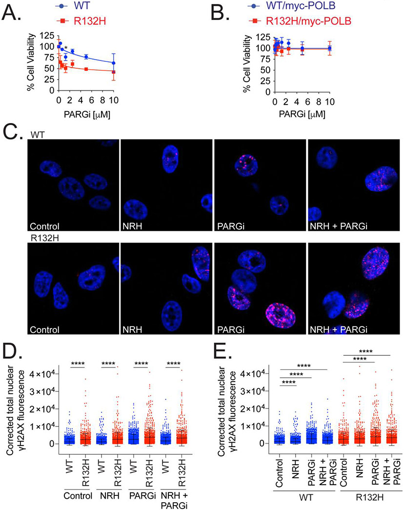
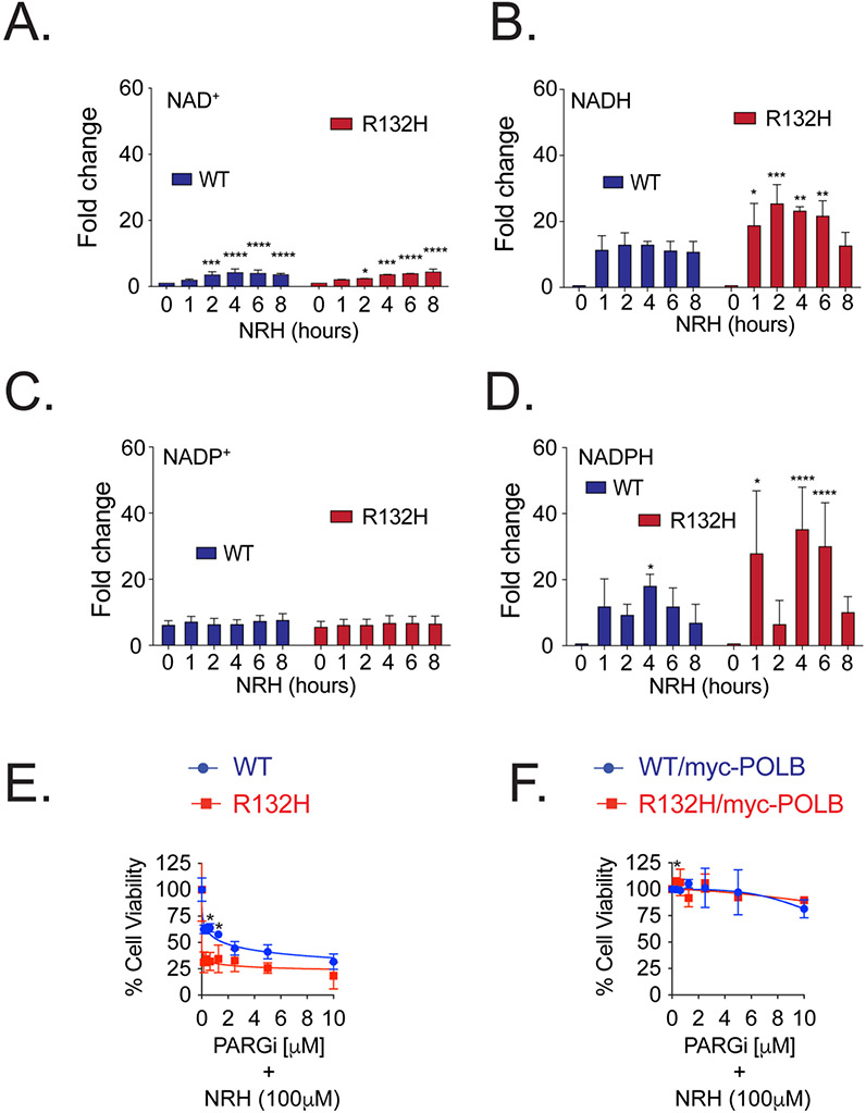
Mutations in isocitrate dehydrogenase isoform 1 (IDH1) are primarily found in secondary glioblastoma (GBM) and low-grade glioma but are rare in primary GBM. The standard treatment for GBM includes radiation combined with temozolomide, an alkylating agent. Fortunately, IDH1 mutant gliomas are sensitive to this treatment, resulting in a more favorable prognosis. However, it's estimated that up to 75 % of IDH1 mutant gliomas will progress to WHO grade IV over time and develop resistance to alkylating agents. Therefore, understanding the mechanism(s) by which IDH1 mutant gliomas confer sensitivity to alkylating agents is crucial for developing targeted chemotherapeutic approaches. The base excision repair (BER) pathway is responsible for repairing most base damage induced by alkylating agents. Defects in this pathway can lead to hypersensitivity to these agents due to unresolved DNA damage. The coordinated assembly and disassembly of BER protein complexes are essential for cell survival and for maintaining genomic integrity following alkylating agent exposure. These complexes rely on poly-ADP-ribose formation, an NAD+-dependent post-translational modification synthesized by PARP1 and PARP2 during the BER process. At the lesion site, poly-ADP-ribose facilitates the recruitment of XRCC1. This scaffold protein helps assemble BER proteins like DNA polymerase beta (Polβ), a bifunctional DNA polymerase containing both DNA synthesis and 5'-deoxyribose-phosphate lyase (5'dRP lyase) activity. Here, we confirm that IDH1 mutant glioma cells have defective NAD+ metabolism, but still produce sufficient nuclear NAD+ for robust PARP1 activation and BER complex formation in response to DNA damage. However, the overproduction of 2-hydroxyglutarate, an oncometabolite produced by the IDH1 R132H mutant protein, suppresses BER capacity by reducing Polβ protein levels. This defines a novel mechanism by which the IDH1 mutation in gliomas confers cellular sensitivity to alkylating agents and to inhibitors of the poly-ADP-ribose glycohydrolase, PARG.

Keywords: 2-hydroxyglutarate; Base excision repair; DNA polymerase beta; IDH1; NAD(+) metabolism; PARP1.


Conflict of interest statement

Declaration of Competing Interest R.W.S. is co-founder of Canal House Biosciences, LLC, is on the Scientific Advisory Board, and has an equity interest. Canal House Biosciences was not involved in the preparation of this study nor of this manuscript. The authors state that there is no conflict of interest.

Figures

Figure 1:
Figure 1:. Characterization of U87/IDH1(WT) and U87/IDH1(R132H) cells.
A) NAD+ analysis in U87/IDH1(WT) cells as compared to U87/IDH1(R132H) cells, n=3 (p*<0.05; Student’s t-test); B) NADH analysis in U87/IDH1(WT) cells as compared to U87/IDH1(R132H) cells, n=3 (p*<0.05; Student’s t-test); C) NADPH analysis in U87/IDH1(WT) cells as compared to U87/IDH1(R132H) cells, n=3 (p*<0.05; Student’s t-test); D) NADP+ analysis in U87/IDH1(WT) cells as compared to U87/IDH1(R132H) cells, n=3 (ns, not significant; Student’s t-test); E) Immunoblot analysis of MESH1 and NOCT protein expression in whole cell lysates from U87/IDH1(WT) cells compared to U87/IDH1(R132H) cells, PCNA as a loading control; F) Quantitative analysis of NOCT protein expression by immunoblot, normalized to PCNA. Each data point represents the average of three biological replicates (p*<0.05; Student’s t-test); G) Quantitative analysis of MESH1 protein expression by immunoblot, normalized to PCNA, Each data point represents the average of three biological replicates (p*<0.05; Student’s t-test); H) Graphic demonstrating an overview of NAD(P)+/NAD(P)H metabolism: NADK phosphorylates NAD+ to form NADP+, IDH1 reduces NADP+ to NADPH, IDH1 mutations oxidize NADPH to NADP+, NOCT and MESH1 phosphatases remove the phosphate group from NADPH, and also, NOCT functions to remove the phosphate group from NADP+ (created using BioRender).
Figure 2:
Figure 2:. Polβ protein expression is suppressed in IDH1 mutant cells.
A) XRCC1-EGFP time to peak (seconds), following micro-irradiation laser-induced (355nm) DNA damage, as determined in MIDAS; B) EGFP-Polβ time to peak (seconds), following micro-irradiation laser-induced (355nm) DNA damage, as determined in MIDAS; C) Immunoblot analysis of whole cell lysates from U87/IDH1(WT) and U87/IDH1(R132H) cells to probe for BER proteins; D) Quantitative analysis of immunoblot expression for Polβ, normalized to Histone H3, n=4 (p<***0.001; Paired Student’s t-test); E) Quantitative analysis of immunoblot expression for Polβ, normalized to actin, and normalized to PCNA, of whole cell lysates from U87/IDH1(WT) and U87/IDH1(R132H) cells treated with media alone or with D-2-HG (1 mM) for 2 hours; n=2 (ns, not significant; p<**0.01, p<***0.001; Student’s t test).
Figure 3:
Figure 3:. Overexpression of Polβ in IDH1 mutant glioma cells induces an MMS and MNNG resistant phenotype.
A) Cell viability assay of U87/IDH1(WT) and U87/IDH1(R132H) cells treated with MMS for 120 hours, as indicated, n=3 (comparison of WT and R132H at each dose: no stars = not significant, p<*0.05, p<**0.01, p<***0.001; Paired Student’s t-test); B) Cell viability assay of U87/IDH1(WT) and U87/IDH1(R132H) cells treated with MNNG for 120 hours, as indicated, n=3 (comparison of WT and R132H at each dose: no stars = not significant, p<*0.05, p<**0.01; Paired Student’s t-test); C) Cell viability assay of U87/IDH1(WT)/myc-PolB and U87/IDH1(R132H)/myc-PolB cells treated with MMS for 120 hours, as indicated, n=2 (comparison of WT/myc-PolB and R132H/myc-PolB at each dose: no stars = not significant; Paired Student’s t-test); D) Cell viability assay of U87/IDH1(WT)/myc-PolB and U87/IDH1(R132H)/myc-PolB cells treated with MNNG for 120 hours, as indicated, n=2 (comparison of WT/myc-PolB and R132H/myc-PolB at each dose: no stars = not significant; Paired Student’s t-test).
Figure 4.
Figure 4.. IDH1 mutant cells deficient in Polβ show increased sensitivity to PARGi.
A) Cell viability assay of U87/IDH1(WT) and U87/IDH1(R132H) cells treated with PARGi as indicated, n=2 (comparison of WT and R132H at each dose: no stars = not significant, p<*0.05; Paired Student’s t-test); B) Cell viability assay of U87/IDH1(WT)/myc-PolB cells and U87/IDH1(R132H)/myc-PolB cells treated with PARGi as indicated, n=2 (comparison of WT/myc-PolB and R132H/myc-PolB at each dose: no stars = not significant; Paired Student’s t-test); C) Influence of IDH1 mutation on γH2AX formation upon PARG inhibition: Confocal micrographs of U87/IDH1(WT) and U87/IDH1(R132H) cells exposed to DMSO (control), to NRH (100μM) and/or to PARGi (5μM), for 24 hours. Nuclear staining (DAPI, Blue) and γH2AX (Red) are shown; D) & E) Quantification of corrected nuclear fluorescence of γH2AX in U87/IDH1(WT) and U87/IDH1(R132H) cells exposed to DMSO (control), to NRH (100μM) and/or to PARGi (5μM), for 24 hours. The data presented are from four independent experiments where at least two hundred nuclei per treatment type were analyzed for their fluorescence signal; D) For every treatment type, the data are plotted to demonstrate the difference between U87/IDH1(WT) and U87/IDH1(R132H) cells, n=4, >200 cells per each condition, (p<****0.001; Mann-Whitney, two-tailed); E) The data are plotted to demonstrate the influence of treatment within each specific cell line, n=4, >200 cells per each condition, (p<****0.001; Mann-Whitney, two-tailed).
Figure 5.
Figure 5.. PARGi induced cell death is enhanced by NRH in IDH1 mutant cells.
A) NAD+ analysis of U87/IDH1(WT) and U87/IDH1(R132H) cells following treatment with NRH (100μM) for the times indicated; n=2 (comparison to untreated: no stars = not significant, p<*0.05, p<***0.001, p<****0.001; Two-way ANOVA); B) NADH analysis of U87/IDH1(WT) and U87/IDH1(R132H) cells following treatment with NRH (100μM) for the times indicated; n=2 (comparison to untreated: no stars = not significant, p<*0.05, p<**0.01, p<***0.001; Two-way ANOVA); C) NADP+ analysis of U87/IDH1(WT) and U87/IDH1(R132H) cells following treatment with NRH (100μM) for the times indicated n=2 (comparison to untreated: no stars = not significant; Two-way ANOVA); D) NADPH analysis of U87/IDH1(WT) and U87/IDH1(R132H) cells following treatment with NRH (100μM) for the times indicated; n=2 (comparison to untreated: no stars = not significant, p<*0.05, p<****0.0001; Two-way ANOVA); E) Cell viability assay following co-treatment with NRH (100μM) and PARGi (for the doses indicated) for 120 hours in U87/IDH1(WT) cells and U87/IDH1(R132H) cells, n=2 (comparison of WT and R132H at each dose: no stars = not significant, p<*0.05; Paired Student’s t-test); F) Cell viability assay following co-treatment with NRH (100μM) and PARGi (for the doses indicated) for 120 hours in U87/IDH1(WT)/myc-PolB cells and U87/IDH1(R132H)/myc-PolB cells, n=2 (comparison of WT/myc-PolB and R132H/myc-PolB at each dose: no stars = not significant, p<*0.05; Paired Student’s t-test).

Similar articles

Cited by

References

    1. Kaminska B, Czapski B, Guzik R, Krol SK, Gielniewski B, Consequences of IDH1/2 Mutations in Gliomas and an Assessment of Inhibitors Targeting Mutated IDH Proteins, Molecules, 24 (2019). - PMC - PubMed
    1. Guo J, Zhang R, Yang Z, Duan Z, Yin D, Zhou Y, Biological Roles and Therapeutic Applications of IDH2 Mutations in Human Cancer, Frontiers in oncology, 11 (2021) 644857. - PMC - PubMed
    1. Yan H, Parsons DW, Jin G, McLendon R, Rasheed BA, Yuan W, Kos I, Batinic-Haberle I, Jones S, Riggins GJ, Friedman H, Friedman A, Reardon D, Herndon J, Kinzler KW, Velculescu VE, Vogelstein B, Bigner DD, IDH1 and IDH2 mutations in gliomas, N Engl J Med, 360 (2009) 765–773. - PMC - PubMed
    1. Louis DN, Perry A, Wesseling P, Brat DJ, Cree IA, Figarella-Branger D, Hawkins C, Ng HK, Pfister SM, Reifenberger G, Soffietti R, von Deimling A, Ellison DW, The 2021 WHO Classification of Tumors of the Central Nervous System: a summary, Neuro-oncology, 23 (2021) 1231–1251. - PMC - PubMed
    1. Sun X, Turcan S, From Laboratory Studies to Clinical Trials: Temozolomide Use in IDH-Mutant Gliomas, Cells, 10 (2021). - PMC - PubMed

MeSH terms

LinkOut - more resources