Clinical Trial
. 2024 Jul 16;5(7):101615.
doi: 10.1016/j.xcrm.2024.101615. Epub 2024 Jun 18.

Neoadjuvant sintilimab plus chemotherapy in EGFR-mutant NSCLC: Phase 2 trial interim results (NEOTIDE/CTONG2104)

Affiliations
Clinical Trial

Neoadjuvant sintilimab plus chemotherapy in EGFR-mutant NSCLC: Phase 2 trial interim results (NEOTIDE/CTONG2104)

Chao Zhang et al. Cell Rep Med. .

Abstract

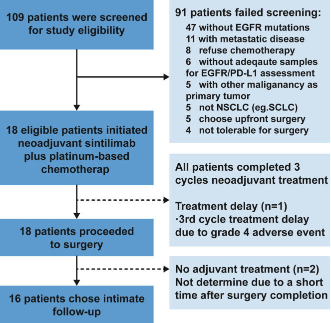
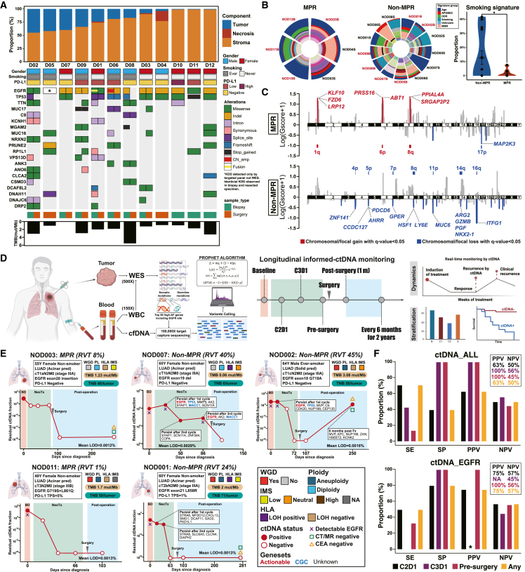
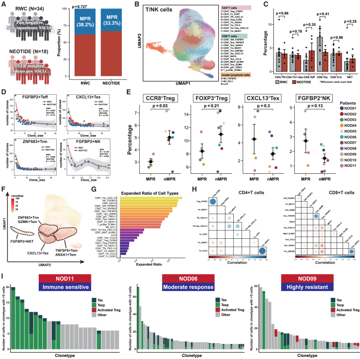
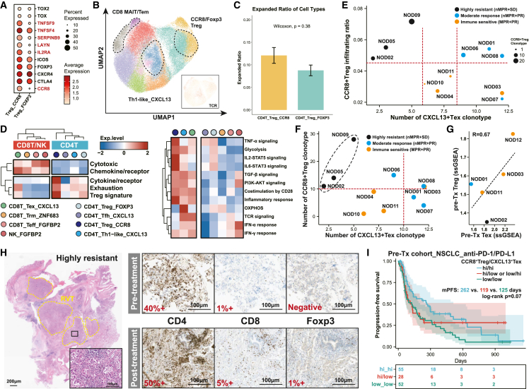
The clinical efficacy of neoadjuvant immunotherapy plus chemotherapy remains elusive in localized epidermal growth factor receptor (EGFR)-mutant non-small cell lung cancer (NSCLC). Here, we report interim results of a Simon's two-stage design, phase 2 trial using neoadjuvant sintilimab with carboplatin and nab-paclitaxel in resectable EGFR-mutant NSCLC. All 18 patients undergo radical surgery, with one patient experiencing surgery delay. Fourteen patients exhibit confirmed radiological response, with 44% achieving major pathological response (MPR) and no pathological complete response (pCR). Similar genomic alterations are observed before and after treatment without influencing the efficacy of subsequent EGFR-tyrosine kinase inhibitors (TKIs) in vitro. Infiltration and T cell receptor (TCR) clonal expansion of CCR8+ regulatory T (Treg)hi/CXCL13+ exhausted T (Tex)lo cells define a subtype of EGFR-mutant NSCLC highly resistant to immunotherapy, with the phenotype potentially serving as a promising signature to predict immunotherapy efficacy. Informed circulating tumor DNA (ctDNA) detection in EGFR-mutant NSCLC could help identify patients nonresponsive to neoadjuvant immunochemotherapy. These findings provide supportive data for the utilization of neoadjuvant immunochemotherapy and insight into immune resistance in EGFR-mutant NSCLC.


Conflict of interest statement

Declaration of interests W.-Z.Z.. received speech honoraria from AstraZeneca, Roche, Eli Lilly, and Pfizer outside the submitted work. Z.-M.Z. is a founder of Analytical BioSciences. Y.-L.W. received research funding from Roche and speech honoraria from AstraZeneca, Roche, Eli Lilly, Pfizer, and Sanofi, and he was a research consultant for AstraZeneca. Q.Z. reports honoraria from AstraZeneca, Boehringer Ingelheim, BMS, Eli Lilly, MSD, Pfizer, Roche, and Sanofi outside the submitted work.

Figures

None
Graphical abstract
Figure 1
Figure 1
Consort diagram and treatment disposition 109 patients in total were screened for study eligibility, and 91 of them did not meet the inclusion criteria due to absence of EGFR mutations, confirmed metastatic disease, etc. 18 enrolled patients had completed three cycles of neoadjuvant sintilimab plus chemotherapy and received radical surgery. 16 patients chose intimate follow-up without adjuvant targeted therapy, while the other two patients did not determine postoperative treatment at data cutoff.
Figure 2
Figure 2
Clinicopathological features and preliminary response (A) Study treatment and representative radiological response before and after neoadjuvant sintilimab plus nab-PC through PET-CT. (B) Individual clinicopathological features and pathological response. The black horizontal line indicates the threshold for MPR patients. (C) Representative pathological response and stratified response patterns in terms of radiological and pathological response, including the highly resistant group (n = 2), moderate response group (n = 2), and immune-sensitive group (n = 2). The yellow dotted lines circle the tumor area. PET-CT, positron emission tomography and computed tomography; MPR, major pathological response; PR, partial response; PD-L1, programmed cell death protein ligand-1; EGFR, epidermal growth factor receptor.
Figure 3
Figure 3
Genomic features of tumor specimens and longitudinal monitoring of informed MRD detection (A) WES analysis of pre- and post-treatment tumor specimens along with specific pathological response assessment. (B) Mutational signature analysis and comparison of smoking signature between MPR and nMPR patients. Dominant signature groups are specifically annotated. Proportions of smoking signature with different response groups were compared using Student’s t test. (C) Comparison of focal and chromosomal copy number variants (CNVs) between MPR and nMPR patients. Gscore was used to quantify the value, and bar plots indicate the relative proportion of significant focal CNVs within various chromosomes. Chromosomal/focal gain and chromosomal/focal loss with q < 0.05 are presented in red and blue, respectively. (D) Schematic of tumor-informed MRD and exploratory design. (E) Representative dynamic changes of tumor-informed MRD before and after neoadjuvant immunochemotherapy along with individual genomic and immunological features. Specifically, the red dots indicate positive ctDNA regarding the PROPHET algorithm, while red circles indicate negative ctDNA with a fraction of no more than 10−6. (F) Performance of longitudinal MRD detection for pathological response. Sensitivity, specificity, NPV, and PPV were used to determine the performance of MRD. Different colors represent different time points of MRD detection. Asterisks indicate no EGFR mutations detected at this time point, which led a to PPV of 0/0. TMB, tumor mutation burden; RVT, residual viable tumor; WES, whole-exome sequencing; WBC, whole blood cell; MRD, minimal residual disease; SE, sensitivity; SP, specificity; PPV, positive predictive value; NPV, negative predictive value; WGD, whole-genome doubling; IMS, immune status; HLA-LOH, human leukocyte antigen loss of heterozygosity; CGC, cancer gene census.
Figure 4
Figure 4
Differential TME phenotype of distinct pathological response in EGFR-mutant NSCLC (A) Comparison of MPR (excluding metastatic lymph nodes) between the NEOTIDE and real-world cohorts. (B) Reclustered T/NK cells and cell type annotation. (C) Comparison of different T/NK cell subsets between the NEOTIDE and real-world cohorts. Wilcoxon rank-sum test was used to quantify the significance. (D) Clonal analysis of different reactive cell subsets including FGFBP2+ Teff/NK cells, CXCL13+ Tex cells, and ZNF683+ Trm cells between the NEOTIDE and real-world cohorts. (E) Percentages of reactive cell subsets between MPR and nMPR patients in the NEOTIDE cohort. Each colored dot represents different individuals, and Fisher’s exact test was used to quantify the significance. (F) Clonal expansion across all T cell subtypes through uniform manifold approximation and projection (UMAP). Clone size is noted, and cells of higher clonal expansion are marked in deep red. Dashed lines indicate T cell subsets of higher clonal expansion. (G) Bar plot of expanded ratio across different T cell subtypes. (H) Pearson correlation of clonal expansion among different T cell subtypes. Dots with dark blue indicate positive correlation, while dark red indicates the opposite. (I) Representative individual clonal expansion regarding terminal Tex, precursor Tex, and activated Treg cells among different treatment responses.
Figure 5
Figure 5
CCR8+ Treg cells and CXCL13+ Tex cells as potentially predictive biomarkers for immunotherapy (A) Transcriptional difference between CD4+Foxp3+ Treg cells and CCR8+ Treg cells in terms of suppressive and inflammatory genes. (B) UMAP of CD4+ T cell clusters and corresponding clonal expansion of CXCL13+ Th1-like, CCR8+ Treg cells and FOPX3+ Treg cells. (C) Difference of expanded ratio between CCR8+Treg and FOXP3+Treg. Wilcoxon was used to calculate the significance. (D) Heatmap of enriched markers and functions among different CD4 and CD8 T subsets. Expression of specific markers or functions was normalized and underwent unsupervised clustering. (E) Correlation of CCR8+ Treg cell infiltration ratio and clonal expansion of CXCL13+ Tex cells in patients exhibiting diverse response patterns. (F) Scatterplot of TCR clonotype for CCR8+ Treg cells and CXCL13+ Tex cells among different response patterns, indicated by diverse colored dots. Red dotted lines represented the artificially defined cutoff to indicate high TCR expansion. The black dotted circles represented tumors with the worst response to immunochemotherapy. (G) Degree of infiltrating CCR8+ Treg cells and CXCL13+ Tex cells, evaluated through single-sample gene set enrichment analysis (ssGSEA) in paired pre-treatment specimens. Correlation was analyzed by Pearson’s correlation. (H) Representative immunohistochemistry of CD4, CD8, and FOXP3 along with corresponding HE staining in a patient exhibiting highly resistant to immunochemotherapy. (I) Comparison of survival after PD-1/PD-L1 inhibitors in external NSCLC and melanoma cohorts. Log rank p value was use to estimate the significance among groups.
Figure 6
Figure 6
Extrinsic and intrinsic response mechanisms of residual tumors (A) Microscopic pathological features through H&E staining among different response patterns. The green dotted circles indicate infiltrating immune cells, and the white dotted circles indicate tumor area. (B) Immunohistochemistry of CD3 and CD8 staining between different response patterns. (C) Schematic of diverse infiltration patterns within the tumor bed and surroundings. Proportions of different infiltrating patterns were calculated in terms of response patterns. Significance was analyzed using Fisher’s exact test. (D) WES-based HLA analysis and immunohistochemistry of HLA-A staining. A bar plot was used to compare proportions of HLA-LOH among different response patterns. (E) Reclustering of epithelial cells and CopyKAT analysis. (F) Expression levels of immune checkpoints and tumor-associated antigen for residual tumors among groups. p values of the top, intermediate, and low row represented MR vs. IS, IS vs. HR, and HR vs. MR, respectively. ∗p < 0.05, ∗∗p < 0.01, ∗∗∗p < 0.001, ∗∗∗∗p < 0.0001. (G) Differential genes expressed in the HR and MR/IS groups. Cancer- or immune-relevant genes are specifically annotated. (H) Proportion comparison of selected myeloid subsets between the NEOTIDE and real-world cohorts. Colored dots represent different treatment responses. Student’s t test was used to measure the significance. (I) CellphoneDB analysis of selected ligand-receptor interactions between tumor cells and myeloid subsets. (J) Immunohistochemistry of CD163 staining within the tumor bed.

References

    1. Lynch T.J., Bell D.W., Sordella R., Gurubhagavatula S., Okimoto R.A., Brannigan B.W., Harris P.L., Haserlat S.M., Supko J.G., Haluska F.G., et al. Activating mutations in the epidermal growth factor receptor underlying responsiveness of non-small-cell lung cancer to gefitinib. N. Engl. J. Med. 2004;350:2129–2139. doi: 10.1056/NEJMoa040938. - DOI - PubMed
    1. Shi Y., Au J.S.K., Thongprasert S., Srinivasan S., Tsai C.M., Khoa M.T., Heeroma K., Itoh Y., Cornelio G., Yang P.C. A prospective, molecular epidemiology study of EGFR mutations in Asian patients with advanced non-small-cell lung cancer of adenocarcinoma histology (PIONEER) J. Thorac. Oncol. 2014;9:154–162. doi: 10.1097/jto.0000000000000033. - DOI - PMC - PubMed
    1. Mok T.S., Wu Y.L., Thongprasert S., Yang C.H., Chu D.T., Saijo N., Sunpaweravong P., Han B., Margono B., Ichinose Y., et al. Gefitinib or carboplatin-paclitaxel in pulmonary adenocarcinoma. N. Engl. J. Med. 2009;361:947–957. doi: 10.1056/NEJMoa0810699. - DOI - PubMed
    1. Wu Y.L., Cheng Y., Zhou X., Lee K.H., Nakagawa K., Niho S., Tsuji F., Linke R., Rosell R., Corral J., et al. Dacomitinib versus gefitinib as first-line treatment for patients with EGFR-mutation-positive non-small-cell lung cancer (ARCHER 1050): a randomised, open-label, phase 3 trial. Lancet Oncol. 2017;18:1454–1466. doi: 10.1016/s1470-2045(17)30608-3. - DOI - PubMed
    1. Soria J.C., Ohe Y., Vansteenkiste J., Reungwetwattana T., Chewaskulyong B., Lee K.H., Dechaphunkul A., Imamura F., Nogami N., Kurata T., et al. Osimertinib in Untreated EGFR-Mutated Advanced Non-Small-Cell Lung Cancer. N. Engl. J. Med. 2018;378:113–125. doi: 10.1056/NEJMoa1713137. - DOI - PubMed

Publication types

MeSH terms