. 2024 Aug 21;112(16):2686-2707.e8.
doi: 10.1016/j.neuron.2024.05.023. Epub 2024 Jun 18.

Therapeutic potential of human microglia transplantation in a chimeric model of CSF1R-related leukoencephalopathy

Affiliations

Therapeutic potential of human microglia transplantation in a chimeric model of CSF1R-related leukoencephalopathy

Jean Paul Chadarevian et al. Neuron. .

Abstract

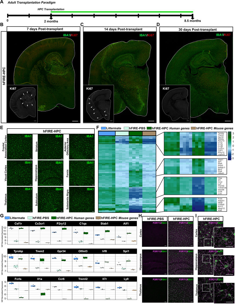
Microglia replacement strategies are increasingly being considered for the treatment of primary microgliopathies like adult-onset leukoencephalopathy with axonal spheroids and pigmented glia (ALSP). However, available mouse models fail to recapitulate the diverse neuropathologies and reduced microglia numbers observed in patients. In this study, we generated a xenotolerant mouse model lacking the fms-intronic regulatory element (FIRE) enhancer within Csf1r, which develops nearly all the hallmark pathologies associated with ALSP. Remarkably, transplantation of human induced pluripotent stem cell (iPSC)-derived microglial (iMG) progenitors restores a homeostatic microglial signature and prevents the development of axonal spheroids, white matter abnormalities, reactive astrocytosis, and brain calcifications. Furthermore, transplantation of CRISPR-corrected ALSP-patient-derived iMG reverses pre-existing spheroids, astrogliosis, and calcification pathologies. Together with the accompanying study by Munro and colleagues, our results demonstrate the utility of FIRE mice to model ALSP and provide compelling evidence that iMG transplantation could offer a promising new therapeutic strategy for ALSP and perhaps other microglia-associated neurological disorders.

Keywords: ALSP; CRISPR correction; CSF1R; FIRE; axonal spheroids; chimera; humanized; leukoencephalopathy; microglia.


Conflict of interest statement

Declaration of interests J.P.C., J.H., R.C.S., S.G. H.D., and M.B.-J. are co-inventors on patent applications filed by the University of California Regents (US 63/169,578) related to genetic modification of cells to confer resistance to CSF1R antagonists and (US 63/388,766) related to transplantation of stem-cell-derived microglia to treat leukodystrophies. M.B.-J. is a co-inventor of patent application WO/2018/160496, related to the differentiation of human pluripotent stem cells into microglia. M.B.-J., S.G., and R.C.S. are co-founders of NovoGlia Inc. Z.K.W. serves as principal investigator (PI) or co-PI on Biohaven Pharmaceuticals, Inc. (BHV4157-206) and Vigil Neuroscience, Inc. (VGL101-01.002, VGL101-01.201, PET tracer development protocol, Csf1r biomarker and repository project, and ultra-high-field MRI in the diagnosis and management of CSF1R-related adult-onset leukoencephalopathy with axonal spheroids and pigmented glia) projects/grants. He serves as co-PI of the Mayo Clinic APDA Center for Advanced Research; as an external advisory board member for Vigil Neuroscience, Inc.; and as a consultant on neurodegenerative medical research for Eli Lilli & Company.

Figures

Figure 1:
Figure 1:. hFIRE mice progressively develop ALSP-related neuropathologies.
(A) hFIRE mice were generated by crossing immune-intact FIRE mice onto the immune-deficient hCSF1+/+/Rag2−/−/il2rg−/y knock-in background and restoring homozygosity for all four alleles. (B-C) Representative confocal imaging of 2-month-old female hCSF1 (B) and hFIRE (C) coronal brain sections for murine microglia (IBA1, green; PU.1, orange); scale bar, 50 μm. (D-F) Quantification of PU.1+/IBA1+ murine microglia within the cortex (D), hippocampus (E), and thalamus (F) of hCSF1 wildtype female littermates and hFIRE mice. Data represented as average number IBA1+/PU1+ microglia per field of view (FOV); P values from unpaired, two-sided t-test. (G-H), Representative immunofluorescence staining of 2-month-old (G) and 8.5-month-old (H) hCSF1 and hFIRE brain sections for astrocytes (GFAP, yellow); scale bar, 50 μm. (I) Quantification of astrogliosis in the thalamus of 2-month-old and 8.5-month-old hCSF1 and hFIRE mice; P values from one-way ANOVA (F3,25=17.55; ****P<0.0001) with Tukey multiple comparisons tests. (J-K) Representative immunofluorescence staining of calcification (Risedronate-647) in 2-month-old (J) and 8.5-month-old (K) hCSF1 and hFIRE brain sections (RIS-647, gray); scale bar, 50 μm. (L) Quantification of RIS+ calcification in the thalamus of 2-month-old and 8.5-month-old hCSF1 and hFIRE mice; P values from one-way ANOVA (F3,25=71.20; ****P<0.0001) with Tukey multiple comparisons tests. (M) Representative confocal imaging of 8.5-month-old hFIRE brain sections stained for neurofilament-positive axonal spheroids (SMI312, green); scale bar, 20 μm. (N) Quantification of SMI312 positive spheroids per hemibrain of 2-month-old and 8.5-month-old hCSF1 and hFIRE mice; P values from one-way ANOVA (F3,25=52.85; ****P<0.0001) with Tukey multiple comparisons tests. For all quantification data is represented as average mean value ±SEM (2-month-old hCSF1, n = 6; 2-month-old hFIRE, n=7; 8.5-month-old hCSF1, n=8; 8.5-month-old hFIRE, n=8 biological replicates). *P<0.05; ****P<0.0001. Comparisons not shown are not significant.
Figure 2:
Figure 2:. Human microglial progenitors efficiently engraft within adult hFIRE brains, restoring canonical microglial gene expression.
(A) Schematic representing adult female hCSF1 and hFIRE mice transplantation paradigm. (B-D) Representative hemibrain confocal stitches of hFIRE mouse showing proliferative expansion and migration of human microglia (IBA1, green; Ki67, red/white) 7 days (B), 14 days (C), and 30 days (D) after stereotactic hippocampal transplantation of HPC microglial progenitors, scale bar, 500μm. (E) Representative 20x confocal imaging of 8.5-month-old hFIRE engrafted with human microglia (IBA1, green); scale bar, 50 μm. (F) Heat map comparing bioinformatically classified mouse or human transcripts from hemibrain samples of PBS-injected littermates (hCSF1, blue; n=6), hFIRE-PBS (light turquoise; n=5), and hFIRE-HPC aligned to human (hFIRE-HPC Human genes, green; n=5) and mouse transcriptomes (hFIRE-HPC Mouse genes, brown; n=5). (G) Examples of fully recovered canonical microglial genes and non-recovered transcripts (also see Figure S2). (H) Representative confocal imaging of hFIRE-PBS and hFIRE-HPC for engrafted microglia (P2RY12, magenta; IBA1, green), scale bar, 50 μm. (I) Representative high power confocal imaging demonstrating highly ramified homeostatic (P2ry12, magenta) human microglia (IBA1, green) in hFIRE-HPC mice 6.5 months after transplantation; scale bar, 20 μm.
Figure 3:
Figure 3:. Microglia progenitor transplantation prevents axonal spheroid formation in hFIRE mice.
(A-B) Representative immunostaining of MAP2- (blue), Degenotag+ (magenta), and SMI312+ (green) spheroids in 8.5-month-old hFIRE-PBS mice; scale bar, 20 μm. (C) Representative confocal microscopy of SMI312+ axonal spheroids exhibiting LAMP1 (red) immunoreactivity in 8.5-month-old hFIRE-PBS mice, scale bar 20 μm. (D) Quantitative Venn diagram of LAMP+, SMI312+, and LAMP1+/SMI312+ spheroids in the hippocampus and fornix of hFIRE-PBS mice (n=8). Representative images of littermate, hFIRE-PBS, and hFIRE-HPC are provided in Figure S3B-D. (E) Representative confocal imaging of axonal spheroids in hFIRE-PBS brain sections exhibiting accumulation of LAMP1 immunoreactivity, amyloid precursor protein (E) (APP; blue), or phosphorylated-Tau (F) (Tau231; yellow). Examples of both myelinated and unmyelinated axonal spheroids are observed (G) (MBP, cyan); scale bar, 20 μm. (H) Confocal imaging of wildtype littermate, hFIRE-PBS, and hFIRE-HPC brain sections immunostained for LAMP1+ and APP+ axonal spheroids; scale bar 100 μm. (I,J) Quantitative Venn diagrams of LAMP1+, APP+, and LAMP1+/APP+ double-labeled axonal spheroids in the hippocampus (I) and fornix (J) of 8.5-month-old hFIRE-PBS mice (n=8). (K,L) Quantification of LAMP1+ (K) and APP+ (L) spheroid numbers in the hippocampus of littermate, hFIRE-PBS, and hFIRE-HPC mice; P values from one-way ANOVA (LAMP1+: F2,23=76.17; ****P<0.0001) (APP+: F2,23=78.51; ****P<0.0001) with Tukey multiple comparisons tests. (M,N) Quantification of number of LAMP1+ (M) and APP+ (N) spheroids in the fimbria fornix of littermate, hFIRE-PBS, and hFIRE-HPC mice; P values from one-way ANOVA (LAMP1+: F2,23=67.60; ****P<0.0001) (APP+: F2,23=85.76; ****P<0.0001) with Tukey multiple comparisons tests. Data represented as average mean value ±SEM (Littermates, n=9; hFIRE-PBS, n=8; hFIRE-HPC, n=9). ns=not significant; **P<0.01; ***P<0.001; ****P<0.0001.
Figure 4:
Figure 4:. Microglia engraftment prevents ALSP-related neuropathologies in hFIRE mice.
(A-C) Representative immunostaining for Risedronate+ calcifications (A) (RIS, gray), astrogliosis (B) (GFAP, yellow), and osteopontin (C) accumulation (OPN, red) in hFIRE-PBS and hFIRE-HPC mice, scale bar 50 μm. (D) Confocal imaging of calcification (white), osteopontin (red), and astrogliosis (yellow) pathologies within the thalamus of hFIRE-PBS mice, scale bar 50 μm. (E-G) Quantification of RIS+ calcification (E), astrogliosis (F), and OPN accumulation (G) in the thalamus of 8.5-month-old littermate, hFIRE-PBS, and hFIRE-HPC mice. Representative images of littermate controls provided in Figure S4; P values from one-way ANOVA (calcification: F2,24=7.805; **P=0.0024) (astrogliosis: F2,24=13.91; ****P<0.0001) (OPN accumulation: F2,24=18.65; ****P<0.0001) with Tukey multiple comparisons tests. (H) Simple linear regression plotted between OPN accumulation and astrogliosis levels (R2=0.57256, *P=0.0183) in thalamus of hFIRE-PBS mice. (I) Simple linear regression between OPN accumulation and RIS+ calcification (R2=0.9578, ****P<0.0001) in the thalamus of hFIRE-PBS mice. (J-K) ELISA quantification of GFAP levels in soluble brain samples (J) and plasma (K); P values from one-way ANOVA (brain: F2,24=9.053; **P=0.0012) (plasma: F2,24=11.98; ***P=0.0002) with Tukey multiple comparisons tests. (L) Simple linear regression plotted between levels of GFAP in soluble brain extracts and plasma (R2=0.1884, *P=0.0183) of hCSF1-PBS littermates (blue), hFIRE-PBS (light turquoise), hFIRE-GFP (green) mice. (M) Biochemical quantification of MCP-1 levels in soluble brain extracts; P values from one-way ANOVA (F2,24=11.67; ***P=0.0003) with Tukey multiple comparisons tests. (N-O) Simple linear regression between levels of MCP-1 in soluble brain extracts and GFAP levels in the brain (N) (R2=0.1656, *P=0.0352) and plasma (O) (R2=0.6257, ****P<0.0001) of hCSF1-PBS littermates (blue), hFIRE-PBS (light turquoise), hFIRE-GFP mice (green). (P) ELISA quantification of plasma GFAP levels in hCSF1 and hFIRE mice at 1–2 months of age (n=10–11), 4–5 months (n=20), and 7–8 months (n=9); P values from one-way ANOVA (F5,73=35.96; ****P<0.0001) with Tukey multiple comparisons tests. Comparisons not shown are not significant. Data represented as average mean value ±SEM (Littermates, n = 9; hFIRE-PBS, n=9; hFIRE-HPC, n=9). ns=not significant; **P<0.01, ***P<0.001, and ****P<0.0001.
Figure 5:
Figure 5:. Microglia engraftment prevents thalamic microhemorrhages and protects against neuronal and synaptic loss in hFIRE mice.
(A) Representative immunostaining for calcification (RIS, gray) and amyloid precursor protein (APP, blue), scale bar 50 μm. (B) Quantification of amyloid precursor protein in the thalamus of 8.5-month-old littermates, hFIRE-PBS, and hFIRE-HPC. P values from one-way ANOVA with Tukey multiple comparisons tests. (C) Representative imaging of Prussian blue positive microbleeds, scale bar 50 μm. (D) Quantification of Prussian blue in the thalamus of 8.5-month-old littermates, hFIRE-PBS, and hFIRE-HPC; P values from one-way ANOVA (F2,24=44.52; ****P<0001) with Tukey multiple comparisons tests. (E) Thalamic microhemorrhages in hFIRE-PBS mice exhibit diffuse parenchymal accumulation of the plasma protein albumin (purple) outside of the vasculature (CD31, green) that is prevented by microglia (IBA1, white) transplantation in hFIRE-HPC mice, scale bar 50 μm. (F) Representative confocal microscopy of NeuN+ neurons (cyan) in 8.5-month hFIRE-PBS and hFIRE-HPC transplanted hemibrains, scale bar 500μm. (G-I) Quantification of NeuN+ neurons in the visual cortex (G), hippocampus (H), and thalamus (I) of 8.5-month littermates, hFIRE-PBS, and hFIRE-HPC mice; P values from one-way ANOVA (cortex: F2,24=2.164; ns=0.1368) (hippocampus: F2,24=0.7322; ns=0.4913) (thalamus: F2,24=29.77; ****P<0.0001) with Tukey multiple comparisons tests. (J-L) Quantification of neuron specific enolase (J), post-synaptic density protein 95 (K), and synaptic vesicle glycoprotein 2A (L) levels in soluble brain extracts; P values from one-way ANOVA (NSE: F2,24=53944; **P=0.0080) (PSD 95: F2,24=8.072; **P=0.0021) (SV2A: F2,24=2.614; ns) with Tukey multiple comparisons tests. Data represented as average mean value ±SEM (Littermates, n = 9; hFIRE-PBS, n=9; hFIRE-HPC, n=9). ns=not significant; *P<0.05, **P<0.01, ***P<0.001, and ****P<0.0001.
Figure 6:
Figure 6:. Microglia engraftment prevents myelin-related abnormalities in hFIRE mice.
(A) Representative confocal imaging for 405-autofluorescence (blue) and lipid-enriched myelin (FluoroMyelin Red) of 8.5-month-old Littermates, hFIRE-PBS, and hFIRE-HPC mice; hemibrain scale bar 500μm, fornix scale bar 50μm. (B-C) Quantification of FluoroMyelin (B) and autofluorescence (C) in the fimbria fornix of 8.5-month-old; P values from one-way ANOVA (FluoroMyelin F2,24=15.93; ****P<0.0001) (Autofluorescence: F2,24=47.98; ****P<0.0001) with Tukey multiple comparisons tests. (D) Biochemical quantification of myelin basic protein levels in soluble brain samples of 8.5-month-old mice; P values from one-way ANOVA (F2,24=70.42; ****P<0.0001) with Tukey multiple comparisons tests. (E) Simple linear regression model plotted between quantified autofluorescence mean intensity of fimbria fornix and soluble MBP levels of brain samples (R2=0.7508, ****P<0.0001) of 8.5-month-old littermates, hFIRE-PBS, and hFIRE-HPC mice. (F) Representative immunostaining of SERPINA3N (teal) within the fimbria fornix; scale bar, 100μm. (G-H) Quantification of SERPINA3N count (G) and expression (H) in fimbria fornix of 8.5-month-old mice; P values from one-way ANOVA (count: F2,24=32.02; ****P<0.0001) (sum mean intensity: F2,24=25.39; ****P<0.0001) with Tukey multiple comparisons tests. (I) Representative immunostaining of oligodendrocytes (OLIG2, green) of the fimbria fornix; scale bar, 50μm. (J-K) Quantification of OLIG2 count (J) and expression (K) in fimbria fornix of 8.5-month-old mice; P values from one-way ANOVA (count: F2,24=0.4403; ns=0.6489) (sum mean intensity: F2,24=1.310; ns=0.2885) with Tukey multiple comparisons tests. Data represented as average mean value ±SEM (Littermates, n = 9; hFIRE-PBS, n=9; hFIRE-HPC, n=9). ns=not significant; **P<0.01, ***P<0.001, and ****P<0.0001.
Figure 7:
Figure 7:. CRISPR correction rescues proliferative deficiencies in ALSP patient-derived microglia.
(A) Diagram of CRISPR-targeted mutant sequence within CSF1R. (B) Chromatograms of L786S-Het and L786L-corrected CSF1R iPSC clones following CRISPR/Cas-9 editing. (C) Quantification of L786S-Het (purple) and L786L-corrected (green) iPSC-microglia (iMG) confluency across 48 h of culture in complete iMG media; P values from repeated two-way ANOVA (F1,392=2000; ****P<0.0001) with Sidak’s multiple comparisons tests. Data represented as average normalized confluency. Error bars, SEM. (D) Schematic representing transplantation paradigm of L786S-Het and L786L-corrected iMG transplantation into 4-month-old hFIRE mice. (E) Representative confocal imaging of engrafted hemibrains with mutant L786S and corrected L786L human microglia (xMG; IBA1, green; Ku80, magenta). (F) 6-weeks post-transplantation the number of Ku80/IBA1 double positive human microglia within half brain sections was quantified; P value from unpaired, two-sided t-test (t(4)=16.40; ****P<0.0001). (G-H), Area of microglia engraftment within representative cortical (G) and hippocampal (H) fields; P values from unpaired, two-sided t-test (cortex: t(4)=34.24; ****P<0.0001) (hippocampus: t(4)=5.188; **P=0.0066). (I-J) Higher power view of mutant (I) and corrected (J) xMG within the hippocampus co-labeled with IBA1 (green) and the homeostatic microglia marker P2RY12 (orange). (K-L) Quantification of hippocampal IBA1 (K) and P2RY12 (L) average mean intensity within mutant and corrected microglia; P values from unpaired, two-sided t-test (IBA1: t(4)=0.9525; ns=0.9525) (P2RY12: t(4)=3.634; *P=0.0221). Data represented as average mean value ±SEM (L786S-Het xMG, n = 3; L786L-corrected xMG, n=3). ns=not significant; *P<0.05, **P<0.01, and ****P<0.0001.
Figure 8:
Figure 8:. Transplantation of patient-corrected microglia reverses pre-existing ALSP-associated neuropathologies.
(A-B) hFIRE mice were transplanted with L786S mutant or L786L CRISPR-corrected human microglia at 4-months of age and examined six weeks later (5.5-months). Representative immunostaining of axonal spheroids (LAMP1, red) and microglia (IBA1, green) within the CA2/CA3 of the hippocampus 5.5-month-old hFIRE mice transplanted with L786S-Het (A) or L786L-corrected (B) human microglia, scale bar 100 μm. (C) Quantification of LAMP1+ spheroid numbers within the hippocampus of non-transplanted 4-month-old Littermates (blue) and non-transplanted hFIRE mice (light green) in comparison to 5.5-month-old hFIRE mice that were transplanted at 4 months of age with L786S-Het (purple) or L786L-corrected (dark green) microglia. Representative images of littermate controls provided in Figure S9; P values from one-way ANOVA (F3,10=39.83; ****P<0.0001) with Tukey multiple comparisons tests. (D-E) Representative immunostaining of LAMP1+ axonal spheroids and microglia within the fimbria fornix of 5.5-month-old hFIRE mice transplanted with L786S-Het (D) or L786L-corrected (E) human microglia, scale bar 100 μm. (F) Quantification of LAMP1+ spheroid numbers within the fimbria fornix of non-transplanted 4-month-old Littermates and non-transplanted hFIRE mice in comparison to 5.5-month-old hFIRE mice that were transplanted at 4 months of age with L786S-Het or L786L-corrected microglia. Representative images of littermate controls provided in Figure S9; P values from one-way ANOVA (F3,10=47.92; ****P<0.0001) with Tukey multiple comparisons tests. (G-H) Representative immunostaining of astrogliosis (GFAP, yellow) and calcification (RIS, gray) in the thalamus of L786S-Het (G) and L786L-corrected (H) transplanted 5.5-month-old hFIRE mice, scale bar 100μm. (I-J) Quantification of RIS+ calcifications (I) and GFAP+ astrogliosis (J) of non-transplanted 4-month-old Littermates and hFIRE mice and 5.5-month-old L786S-Het and L786L-corrected transplanted hFIRE mice. Representative images of littermate controls provided in Figure S9; P values from one-way ANOVA (GFAP: F3,10=14.44; ***P=0.0006) (RIS: F3,10=22.81; ****P<0.0001) with Tukey multiple comparisons tests. (K-L) Representative immunostaining of calcification (RIS, gray) and osteopontin (OPN, red) accumulation in the thalamus of L786S-Het (K) and L786L-corrected (L) human microglia (Ku80, magenta; IBA1, green) transplanted 5.5-month-old hFIRE mice, scale bar 100 μm. High power scale bar 20 μm. (M) Quantification of OPN accumulation in 4-month-old Littermates and hFIRE mice and 5.5-month-old L786S-Het and L786L-corrected transplanted hFIRE mice; P values from one-way ANOVA (fornix: F3,10=14.36; ***P=0.0006) with Tukey multiple comparisons tests. Data represented as mean value ±SEM (4-month-old Littermates, n = 4; 4-month-old hFIRE, n=4; 5.5-month-old hFIRE-L786S-Het, n=3, 5.5-month-old hFIRE-L786L-corrected, n=3). *P<0.05, **P<0.01, ***P<0.001, and ****P<0.0001. Comparisons not shown are not significant.

Similar articles

Cited by

References

    1. Rademakers R, Baker M, Nicholson AM, Rutherford NJ, Finch N, Soto-Ortolaza A, Lash J, Wider C, Wojtas A, DeJesus-Hernandez M, et al. (2011). Mutations in the colony stimulating factor 1 receptor (CSF1R) gene cause hereditary diffuse leukoencephalopathy with spheroids. Nat Genet 44, 200–205. 10.1038/ng.1027. - DOI - PMC - PubMed
    1. Ishikawa R, Wada K, and Ikeuchi T (2023). Adult-onset leukoencephalopathy with axonal spheroids and pigmental glia with diffuse cerebral microbleeds: case report. Acta Neurol Belg 123, 739–741. 10.1007/s13760-022-02110-z. - DOI - PubMed
    1. Kempthorne L, Yoon H, Madore C, Smith S, Wszolek ZK, Rademakers R, Kim J, Butovsky O, and Dickson DW (2020). Loss of homeostatic microglial phenotype in CSF1R-related Leukoencephalopathy. Acta Neuropathol Commun 8, 72. 10.1186/s40478-020-00947-0. - DOI - PMC - PubMed
    1. Tada M, Konno T, Tezuka T, Miura T, Mezaki N, Okazaki K, Arakawa M, Itoh K, Yamamoto T, Yokoo H, et al. (2016). Characteristic microglial features in patients with hereditary diffuse leukoencephalopathy with spheroids. Ann Neurol 80, 554–565. 10.1002/ana.24754. - DOI - PubMed
    1. Konno T, Yoshida K, Mizuno T, Kawarai T, Tada M, Nozaki H, Ikeda SI, Nishizawa M, Onodera O, Wszolek ZK, and Ikeuchi T (2017). Clinical and genetic characterization of adult-onset leukoencephalopathy with axonal spheroids and pigmented glia associated with CSF1R mutation. Eur J Neurol 24, 37–45. 10.1111/ene.13125. - DOI - PMC - PubMed

MeSH terms

Substances

LinkOut - more resources