. 2024 Jul;300(7):107482.
doi: 10.1016/j.jbc.2024.107482. Epub 2024 Jun 17.

Dissecting the abilities of murine Siglecs to interact with gangliosides

Affiliations

Dissecting the abilities of murine Siglecs to interact with gangliosides

Edward N Schmidt et al. J Biol Chem. 2024 Jul.

Abstract

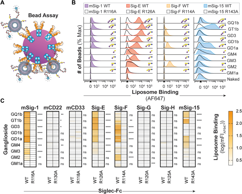
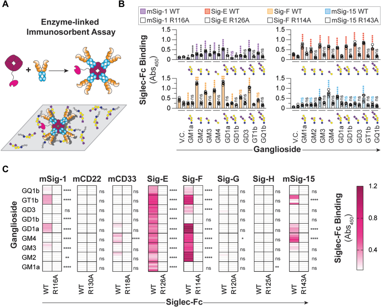
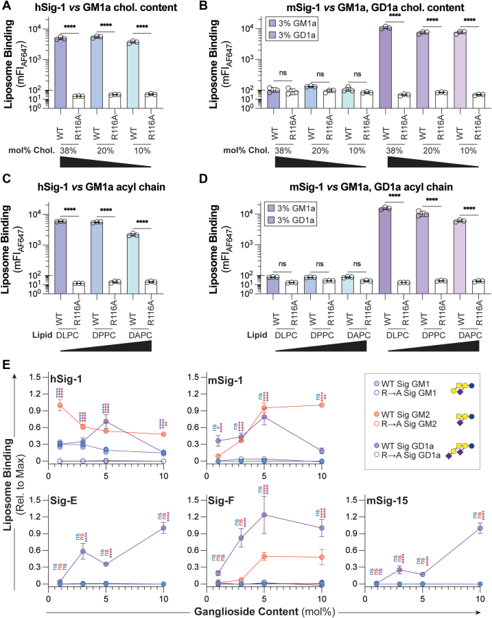
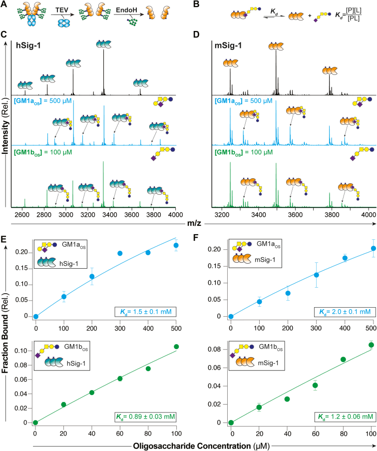
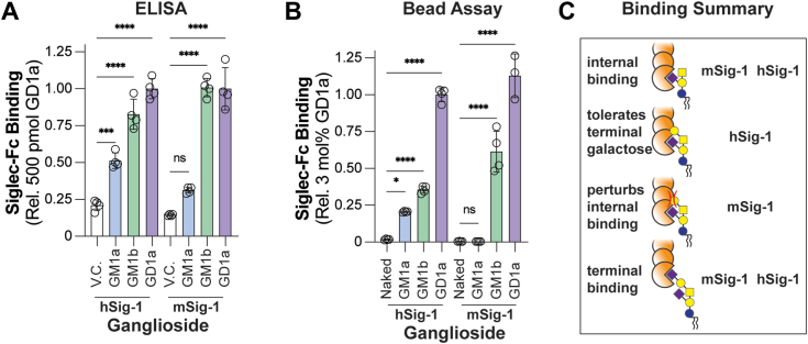
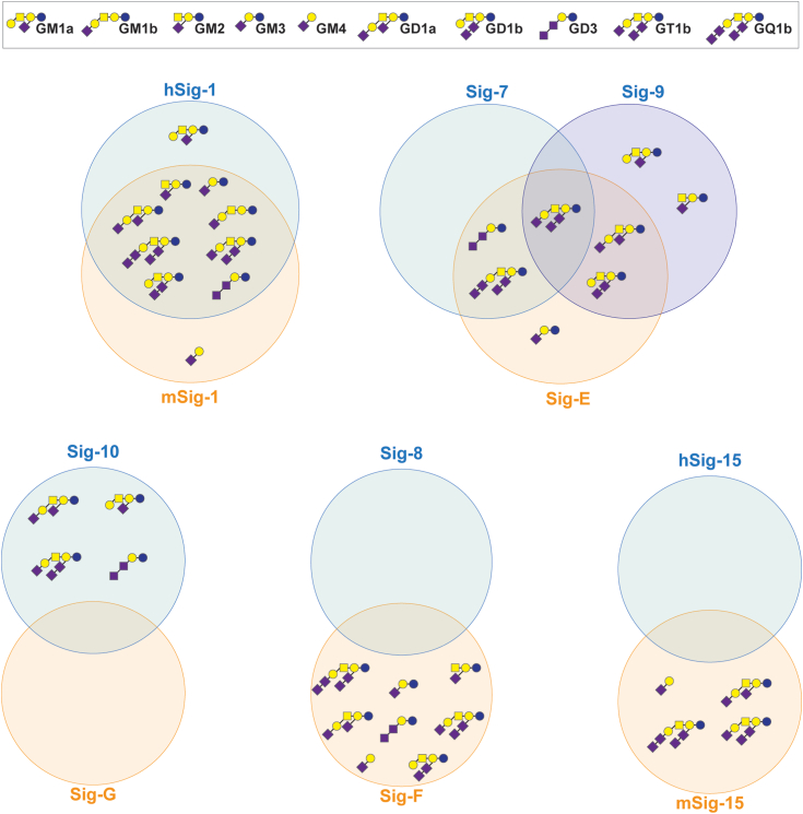
Siglecs are cell surface receptors whose functions are tied to the binding of their sialoglycan ligands. Recently, we developed an optimized liposome formulation and used it to investigate the binding of human Siglecs (hSiglec) against a panel of gangliosides. Animal models, more specifically murine models, are used to understand human biology; however, species-specific differences can complicate the interpretation of the results. Herein, we used our optimized liposome formulation to dissect the interactions between murine Siglecs (mSiglecs) and gangliosides to assess the appropriateness of mSiglecs as a proxy to better understand the biological roles of hSiglec-ganglioside interactions. Using our optimized liposome formulation, we found that ganglioside binding is generally conserved between mice and humans with mSiglec-1, -E, -F, and -15 binding multiple gangliosides like their human counterparts. However, in contrast to the hSiglecs, we observed little to no binding between the mSiglecs and ganglioside GM1a. Detailed analysis of mSiglec-1 interacting with GM1a and its structural isomer, GM1b, suggests that mSiglec-1 preferentially binds α2-3-linked sialic acids presented from the terminal galactose residue. The ability of mSiglecs to interact or not interact with gangliosides, particularly GM1a, has implications for using mice to study neurodegenerative diseases, infections, and cancer, where interactions between Siglecs and glycolipids have been proposed to modulate these human diseases.

Keywords: Siglec; flow cytometry; ganglioside; glycolipid; liposome; mass spectrometry; mouse.


Conflict of interest statement

Conflict of interest The authors declare that they have no conflicts of interest with the contents of this article.

Figures

Figure 1
Figure 1
Interrogation of mSiglecs against ganglioside liposomes.A, schematic representation of the bead assay where the Siglec-Fc, featuring a C-terminal Strep-tag II, is precomplexed with streptavidin-coated microbeads and gangliosides are embedded in fluorescently labeled liposomes and binding is read out via flow cytometry. B, representative flow cytometry histograms of mSiglec-1, Siglec-E, Siglec-F, mSiglec-15, and their corresponding arginine mutants binding to ganglioside liposomes. C, heatmaps summarizing ganglioside liposome binding to mSiglecs. Color is representative of the mean log10(mFIAF647) of each ganglioside liposome subtracted from the log10(mFIAF647) of the same ganglioside against the corresponding arginine mutant from at least three technical replicates. For (C), a one-way ANOVA was used to compare the binding between the WT Siglec and the corresponding arginine mutant. Not Significant (NS), p > 0.05; ∗0.05 > p ≥ 0.01; ∗∗0.01 > p ≥ 0.001; ∗∗∗0.001 > p ≥ 0.0001; ∗∗∗∗p < 0.0001.
Figure 2
Figure 2
Recognition of gangliosides by mSiglecs is affected by presentation.A, schematic representation of the ELISA where Siglec-Fc is precomplexed with Strep-Tactin-HRP and gangliosides are adsorbed to a microplate. B, representative ELISA results of mSiglec-1, Siglec-E, Siglec-F, mSiglec-15, and their corresponding arginine mutants binding to adsorbed gangliosides. C, heatmaps summarizing mSiglec-Fc binding to adsorbed gangliosides. Color is representative of the mean binding of the WT mSiglec-Fc complex to the adsorbed gangliosides liposome subtracted from the corresponding mutant mSiglec-Fc complex binding to the same ganglioside at least four technical replicates. For (B), a one-way ANOVA was used to compare the binding between the WT Siglec–ganglioside interaction and vehicle control well. For (C), a one-way ANOVA was used to compare the binding between the WT Siglec and the corresponding arginine mutant. Not Significant (NS), p > 0.05; ∗0.05 > p ≥ 0.01; ∗∗0.01 > p ≥ 0.001; ∗∗∗∗p < 0.0001. HRP, horseradish peroxidase.
Figure 3
Figure 3
Optimization of liposome formulation parameters for murine leclec-1.A and B, cholesterol content titration of GM1a and GD1a liposomes against h/mSiglec-1 respectively. C and D, acyl chain length titration of GM1a and GD1a liposomes against h/mSiglec-1, respectively. E, GM1a, GM2, and GD1a content titration (1–10 mol% of total lipids in the liposome) against hSiglec-1, mSiglec-1, Siglec-E, Siglec-F, and mSiglec-15, as well as their respective arginine mutants. Data is represented as the mean of at least three technical replicates, and error bars are representative of one SD from the mean. For panels (AD), a one-way ANOVA was used to compare liposome binding between the WT and mutant Siglec at the same cholesterol content or acyl chain length. For (E), a one-way ANOVA was used to compare the binding between the WT Siglec and the corresponding arginine mutant at each mol% of ganglioside. Not Significant (NS), p > 0.05; ∗0.05 > p ≥ 0.01; ∗∗0.01 > p ≥ 0.001; ∗∗∗∗p < 0.0001. DLPC (12-carbon), DPPC (16-carbon), DAPC (20 carbon). Blue-GM1, red-GM2; purple-GD1a; Chol, cholesterol.
Figure 4
Figure 4
Quantifying the interactions between m/hSiglec-1 and the oligosaccharide of GM1a and GM1b by a native mass spectrometry binding assay.A, schematic for the preparation of the Siglec-1 fragment from CHO Lec1 cells to eliminate heterogeneity from N-glycans. B, depiction of the equilibrium between Siglec-bound GM1b oligosaccharide and free oligosaccharide used to determine disassociation constants (Kd). C and D, representative mass spectra for the binding between m/hSiglec-1 fragment to the oligosaccharide of GM1a (500 μM) and GM1b (100 μM). E and F, summary of ganglioside oligosaccharides titrations binding between m/hSiglec-1 fragment to the oligosaccharide of GM1a (100–500 μM) and GM1b (20–100 μM).
Figure 5
Figure 5
Comparative binding of internal and externally linked sialic acids to m/hSiglec-1 in the bead assay and ELISA.A and B, hSiglec-1 and mSiglec-1 binding to GM1a, GM1b, and GD1a in the ELISA and bead assay, respectively. C, proposed model for human mSiglec-1 binding of gangliosides. Data is represented as the mean of at least three technical replicates and error bars are representative of one SD from the mean. For (A) and (B), a one-way ANOVA was used to compare the binding of each ganglioside to either a vehicle control (V.C.) or a naked liposome respectfully. Not Significant (NS); p > 0.05; ∗0.05 > p ≥ 0.01; ∗∗∗0.001 > p ≥ 0.0001; ∗∗∗∗p < 0.0001.
Figure 6
Figure 6
Venn diagram comparison of murine and human Siglec ganglioside binding. Interactions reported are a summary of interactions identified in this study and our precious study (15).

Similar articles

Cited by

  • An ultrasensitive and modular platform to detect Siglec ligands and control immune cell function.
    Jame-Chenarboo Z, Schmidt EN, Crichton M, Takahashi-Yamashiro K, Lima GM, Luna-Dulcey L, Jung J, Ivison S, St Laurent CD, Enterina JR, Lin SY, Sarkar S, John R, Nanjappa SG, Malaker SA, Levings MK, Marth JD, Derda R, Macauley MS. Jame-Chenarboo Z, et al. bioRxiv [Preprint]. 2025 Jun 12:2025.06.10.658684. doi: 10.1101/2025.06.10.658684. bioRxiv. 2025. PMID: 40661511 Free PMC article. Preprint.

References

    1. Perlman R.L. Mouse models of human disease: an evolutionary perspective. Evol. Med. Public Health. 2016;2016:170–176. - PMC - PubMed
    1. Bhattacherjee A., Rodrigues E., Jung J., Luzentales-Simpson M., Enterina J.R., Galleguillos D., et al. Repression of phagocytosis by human CD33 is not conserved with mouse CD33. Commun. Biol. 2019;2:450. - PMC - PubMed
    1. Macauley M.S., Kawasaki N., Peng W., Wang S.H., He Y., Arlian B.M., et al. Unmasking of CD22 co-receptor on germinal center B-cells occurs by alternative mechanisms in mouse and man. J. Biol. Chem. 2015;290:30066–30077. - PMC - PubMed
    1. Eskandari-Sedighi G., Jung J., Macauley M.S. CD33 isoforms in microglia and Alzheimer's disease: friend and foe. Mol. Aspects Med. 2023;90 - PubMed
    1. Cheon D.J., Orsulic S. Mouse models of cancer. Annu. Rev. Pathol. 2011;6:95–119. - PubMed

Publication types

LinkOut - more resources