. 2024 Jul;300(7):107483.
doi: 10.1016/j.jbc.2024.107483. Epub 2024 Jun 17.

Ubiquitination is involved in PKC-mediated degradation of cell surface Kv1.5 channels

Affiliations

Ubiquitination is involved in PKC-mediated degradation of cell surface Kv1.5 channels

Ananya Chakraborty et al. J Biol Chem. 2024 Jul.

Abstract

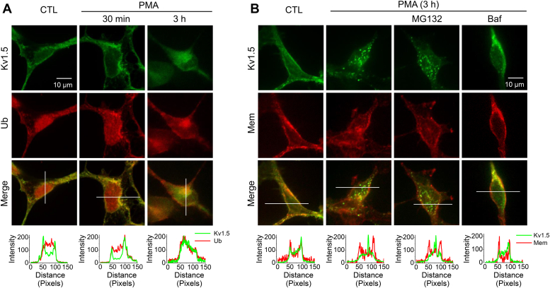
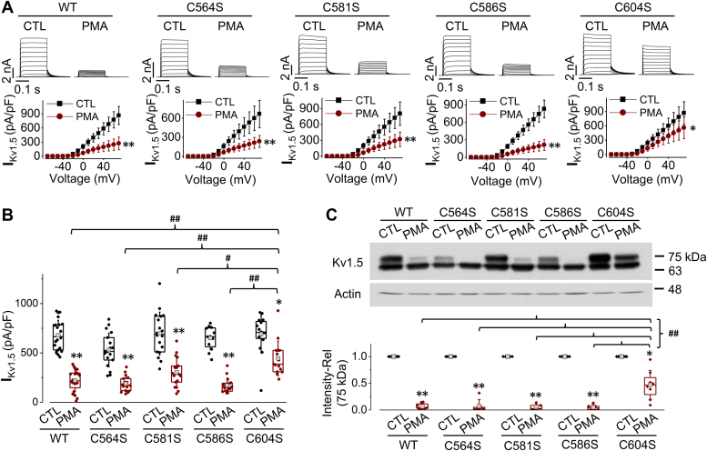
The voltage-gated Kv1.5 potassium channel, conducting the ultra-rapid delayed rectifier K+ current (IKur) in human cells, plays important roles in the repolarization of atrial action potentials and regulation of the vascular tone. We previously reported that activation of protein kinase C (PKC) by phorbol 12-myristate 13-acetate (PMA) induces endocytic degradation of cell-surface Kv1.5 channels, and a point mutation removing the phosphorylation site, T15A, in the N terminus of Kv1.5 abolished the PMA-effect. In the present study, using mutagenesis, patch clamp recording, Western blot analysis, and immunocytochemical staining, we demonstrate that ubiquitination is involved in the PMA-mediated degradation of mature Kv1.5 channels. Since the expression of the Kv1.4 channel is unaffected by PMA treatment, we swapped the N- and/or C-termini between Kv1.5 and Kv1.4. We found that the N-terminus alone did not but both N- and C-termini of Kv1.5 did confer PMA sensitivity to mature Kv1.4 channels, suggesting the involvement of Kv1.5 C-terminus in the channel ubiquitination. Removal of each of the potential ubiquitination residue Lysine at position 536, 565, and 591 by Arginine substitution (K536R, K565R, and K591R) had little effect, but removal of all three Lysine residues with Arginine substitution (3K-R) partially reduced PMA-mediated Kv1.5 degradation. Furthermore, removing the cysteine residue at position 604 by Serine substitution (C604S) drastically reduced PMA-induced channel degradation. Removal of the three Lysines and Cys604 with a quadruple mutation (3K-R/C604S) or a truncation mutation (Δ536) completely abolished the PKC activation-mediated degradation of Kv1.5 channels. These results provide mechanistic insight into PKC activation-mediated Kv1.5 degradation.

Keywords: patch clamp; protein kinase C; ubiquitination; voltage-gated potassium channel.


Conflict of interest statement

Conflict of interest The authors declare that they have no conflicts of interest with the contents of this article.

Figures

Figure 1
Figure 1
Intact intracellular environment is required for PMA-induced reduction in IKv1.5.A, PMA (10 nM, 30 min) application had no effect on IKv1.5 during the whole-cell patch clamp configuration. Representative current traces are shown above summarized current-voltage relationships (n = 6 cells for each group from 3 independent experiments). B, PMA (10 nM, 30 min) application to intact cells reduced IKv1.5. Representative current traces are shown above summarized current-voltage relationships (n = 15 cells for control (CTL), n = 18 cells for PMA treatment from four independent experiments). ∗∗p < 0.01 versus CTL for current amplitudes upon ≥10 mV depolarizing steps. C, relative IKv1.5 continuously recorded with the voltage protocol shown above the current traces every 30 s using perforated-patch whole-cell clamp from cells exposed to PMA (10 nM) in the absence (red) or presence (blue) of the PKC inhibitor sotrastaurin (Sotra, 200 nM). Current traces at time points of ‘a’, ‘b’, ‘c’, and ‘d’ are superimposed for each condition.
Figure 2
Figure 2
Ubiquitination is involved in PMA-induced decrease in IKv1.5and mature channel expression.A, inhibition of ubiquitination by 5 μM TAK-243 (TAK) abolishes the PMA (10 nM, 3 h)-induced reduction in IKv1.5. Representative current traces are shown above summarized current-voltage relationships. ∗∗p < 0.01 versus CTL for current amplitudes upon ≥10 mV depolarizing steps. B, inhibition of ubiquitination by 5 μM TAK-243 abolishes the PMA (10 nM, 3 h)-induced reduction in expression of mature Kv1.5 proteins. β-Actin (42 kDa) was used as a loading control (n = 4). C, effects of PMA treatment for 30 min or 3 h on the expression of Kv1.5 channels in Kv1.5-HEK cells transfected with empty vector (pcDNA3), WT Ub, or mutant Ub, UbKO. D, Co-immunoprecipitation analysis shows that ubiquitination of mature Kv1.5 channels is increased by PMA treatment (10 nM, 30 min) (n = 8). Red arrows indicate ubiquitinated Kv1.5 proteins. CTL, control.
Figure 3
Figure 3
PMA treatment induces Kv1.5 internalization involving Ub via lysosomal degradation.A, confocal images portraying the localization of Kv1.5 (green) and Ub (red) in Kv1.5-HEK cells in control (CTL) as well as in PMA treatment (10 nM) for 30 min or 3 h. B, confocal images portraying the localization of Kv1.5 (green) in cells in control (CTL) and in PMA treatment (10 nM, 3 h) in the absence or presence of proteasomal inhibitor MG132 (10 μM) or lysosomal inhibitor bafilomycin A1 (Baf, 1 μM). Cell membranes (Mem) were labeled with Texas Red X-conjugated wheat germ agglutinin (red). The signal intensity distributions along the white lines were analyzed using ImageJ.
Figure 4
Figure 4
Both N- and C-termini of Kv1.5 are involved in PKC activation-mediated channel degradation.Upper panels: Schematic diagrams of WT Kv1.5 (A), WT Kv1.4 (B), chimeric Kv1.5 with Kv1.4 N-terminus, Kv1.5-Kv1.4NT (C), chimeric Kv1.4 with Kv1.5 N-terminus, Kv1.4-Kv1.5NT (D), chimeric Kv1.4 with Kv1.5 C-terminus, Kv1.4-Kv1.5CT (E), or chimeric Kv1.4 with both Kv1.5 N- and C-termini, Kv1.4-Kv1.5NT+CT (F). Middle and lower panels: Representative current traces and summarized current-voltage relationships showing the effects of 10 nM PMA treatment for 3 h on the channels illustrated in the upper panels (n = 13–27 cells from 3 independent experiments for each group). ∗∗p < 0.01 versus control (CTL) for current amplitudes upon ≥10 mV depolarizing steps. Data are presented as mean ± SD.
Figure 5
Figure 5
Location of Cysteine and Lysine residues on the C-terminus of Kv1.5.A, sequence alignments of the C-terminus of Kv1.4 and Kv1.5 with cysteine and lysine residues investigated in the present study as potential ubiquitination sites. B, illustrative locations of the investigated Lysine and Cysteine residues in Kv1.5 C-terminus.
Figure 6
Figure 6
Role of lysine residues on the C-terminus of Kv1.5 in PMA-induced reduction in IK1.5and mature Kv1.5 channel expression.A, the effects of PMA treatment (10 nM, 3 h) on IKv1.5 of WT Kv1.5, Kv1.5-K536R, Kv1.5-K565R, Kv1.5-K591R, and Kv1.5-3K-R (triple lysines changed to arginines) mutants. Representative current traces and summarized current-voltage relationships are shown. ∗p < 0.05, ∗∗p < 0.01 versus control (CTL) for current amplitudes upon ≥10 mV depolarizing steps. B, peak IKv1.5 density is shown as box plots with mean ± SD in control (CTL) and in PMA treatment (10 nM, 3 h) for WT and mutant channels. The PMA-induced reduction in IKv1.5 of the triple mutant is significantly weakened than that seen in WT Kv1.5 or in single-point mutants. ∗p < 0.05, ∗∗p < 0.01 versus CTL; #p < 0.05, ##p < 0.01 versus indicated groups. C, the effects of PMA treatment (10 nM, 3 h) on the expression of WT Kv1.5, Kv1.5-K536R, Kv1.5-K565R, Kv1.5-K591R, and Kv1.5-3K-R mutants. The intensity of the 75-kDa band in each lane was firstly normalized to the loading control actin (42-kDa) in the same lane, then normalized to its control, and summarized as box plots with mean ± SD beneath the representative Western blot images. n = 3 to 8, ∗p < 0.05, ∗∗p < 0.01 versus CTL; ##p < 0.01 versus indicated groups.
Figure 7
Figure 7
Role of cysteine residues on the C-terminus of Kv1.5 in PMA-induced reduction in IK1.5and mature Kv1.5 channel expression.A, the effects of PMA treatment (10 nM, 3 h) on IKv1.5 of WT Kv1.5, Kv1.5-C564S, Kv1.5-C581S, Kv1.5-C586S, and Kv1.5-C604S mutants. Representative current traces and summarized current-voltage relationships are shown. ∗p < 0.05, ∗∗p < 0.01 versus control (CTL) for current amplitudes upon ≥10 mV depolarizing steps. B, IKv1.5 density is shown as box plots with mean ± SD in control (CTL) and in PMA treatment (10 nM, 3 h) for WT and mutant channels. The PMA-induced reduction in IKv1.5 of Kv1.5-C604S is significantly weakened than that seen in WT Kv1.5 or in other mutant channels. ∗p < 0.05, ∗∗p < 0.01 versus CTL; #p < 0.05, ##p < 0.01 versus indicated groups. C, the effects of PMA treatment (10 nM, 3 h) on the expression of WT Kv1.5, Kv1.5-C564S, Kv1.5-C581S, Kv1.5-C586S, and Kv1.5-C604S mutants. The intensity of the 75-kDa band in each lane was firstly normalized to the loading control actin (42-kDa) in the same lane, then normalized to its control, and summarized as box plots with mean ± SD beneath the representative Western blot images. n = 5 to 8, ∗p < 0.05, ∗∗p < 0.01 versus CTL; ##p < 0.01 versus indicated groups.
Figure 8
Figure 8
Removing all potential ubiquitination sites completely abolishes PMA-induced reduction in IK1.5and mature Kv1.5 channel expression.A, the effects of PMA treatment (10 nM, 3 h) on IKv1.5 of WT Kv1.5, Kv1.5-Δ536 and Kv1.5-3K-R/C604S. Representative current traces and summarized current-voltage relationships are shown. n = 10 to 12 cells for WT Kv1.5, n = 17 to 18 cells for Kv1.5-Δ536, n = 17 to 18 cells for Kv1.5-3K-R/C604S. For each channel, at least 3 independent experiments were performed. ∗∗p < 0.01 versus control (CTL). B, the effects of PMA treatment (10 nM, 3 h) on the expression of WT Kv1.5, Kv1.5-Δ536, and Kv1.5-3K-R/C604S channels. The intensity of the upper band in each lane was firstly normalized to the loading control actin (42-kDa) in the same lane, then normalized to its control, and summarized as box plots with mean ± SD beneath the representative Western blot images. n = 5 to 7, ∗∗p < 0.01 versus CTL; ##p < 0.01 versus WT with PMA. C, co-immunoprecipitation analysis shows that PMA treatment had no effect on the ubiquitination of Kv1.5-Δ536 channels (n = 4). D, immunofluorescence images portraying the localization of Kv1.5 in HEK293 cells stably expressing WT Kv1.5 or Kv1.5-Δ536 after culture with 10 nM PMA for 3 h. Kv1.5 was detected with anti-Kv1.5 primary antibody and Alexa Fluor 488-conjugated secondary antibody (green). Cell membranes (Mem) are labeled with Texas Red X-conjugated wheat germ agglutinin (red). The signal intensity distributions along the white lines were analyzed using ImageJ.

Similar articles

Cited by

References

    1. Albrecht B., Weber K., Pongs O. Characterization of a voltage-activated K-channel gene cluster on human chromosome 12p13. Recept Channels. 1995;3:213–220. - PubMed
    1. Fedida D., Wible B., Wang Z., Fermini B., Faust F., Nattel S., et al. Identity of a novel delayed rectifier current from human heart with a cloned K+ channel current. Circ. Res. 1993;73:210–216. - PubMed
    1. Wang Z., Fermini B., Nattel S. Sustained depolarization-induced outward current in human atrial myocytes: evidence for a novel delayed rectifier K+ current similar to Kv1.5 cloned channel currents. Circ. Res. 1993;73:1061–1076. - PubMed
    1. MacDonald P.E., Wheeler M.B. Voltage-dependent K+ channels in pancreatic beta cells: role, regulation and potential as therapeutic targets. Diabetologia. 2003;46:1046–1062. - PubMed
    1. Kidd M.W., Bulley S., Jaggar J.H. Angiotensin II reduces the surface abundance of Kv1.5 channels in arterial myocytes to stimulate vasoconstriction. J. Physiol. 2017;595:1607–1618. - PMC - PubMed

Publication types

LinkOut - more resources