. 2024 Jul;300(7):107486.
doi: 10.1016/j.jbc.2024.107486. Epub 2024 Jun 18.

Crosstalk of MAP3K1 and EGFR signaling mediates gene-environment interactions that block developmental tissue closure

Affiliations

Crosstalk of MAP3K1 and EGFR signaling mediates gene-environment interactions that block developmental tissue closure

Jingjing Wang et al. J Biol Chem. 2024 Jul.

Abstract

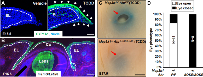
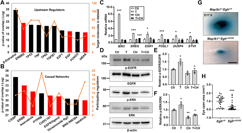
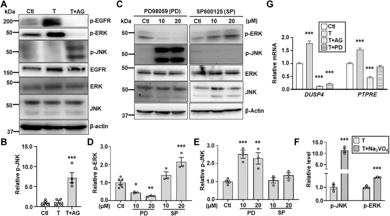
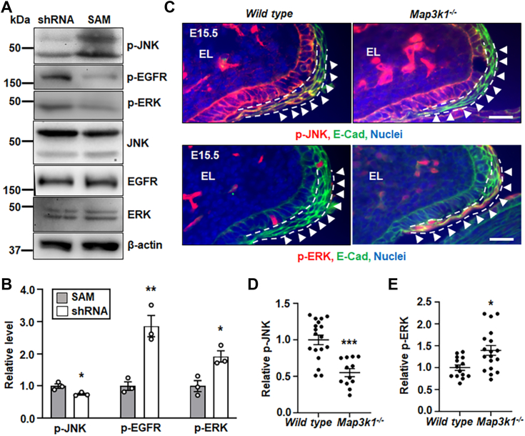
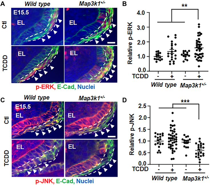
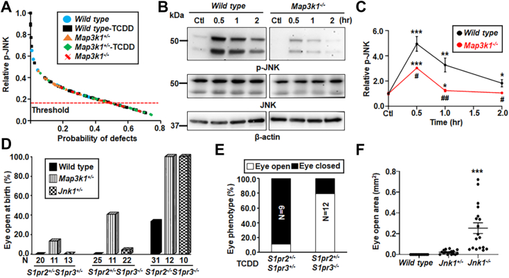
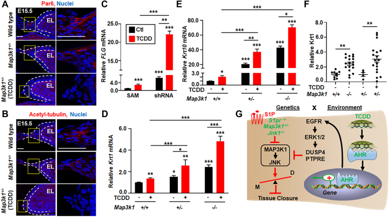
Aberrant regulation of signal transduction pathways can adversely derail biological processes for tissue development. One such process is the embryonic eyelid closure that is dependent on the mitogen-activated protein kinase kinase kinase 1 (MAP3K1). Map3k1 KO in mice results in defective eyelid closure and an autosomal recessive eye-open at birth phenotype. We have shown that in utero exposure to dioxin, a persistent environmental toxicant, induces the same eye defect in Map3k1+/- heterozygous but not WT pups. Here, we explore the mechanisms of the Map3k1 (gene) and dioxin (environment) interactions (GxE) underlying defective eyelid closure. We show that, acting through the aryl hydrocarbon receptor, dioxin activates epidermal growth factor receptor signaling, which in turn depresses MAP3K1-dependent Jun N-terminal kinase (JNK) activity. The dioxin-mediated JNK repression is moderate but is exacerbated by Map3k1 heterozygosity. Therefore, dioxin exposed Map3k1+/- embryonic eyelids have a marked reduction of JNK activity, accelerated differentiation and impeded polarization in the epithelial cells. Knocking out Ahr or Egfr in eyelid epithelium attenuates the open-eye defects in dioxin-treated Map3k1+/- pups, whereas knockout of Jnk1 and S1pr that encodes the sphigosin-1-phosphate (S1P) receptors upstream of the MAP3K1-JNK pathway potentiates the dioxin toxicity. Our novel findings show that the crosstalk of aryl hydrocarbon receptor, epidermal growth factor receptor, and S1P-MAP3K1-JNK pathways determines the outcome of dioxin exposure. Thus, gene mutations targeting these pathways are potential risk factors for the toxicity of environmental chemicals.

Keywords: AHR; EGFR; developmental tissue closure; dioxin; epithelial morphogenesis; gene-environment interactions; the S1PR-MAP3K1-JNK pathway.


Conflict of interest statement

Conflict of interest The authors declare that they have no conflicts of interest with the contents of this article.

Figures

Figure 1
Figure 1
Eyelid epithelial AHR is required for TCDD toxicity in embryonic eyelid closure.A, immunofluorescent staining using anti-CYP1A1 (green) and Hoechst (blue) for nucleus of the eyelids in WT E15.5 embryos with or without TCDD exposure (50 μg/kg body weight) at 11.5 days. Dash lines mark basement membrane that separated the eyelid epithelium and the underneath dermis; white arrows point at CYP1A1 positive staining. B, Hoechst (blue) staining for nucleus of eyes of the E15.5 mTmG/Le-Cre embryos. Cre expressing, i.e., GFP positive, cells were located at the ocular surface epithelium (OSE), but not stroma of eyelid (EL), cornea (Co), and lens. The dotted lines mark the basement membrane that separated the epithelium and the stroma. Pups exposed at E11.5 to TCDD (250 μg/kg body weight) were collected at E17.5, and their eyes (C) were photographed, red arrow, eyelid leading edge, and (D) quantification of eyes displaying open eye phenotype in Map3k1+/−AhrF/F (N = 18) and Map3k1+/−AhrΔOSE/ΔOSE (N = 8) fetuses. N= number of pups of the indicated genotype. The scale bars represent 50 μm in A, 100 μm in B, and 500 μm in C. AHR, aryl hydrocarbon receptor; MAP3K1, mitogen-activated protein kinase kinase kinase 1; OSE, ocular surface epithelium; TCDD, 2,3,7,8-tetrachlorodibenzo-para-dioxin.
Figure 2
Figure 2
The TCDD-AHR axis activates EGFR signaling. The TCDD-AHR axis upregulated genes were subjected to ingenuity pathway analyses to identify (A) top upstream regulators and (B) top causal networks, with relative p-value (bars) and number of participating molecules (orange line). The EGFR signaling relevant upstream regulators and causal networks are labeled as red bars. C, selective gene expression was measured by qRT-PCR using RNA isolated from HaCaT cells treated with vehicle (DMSO, Ctl), TCDD (T, 10 nM), CH223191 (CH, 10 μM), and TCDD plus CH223191 (T+CH). Relative expression was calculated using the housekeeping gene GAPDH as an internal control and compared to that in Ctl set as 1. D, Western blot analyses of the EGFR pathway activity using antibodies for phosphor (p) and total EGFR and ERK, and β-actin as a loading control. Quantification of (E) p-EGFR/EGFR and (F) p-ERK/ERK ratio of the immunoblotting data, with ratio in Ctl set as 1. The TCDD-exposed Map3k1+/−Egfr+/F (N = 13) and Map3k1+/−Egfr+/ΔOSE (N = 14) fetuses were collected at E17.5 and their eyes were (G) photographed and representative images were shown. The scale bar represents 500 μm, and (H) the areas of open eye were measured. Results were shown as mean ± SEM of at least three independent experiments (N ≥ 3). ∗p < 0.05, ∗∗p < 0.01 and ∗∗∗p < 0.001 were considered statistically significant compared to Ctl (in C, E, and F) and TCDD-treated Map3k1+/−Egfr+/F group (in H). AHR, aryl hydrocarbon receptor; DMSO, dimethyl sulfoxide; EGFR, epidermal growth factor receptor; ERK, extracellular signal regulated kinase; MAP3K1, mitogen-activated protein kinase kinase kinase 1; OSE, ocular surface epithelium; qRT-PCR, quantitative reverse transcription PCR; TCDD, 2,3,7,8-tetrachlorodibenzo-para-dioxin.
Figure 3
Figure 3
The EGFR-ERK pathway inhibits JNK activities. HaCaT cells treated with vehicle (DMSO, Ctl) and TCDD (T, 10 nM) in the presence or absence of the EGFR inhibitor, AG1478 (AG, 10 μM), were (A) subjected to immunoblotting for p-EGFR, p-ERK, p-JNK, total JNK, and β-actin, and (B) quantification for p-JNK using β-actin as a loading control. HaCaT cells treated with different concentrations of an ERK inhibitor, PD98059 (PD), and a JNK inhibitor, SP600125 (SP) were (C) examined by immunoblotting for p-ERK, p-JNK, and β-actin, and values of (D) p-ERK and (E) p-JNK were quantified and compared to that of β-actin. F, p-JNK, p-ERK, and β-actin were examined by immunoblotting in lysates of HaCaT cells treated with TCDD (T, 10 nM) in the presence and absence of phosphatase inhibitor Na3VO4 (0.5 mM). Values of relative p-JNK and p-ERK were quantified using β-actin. G, expression of DUSP4 and PTPRE was examined by qRT-PCR in HaCaT cells treated with vehicle (DMSO, Ctl) and TCDD (T, 10 nM) in the presence and absence of 10 μM AG and PD. Relative mRNA of DUSP4 and PTPRE was calculated using GAPDH as an internal control. Value of Ctl sample was set as 1. Data were shown as mean ± SEM of at least three independent experiments (N ≥ 3). ∗p < 0.05, ∗∗p < 0.01 and ∗∗∗p < 0.001 were considered significantly different compared to Ctl (in B, D, E, and G) and TCDD-treated cells (in F). DMSO, dimethyl sulfoxide; DUSP4, dual specificity phosphatase 4; EGFR, epidermal growth factor receptor; ERK, extracellular signal regulated kinase; JNK, Jun N-terminal kinase; PTPRE, protein tyrosine phosphatase epsilon; qRT-PCR, quantitative reverse transcription PCR; TCDD, 2,3,7,8-tetrachlorodibenzo-para-dioxin.
Figure 4
Figure 4
The MAP3K1-JNK and EGFR-ERK pathways inhibit each other. Lysates of the shRNA HaCaT and SAM HaCaT cells were examined by Western blotting for (A) p-JNK, p-EGFR, p-ERK, and β-actin, and (B) quantification of p-JNK, p-EGFR, and p-ERK using β-actin as a loading control. Levels in SAM HaCaT were set as 1. Data represented three independent experiments (N = 3) and were shown as mean ± SEM. C, immunofluorescence staining of WT and Map3k1−/− E15.5 embryonic eyelids with anti-p-JNK (red, top panels) and anti-p-ERK (red, bottom panels), costained with anti-E-cadherin (green) that marks epithelial membrane, and Hoechst (blue) labels nuclei. Representative images were shown, the scale bars represent 50 μm. The (D) p-JNK, and (E) p-ERK, in the suprabasal epithelial cells, marked with dash lines, of the eyelid leading edge (arrowheads) were quantified and compared to that in WT, set as 1. At least three sections (N ≥ 3) per embryo and three embryos (N ≥ 3) of each genotype from different litters were examined. Data were shown as mean ± SEM. ∗p < 0.05, ∗∗p < 0.01 and ∗∗∗p < 0.001 were considered significantly different from SAM cells (in B) and WT embryos (in D and E). EGFR, epidermal growth factor receptor; ERK, extracellular signal regulated kinase; JNK, Jun N-terminal kinase; MAP3K1, mitogen-activated protein kinase kinase kinase 1; SAM, synergistic activation mediator.
Figure 5
Figure 5
TCDD plus Map3k1 heterozygosity tilt the balance of JNK and ERK. Eyelid tissues of WT and Map3k1+/− E15.5 embryos with or without TCDD (50 μg/kg body weight) exposure were subjected to immunohistochemistry for (A and B) p-ERK (red) and (C and D) p-JNK (red). E-cadherin (green) and Hoechst (blue) were markers of epithelial membrane and nuclei, respectively. Representative images captured with a fluorescent microscopy were shown. The (B) p-ERK and (D) p-JNK in the suprabasal epithelium of the eyelid leading edge (arrowheads), marked with dash lines, were quantified. At least three sections (N ≥ 3) per embryo and three embryos (N ≥ 3) per genotype/treatment conditions were examined. Results were shown as mean ± SEM. ∗∗p < 0.01, ∗∗∗p < 0.001 represents significantly different from the unexposed groups and TCDD-treated WT group (in B and D). The scale bar represent 50 μm (A and C). ERK, extracellular signal regulated kinase; JNK, Jun N-terminal kinase; MAP3K1, mitogen-activated protein kinase kinase kinase 1; TCDD, 2,3,7,8-tetrachlorodibenzo-para-dioxin.
Figure 6
Figure 6
Crosstalk of TCDD and the S1PR-MAP3K1-JNK1 pathway for embryonic eyelid closure.A, the levels of p-JNK in the eyelid epithelium were collected from E15.5 embryos of different genetic and exposure conditions as indicated. Data were analyzed by ImageJ software, and the relationship between the relative p-JNK level and the probability of the open-eye defects was calculated using a logistic regression model based on the binary outcome of eyelid closure, along with ROC analysis. Probability of defect was significant higher when p-JNK was below 0.17. B, The WT and Map3k1−/− keratinocytes treated with vehicle (DMSO, Ctl) or 20 μM S1P for 0.5 to 2 h and p-JNK and β-actin were examined with Western blotting. C, quantification of p-JNK using β-actin as a loading control. Data are mean ± SEM of at least three independent experiments (N ≥ 3). The S1pr2-and S1pr3-compound mutant pups under WT, Map3k1+/− and Jnk1+/− genetic backgrounds as indicated (D) without and (E) with in utero exposure to 50 μg/kg TCDD on E11.5 were collected at E17.5. The pups were examined for the eyelid open/close. N = number of pups of the indicated genotype. F, the open eye areas were measured in E17.5 WT, Jnk1+/− and Jnk1−/− fetuses exposed to TCDD (50 μg/kg body weight) at E11.5. At least 8 pups (N ≥ 8) under each condition as indicated were analyzed. Values in Ctl were set as 1. ∗p < 0.05, ∗∗p < 0.01, ∗∗∗p < 0.001 were significantly different from Ctl of the same genotype (in C) and TCDD exposed WT (in F). #p < 0.05, ##p < 0.01 was significantly different between genotypes under the same treatment condition (in C). DMSO, dimethyl sulfoxide; JNK, Jun N-terminal kinase; MAP3K1, mitogen-activated protein kinase kinase kinase 1; ROC, receiver operating characteristic; S1P, sphigosin-1-phosphate; TCDD, 2,3,7,8-tetrachlorodibenzo-para-dioxin.
Figure 7
Figure 7
TCDD and Map3k1 loss-of-function disrupt cell polarity and potentiate epithelial terminal differentiation. Eyelids of E15.5 WT, Map3k1+/−, and TCDD treated Map3k1+/− embryos were subjected to immunohistochemistry using (A) anti-Par6 and (B) anti-acetylated-tubulin, detecting apical epithelial polarity and Hoechst labeling nuclei. Dash lines mark the eyelid epithelium. The scale bar represents 50 μm. C, the expression of Filaggrin (FLG), a marker of terminally differentiated keratinocytesin the granular and cornified epidermis, examined by qRT-PCR in shRNA HaCaT and SAM HaCaT treated with 10 nM TCDD or vehicle (DMSO, Ctl). The relative FLG mRNA was calculated using the housekeeping gene GAPDH as an internal control. FLG levels in SAM (Ctl) were set as 1, expression of (D) Krt1 and (E) Krt10, markers of the differentiating suprabasal keratinocytes, were examined in WT, Map3k1+/−, and Map3k1−/− keratinocytes treated with 10 nM TCDD or vehicle (DMSO). Relative expression was calculated using the housekeeping gene GAPDH as an internal control and compared to levels in WT keratinocytes set as 1. F, the E15.5 embryos of WT, Map3k1−/−, and Map3k1+/− with or without TCDD exposure were examined by immunohistochemistry for Krt1. Staining signals were quantified, and relative expression was calculated. Data are mean ± SEM of at least three independent experiments (N ≥ 3) or at least three eye sections (N ≥ 3) per embryo and three embryos (N ≥ 3) from different litters. ∗p < 0.05, ∗∗p < 0.01, ∗∗∗p < 0.001 were considered statistically significant compared to WT and SAM untreated or as indicated. G, graphic illustration of the proposed GxE interactions model in eyelid closure defects. The environmental factor TCDD activates the AHR to induce gene expression and activate the EGFR-ERK pathways. EGFR signaling in turn induces phosphatases to inhibit the S1PR-MAP3K1-JNK pathway. This effect of TCDD is trivial and insufficient to induce the eyelid phenotype. However, in the presence of gene mutations, i.e., S1pr2/3−/−, Map3k1+/− and Jnk1−/−, which also slightly attenuate MAP3K1-JNK signaling, the effect of TCDD is largely amplified. As the results, the GxE interactions significantly inhibit JNK, accelerate differentiation (D) and impede morphogenesis (M) of the epithelium, leading to defective tissue closure. AHR, aryl hydrocarbon receptor; DMSO, dimethyl sulfoxide; EGFR, epidermal growth factor receptor; ERK, extracellular signal regulated kinase; JNK, Jun N-terminal kinase; Krt1, keratin 1; MAP3K1, mitogen-activated protein kinase kinase kinase 1; qRT-PCR, quantitative reverse transcription PCR; S1P, sphigosin-1-phosphate; SAM, synergistic activation mediator; TCDD, 2,3,7,8-tetrachlorodibenzo-para-dioxin.

Similar articles

Cited by

References

    1. Kuida K., Boucher D.M. Functions of MAP kinases: insights from gene-targeting studies. J. Biochem. 2004;135:653–656. - PubMed
    1. Hagemann C., Blank J.L. The ups and downs of MEK kinase interactions. Cell Signal. 2001;13:863–875. - PubMed
    1. Uhlik M.T., Abell A.N., Cuevas B.D., Nakamura K., Johnson G.L. Wiring diagrams of MAPK regulation by MEKK1, 2, and 3. Biochem. Cell Biol. 2004;82:658–663. - PubMed
    1. Craig E.A., Stevens M.V., Vaillancourt R.R., Camenisch T.D. MAP3Ks as central regulators of cell fate during development. Dev. Dyn. 2008;237:3102–3114. - PubMed
    1. Wang J., Kimura E., Mongan M., Xia Y. Genetic control of MAP3K1 in eye development and sex differentiation. Cells. 2021;11:34. - PMC - PubMed

Publication types

MeSH terms

LinkOut - more resources