

The relative brain signal variability increases in the behavioral variant of frontotemporal dementia and Alzheimer's disease but not in schizophrenia
- PMID: 38897598
- PMCID: PMC11574935
- DOI: 10.1177/0271678X241262583
The relative brain signal variability increases in the behavioral variant of frontotemporal dementia and Alzheimer's disease but not in schizophrenia
Abstract
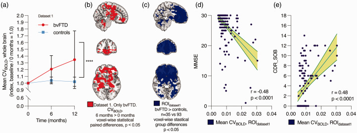
Overlapping symptoms between Alzheimer's disease (AD), behavioral variant of frontotemporal dementia (bvFTD), and schizophrenia (SZ) can lead to misdiagnosis and delays in appropriate treatment, especially in cases of early-onset dementia. To determine the potential of brain signal variability as a diagnostic tool, we assessed the coefficient of variation of the BOLD signal (CVBOLD) in 234 participants spanning bvFTD (n = 53), AD (n = 17), SZ (n = 23), and controls (n = 141). All underwent functional and structural MRI scans. Data unveiled a notable increase in CVBOLD in bvFTD patients across both datasets (local and international, p < 0.05), revealing an association with clinical scores (CDR and MMSE, r = 0.46 and r = -0.48, p < 0.0001). While SZ and control group demonstrated no significant differences, a comparative analysis between AD and bvFTD patients spotlighted elevated CVBOLD in the frontopolar cortices for the latter (p < 0.05). Furthermore, CVBOLD not only presented excellent diagnostic accuracy for bvFTD (AUC 0.78-0.95) but also showcased longitudinal repeatability. During a one-year follow-up, the CVBOLD levels increased by an average of 35% in the bvFTD group, compared to a 2% increase in the control group (p < 0.05). Our findings suggest that CVBOLD holds promise as a biomarker for bvFTD, offering potential for monitoring disease progression and differentiating bvFTD from AD and SZ.
Keywords: Alzheimer’s disease; brain signal variability; frontotemporal dementia; functional MRI; schizophrenia.
Conflict of interest statement
Declaration of conflicting interestsThe author(s) declared no potential conflicts of interest with respect to the research, authorship, and/or publication of this article.
Figures


Similar articles
-
Single Subject Classification of Alzheimer's Disease and Behavioral Variant Frontotemporal Dementia Using Anatomical, Diffusion Tensor, and Resting-State Functional Magnetic Resonance Imaging.J Alzheimers Dis. 2018;62(4):1827-1839. doi: 10.3233/JAD-170893. J Alzheimers Dis. 2018. PMID: 29614652
-
Exploring Links Between Psychosis and Frontotemporal Dementia Using Multimodal Machine Learning: Dementia Praecox Revisited.JAMA Psychiatry. 2022 Sep 1;79(9):907-919. doi: 10.1001/jamapsychiatry.2022.2075. JAMA Psychiatry. 2022. PMID: 35921104 Free PMC article.
-
A Longitudinal Study on Resting State Functional Connectivity in Behavioral Variant Frontotemporal Dementia and Alzheimer's Disease.J Alzheimers Dis. 2017;55(2):521-537. doi: 10.3233/JAD-150695. J Alzheimers Dis. 2017. PMID: 27662284
-
Exploring quantitative group-wise differentiation of Alzheimer's disease and behavioural variant frontotemporal dementia using tract-specific microstructural white matter and functional connectivity measures at multiple time points.Eur Radiol. 2019 Oct;29(10):5148-5159. doi: 10.1007/s00330-019-06061-7. Epub 2019 Mar 11. Eur Radiol. 2019. PMID: 30859283 Free PMC article.
-
Frontal variant of Alzheimer's disease with asymmetric presentation mimicking frontotemporal dementia: Case report and literature review.Brain Behav. 2020 Mar;10(3):e01548. doi: 10.1002/brb3.1548. Epub 2020 Jan 27. Brain Behav. 2020. PMID: 31989779 Free PMC article. Review.
Cited by
-
Ultrafast Imaging of Physiological Brain Pulsations With Magnetic Resonance Encephalography-From Noise to Predictive Clinical Biomarker.NMR Biomed. 2025 Aug;38(8):e70092. doi: 10.1002/nbm.70092. NMR Biomed. 2025. PMID: 40625063 Free PMC article. Review.
-
Orexin effect on physiological pulsations of the human brain.Proc Natl Acad Sci U S A. 2025 Aug 5;122(31):e2501578122. doi: 10.1073/pnas.2501578122. Epub 2025 Aug 1. Proc Natl Acad Sci U S A. 2025. PMID: 40748959 Free PMC article.
References
MeSH terms
LinkOut - more resources
Full Text Sources
Medical
