

Unusual Presentation of Double-seronegative Myasthenia Gravis with Positive Anti-LRP4 Antibody: Diagnostic Utility of a Videofluoroscopic Swallowing Study
- PMID: 38897960
- PMCID: PMC11802234
- DOI: 10.2169/internalmedicine.3348-23
Unusual Presentation of Double-seronegative Myasthenia Gravis with Positive Anti-LRP4 Antibody: Diagnostic Utility of a Videofluoroscopic Swallowing Study
Abstract
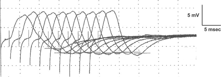
An 86-year-old woman was admitted to our hospital with cryptogenic progressive dyspnea and dysphagia following a tracheostomy procedure 4 months prior to presentation. She exhibited fluctuating diplopia, bilateral vocal fold paralysis, normal nerve test results, negative findings for serum anti-acetylcholine receptor and anti-muscle-specific kinase antibodies, and positive findings for anti-LDL receptor-related protein 4 (LRP4). A videofluoroscopic swallowing study (VFSS) with edrophonium revealed an improvement in bulbar paralysis. Consequently, the patient was diagnosed with double-seronegative myasthenia gravis (DSN-MG) and began immunomodulatory therapy. This case emphasizes the diagnostic challenges of bulbar-type DSN-MG and underscores the value of a VFSS with edrophonium for diagnosing this condition.
Keywords: anti-LDL receptor-related protein 4; bulbar paralysis; double seronegative myasthenia gravis; dysphagia; videofluoroscopic swallowing study; vocal fold paralysis.
Conflict of interest statement
The authors state that they have no Conflict of Interest (COI).
Figures


Similar articles
-
Flow Cytofluorimetric Analysis of Anti-LRP4 (LDL Receptor-Related Protein 4) Autoantibodies in Italian Patients with Myasthenia Gravis.PLoS One. 2015 Aug 18;10(8):e0135378. doi: 10.1371/journal.pone.0135378. eCollection 2015. PLoS One. 2015. PMID: 26284792 Free PMC article.
-
Anti-LRP4 autoantibodies in AChR- and MuSK-antibody-negative myasthenia gravis.J Neurol. 2012 Mar;259(3):427-35. doi: 10.1007/s00415-011-6194-7. Epub 2011 Aug 5. J Neurol. 2012. PMID: 21814823
-
Autoantibodies to Low-Density Lipoprotein Receptor-Related Protein 4 in Double Seronegative Myasthenia Gravis: A Systematic Review.Can J Neurol Sci. 2018 Jan;45(1):62-67. doi: 10.1017/cjn.2017.253. Can J Neurol Sci. 2018. PMID: 29334041
-
Anti-LRP4 autoantibodies in Chinese patients with myasthenia gravis.Muscle Nerve. 2017 Nov;56(5):938-942. doi: 10.1002/mus.25591. Epub 2017 Apr 8. Muscle Nerve. 2017. PMID: 28120340
-
[Novel autoantibodies in myasthenia gravis].Nihon Rinsho. 2013 May;71(5):876-80. Nihon Rinsho. 2013. PMID: 23777098 Review. Japanese.
References
-
- Gilhus NE, Verschuuren JJ. Myasthenia gravis: subgroup classification and therapeutic strategies. Lancet Neurol 14: 1023-1036, 2015. - PubMed
-
- Higuchi O, Hamuro J, Motomura M, Yamanashi Y. Autoantibodies to low-density lipoprotein receptor-related protein 4 in myasthenia gravis. Ann Neurol 69: 418-422, 2011. - PubMed
-
- Zisimopoulou P, Evangelakou P, Tzartos J, et al. . A comprehensive analysis of the epidemiology and clinical characteristics of anti-LRP4 in myasthenia gravis. J Autoimmun 52: 139-145, 2014. - PubMed
-
- Li M, Han J, Zhang Y, et al. . Clinical analysis of Chinese anti-low-density-lipoprotein-receptor-associated protein 4 antibodies in patients with myasthenia gravis. Eur J Neurol 26: 1296-e84, 2019. - PubMed
Publication types
MeSH terms
Substances
LinkOut - more resources
Full Text Sources
Medical
Miscellaneous