. 2024 Jun 20;16(1):48.
doi: 10.1038/s41368-024-00302-2.

Exosomal miR-17-5p derived from epithelial cells is involved in aberrant epithelium-fibroblast crosstalk and induces the development of oral submucosal fibrosis

Affiliations

Exosomal miR-17-5p derived from epithelial cells is involved in aberrant epithelium-fibroblast crosstalk and induces the development of oral submucosal fibrosis

Changqing Xie et al. Int J Oral Sci. .

Abstract

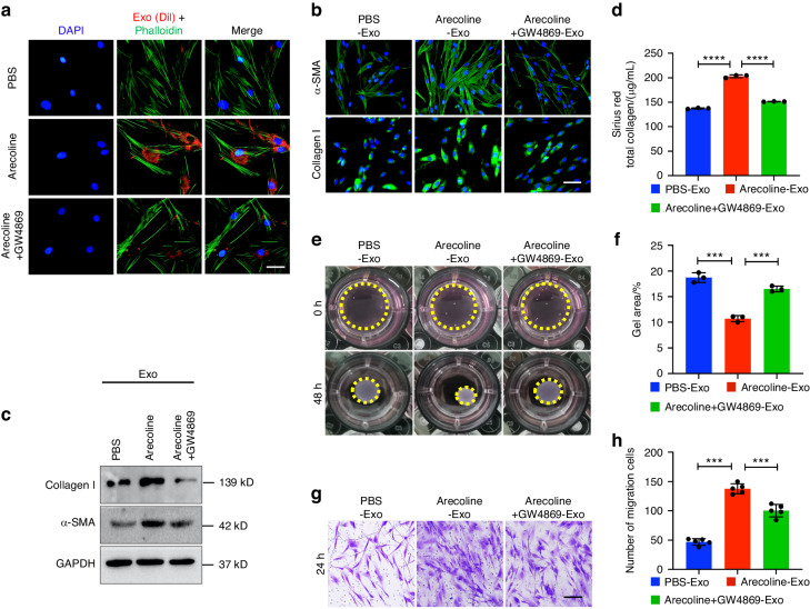
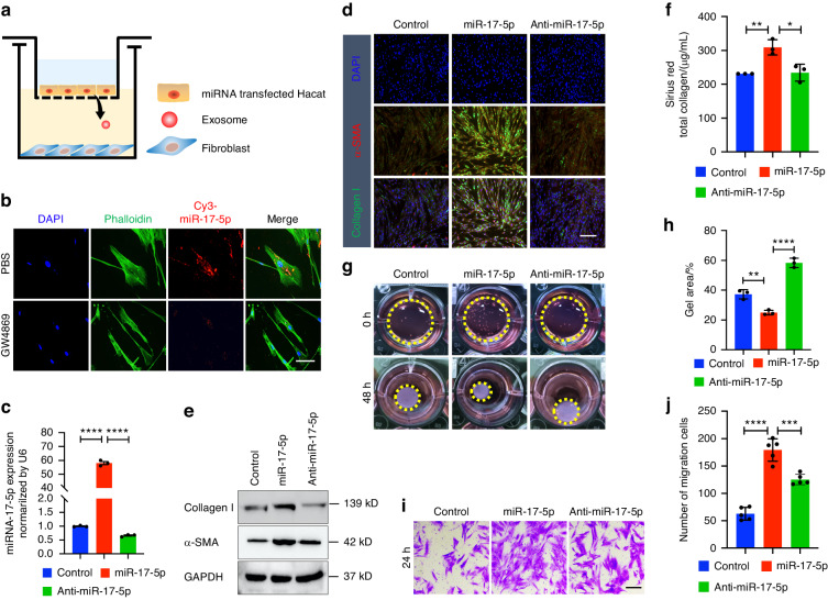
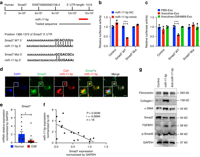
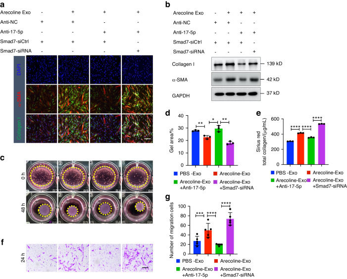
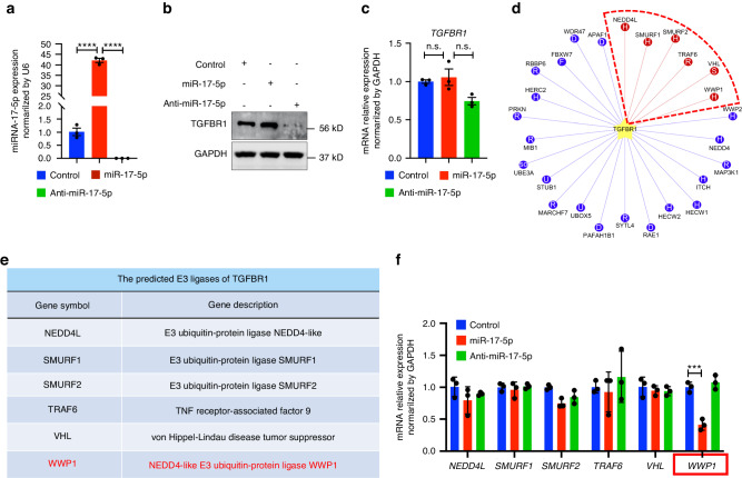
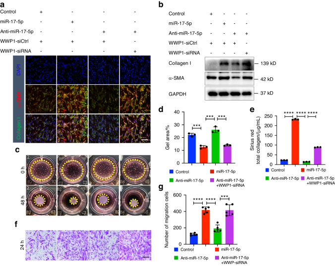
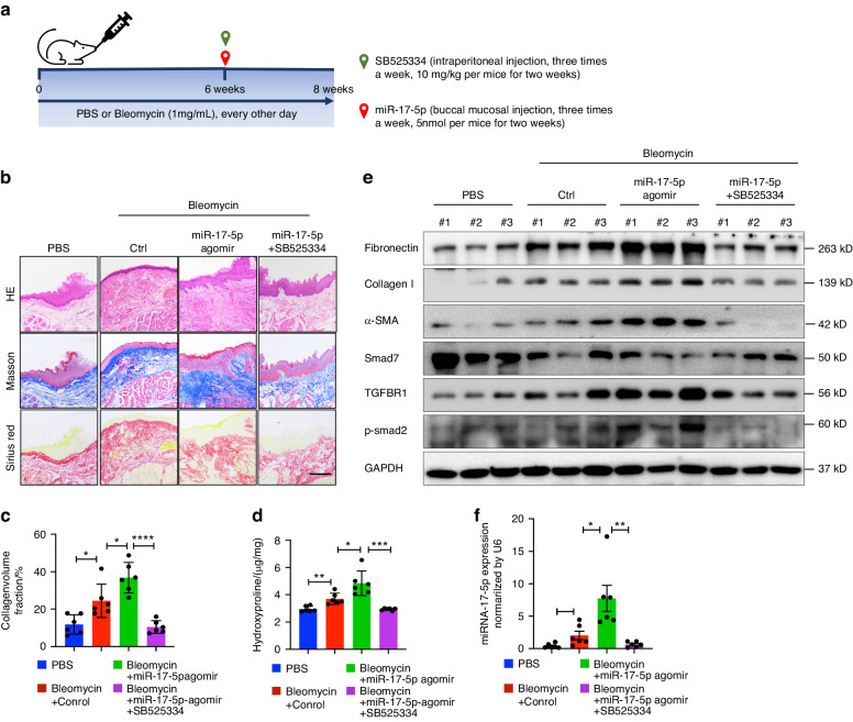
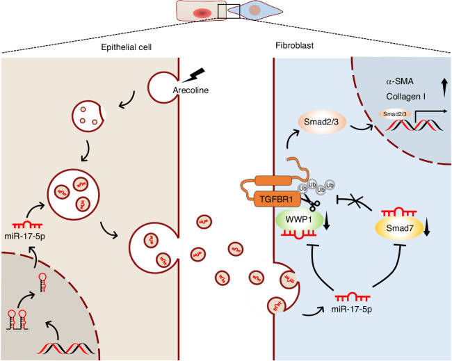
Oral submucous fibrosis (OSF) is a chronic and inflammatory mucosal disease caused by betel quid chewing, which belongs to oral potentially malignant disorders. Abnormal fibroblast differentiation leading to disordered collagen metabolism is the core process underlying OSF development. The epithelium, which is the first line of defense against the external environment, can convert external signals into pathological signals and participate in the remodeling of the fibrotic microenvironment. However, the specific mechanisms by which the epithelium drives fibroblast differentiation remain unclear. In this study, we found that Arecoline-exposed epithelium communicated with the fibrotic microenvironment by secreting exosomes. MiR-17-5p was encapsulated in epithelial cell-derived exosomes and absorbed by fibroblasts, where it promoted cell secretion, contraction, migration and fibrogenic marker (α-SMA and collagen type I) expression. The underlying molecular mechanism involved miR-17-5p targeting Smad7 and suppressing the degradation of TGF-β receptor 1 (TGFBR1) through the E3 ubiquitination ligase WWP1, thus facilitating downstream TGF-β pathway signaling. Treatment of fibroblasts with an inhibitor of miR-17-5p reversed the contraction and migration phenotypes induced by epithelial-derived exosomes. Exosomal miR-17-5p was confirmed to function as a key regulator of the phenotypic transformation of fibroblasts. In conclusion, we demonstrated that Arecoline triggers aberrant epithelium-fibroblast crosstalk and identified that epithelial cell-derived miR-17-5p mediates fibroblast differentiation through the classical TGF-β fibrotic pathway, which provided a new perspective and strategy for the diagnosis and treatment of OSF.


Conflict of interest statement

The authors declare no competing interests.

Figures

Fig. 1
Fig. 1
Epithelial cell-derived exosomes are transported to the primary fibroblasts, which promotes the myofibroblast differentiation phenotype. PBS-Exo (exosomes derived from epithelial cells treated with PBS), Arecoline-Exo (exosomes derived from epithelial cells treated with Arecoline), Arecoline+GW4869-Exo (exosomes derived from epithelial cells treated with Arecoline and GW4869). a Fibroblasts was incubated with DiI-labeled (red) exosomes from epithelial cells (50 μg) for 30 min and fixed for fluorescence staining. Nuclei were stained with DAPI (blue). Scale bars, 100 μm. b Immunostaining for fibrotic markers collagen type I and α-SMA in primary fibroblast with PBS-, Arecoline- and Arecoline+GW4869-Exo was evaluated by microscopy. DAPI (blue) was used for nuclear staining. Scale bar, 100 μm. c Immunoblotting of collagen type I and α-SMA by PBS-, Arecoline- and Arecoline+GW4869-Exo in primary fibroblasts. Sirius Red total collagen assay (d), collagen contraction assay (e, f) and transwell migration assay (g, h) were determined for fibroblast. n = 3–5 technical replicates, representative of two or three assays. Scale bars, 100 μm. Statistics: mean ± SEM, unpaired, one-way ANOVA (d, f, h), ***P < 0.001, ****P < 0.000 1
Fig. 2
Fig. 2
Arecoline induced the expression of miR-17-5p in epithelial cell exosomes. PBS-Exo (exosomes derived from epithelial cells treated with PBS), Arecoline-Exo (exosomes derived from epithelial cells treated with Arecoline), Arecoline+GW4869-Exo (exosomes derived from epithelial cells treated with Arecoline and GW4869), U6 was used as an internal control for qRT-PCR. The exosomes RNA–seq (a) and volcano plot (b) identified top 8 up-regulated miRNAs (change 1.5-fold) of 177 expressed differential genes in PBS-Exo and Arecoline-Exo. c qRT-PCR validation of exosomes miRNAs from PBS-Exo and Arecoline-Exo. The levels of miR-17-5p in epithelial cells (d) and exosomes (e) from its cell culture medium treated with PBS and Arecoline were determined by qRT-PCR. f The levels of miR-17-5p in epithelial cells treated with PBS, Arecoline and Arecoline+GW4869 were determined by qRT-PCR. g The levels of miR-17-5p in fibroblast after 48 h co-culturing with PBS-Exo, Arecoline-Exo and Arecoline+GW4869-Exo. h The levels of miR-17-5p in normal (n = 12) or OSF (n = 17) tissues. i miR-17-5p expression in normal (n = 3) and cardiac fibrosis patients (n = 3) from a public dataset (GSE196421). j miR-17 expression in normal (n = 4) and diabetic nephropathy kidney fibrosis patients (n = 12) from a public dataset (GSE51674). Statistics: mean ± SEM, unpaired, two-tailed Student’s t test (c, h, i, j) or one-way ANOVA (dg), *P < 0.05, **P < 0.01, ***P < 0.001, ****P < 0.000 1
Fig. 3
Fig. 3
Exosomes derived from epithelial cells transfer miR-17-5p to fibroblast, which promotes myofibroblast differentiation. Control (epithelial cells transfected negative control), miR-17-5p (epithelial cells transfected mimic), Anti-miR-17-5p (epithelial cells transfected inhibitor), U6 or GAPDH was used as an internal control for qRT-PCR. a A transwell co-culture cell model with transfected epithelial cells (top well) and fibroblasts (bottom well). A 0.4-μm porous membrane is between the 2 wells, allowing the transmission of exosomes, but inhibiting direct contact between cells. b A co-culture assay to study the miRNA cargo from epithelial cells to fibroblasts. The epithelial cells were transfected with a Cy3-labeled miR-17-5p (red) and then treated with PBS or GW4869. Nuclei were counterstained with DAPI (blue). Scale bar: 50 μm. c The expression level of miR-17-5p in fibroblasts after 48 h co-culturing with epithelial cells transfected with miR-17-5p or anti-miR-17-5p. Immunostaining (d) and (e) Immunoblotting detection of collagen type I and α-SMA expression in fibroblast after 48 h co-culturing with epithelial cells. Scale bars, 100 μm. Sirius Red total collagen assay (f), collagen contraction assay (g, h) and transwell migration assay (i, j) were determined for fibroblast. n = 3–5 technical replicates, representative of two or three assays. Scale bars, 100 μm. Statistics: mean ± SEM, one-way ANOVA, *P < 0.05, **P < 0.01, ***P < 0.001, ****P < 0.000 1
Fig. 4
Fig. 4
Exosomal miR-17-5p directly targets Smad7 in fibroblasts. a Schematic miR-17-5p predicted binding sites in the 3’ UTRs of Smad7 and the sequence of mutant UTRs. b Luciferase reporter assay was performed on HEK293T after co-transfected with the miR-NC or miR-17-5p mimic and the Smad7-wt plasmid or Smad7-mut plasmid. c Luciferase reporter was carried out in HEK293T transfected with Smad7-wt plasmid or Smad7-mut plasmid, then incubated with exosomes (50 μg/mL) derived from PBS-, Arecoline- and Arecoline+GW4869- treated epithelial cells for 48 h. d After transfection miR-17-5p in fibroblast, Immunostaining detection of miR-17-5p (red) and Smad7 (green) expression. Scale bars, 100 μm. e The expression level of Smad7 in normal (n = 12) and OSF (n = 17) tissues detected by qRT-PCR. f The relationship between relative Smad7 expression normalized to GAPDH and miR-17-5p expression normalized to U6. g Immunoblotting of Smad7, fibrotic markers and TGF-β path pathway. Statistics: mean ± SEM, unpaired, two-tailed Student’s t test, *P < 0.05, ***P < 0.001
Fig. 5
Fig. 5
Exosomal miR-17-5p derived from Arecoline-treated epithelium confers fibroblast activation through Smad7. Fibroblasts were incubated with exosomes derived from PBS and Arecoline-treated epithelium, then transfection of Anti-15-5p or Smad7-siRNA for 48 h. Immunostaining (a) and Immunoblotting (b) detection of collagen type I and α-SMA expression in fibroblast. Scale bars, 100 μm. Collagen contraction assay (c, d), Sirius Red total collagen assay (e) and transwell migration assay (f, g) were determined for fibroblast. n = 3–5 technical replicates, representative of two or three assays. Statistics: mean ± SEM, one-way ANOVA, *P < 0.05, **P < 0.01, ***P < 0.001, ****P < 0.000 1
Fig. 6
Fig. 6
miR-17-5p inhibits the expression of E3 ubiquitin-protein ligase WWP1. a After transfected with miR-17-5p or anti-miR-17-5p for 48 h, the expression of miR-17-5p in fibroblasts was detected by qRT-PCR. Immunoblotting (b) and qRT-PCR (c) detection of TGFBR1 expression in fibroblasts transfected by miR-17-5p and anti-miR-17-5p. d UbiBrowser 2.0 online site predicts E3 ubiquitin ligase with TGFBR1 as substrate. e The annotation list for the predicted E3 ligases of TGFBR1. f qRT-PCR detection of E3 ubiquitin ligase (NEDD4L, SMURF1, SMURF2, TRAF6, VHL, WWP1) expression level in fibroblast transfected by miR-17-5p and anti-miR-17-5p. Statistics: mean ± SEM, one-way ANOVA, n.s.: no significance, ****P < 0.000 1
Fig. 7
Fig. 7
miR-17-5p inhibits WWP1-mediated degradation of TGFBR1 protein in fibroblasts. a Fibroblast transfected with miR-17-5p then incubated with cycloheximide (CHX, 100 μg/mL) for indicated time points. The protein levels of WWP1 and TGFBR1 were determined at indicated time points by Immunoblotting. b Fibroblasts transfected with miR-17-5p and anti-miR-17-5p then incubated with MG-132 (10 μM, 6 h). The protein levels of TGFBR1 were determined by Immunoblotting. c The expression localization of WWP1 (green) and TGFBR1 (red) in fibroblast was detected by Immunostaining. Scale bar: 50 mm. d, e Co-IP and Immunoblotting detected the interaction between WWP1 and TGFBR1 in fibroblast. f Co-IP and Immunoblotting detected the ubiquitination of TGFBR1 mediated by WWP1 and miR-17-5p overexpressed. g, h The expression of WWP1 and TGFBR1 protein levels in miR-17-5p overexpressed or inhibited in fibroblast was detected by Immunoblotting
Fig. 8
Fig. 8
Down-regulated WWP1 expression can promote myofibroblast transformation. Fibroblasts were transfection of miR-17-5p, anti-15-5p or WWP1-siRNA for 48 h. Immunostaining (a) and Immunoblotting (b) detection of collagen type I and α-SMA expression in fibroblast. Scale bars, 100 μm. Collagen contraction assay (c, d), Sirius Red total collagen assay (e) and transwell migration assay (f, g) were determined for fibroblast. n = 3–5 technical replicates, representative of two or three assays. Statistics: mean ± SEM, one-way ANOVA, ***P < 0.001, ****P < 0.000 1
Fig. 9
Fig. 9
miR-17-5p induces collagen accumulation and promotes the progression of OSF by TGF-β in vivo. a Schematic overview of mouse experimental design. Mice were injected with 30 μL PBS or Bleomycin into the bilaterally buccal mucosa every other day for six weeks. Then, mice were simultaneously treated with miR-NC (Ctrl), miR-17-5p agomir and SB525334 for another two weeks. b HE, Masson and Sirius red staining of mice oral mucosa tissues. Scale bar, 200 μm. Quantification of Masson staining for collagen fraction. Scale bar, 200 μm. d Hydroxyproline content of mice oral mucosa tissues. c Quantification of Masson staining for skin tissues collagen fraction, n = 6 for each group. e Immunoblotting of fibrotic markers (Fibronectin, Collagen type I and α-SMA), Smad7, TGFBR1 and p-Smad2 pathway. f The levels of miR-17-5p of mice blood-derived exosomes. Statistics: mean ± SEM, n = 6 for each group, one-way ANOVA, *P < 0.05, **P < 0.01, ***P < 0.001, ****P < 0.000 1
Fig. 10
Fig. 10
Working model showing that the Arecoline induces epithelial cells to release mir-17-5p enveloped exosomes, which could promote collagen contraction, secretion and migration after being absorbed by fibroblast. Mechanistically, it was demonstrated that miR-17-5p targets Smad7 and maintains TGFBR1 expression by inhibiting its E3 ubiquitination ligase WWP1, which promotes the expression of fibrosis hallmark genes

Similar articles

Cited by

References

    1. Wynn TA, Ramalingam TR. Mechanisms of fibrosis: Therapeutic translation for fibrotic disease. Nat. Med. 2012;18:1028–1040. doi: 10.1038/nm.2807. - DOI - PMC - PubMed
    1. Zhao M, et al. Targeting fibrosis, mechanisms and cilinical trials. Signal Transduct Target Ther. 2022;7:206. doi: 10.1038/s41392-022-01070-3. - DOI - PMC - PubMed
    1. Henderson NC, Rieder F, Wynn TA. Fibrosis: From mechanisms to medicines. Nature. 2020;587:555–566. doi: 10.1038/s41586-020-2938-9. - DOI - PMC - PubMed
    1. Yuwanati M, et al. Prevalence of oral submucous fibrosis among areca nut chewers: A systematic review and meta-analysis. Oral Dis. 2023;29:1920–1926. doi: 10.1111/odi.14235. - DOI - PubMed
    1. Qin X, et al. Oral submucous fibrosis: Etiological mechanism, malignant transformation, therapeutic approaches and targets. Int. J. Mol. Sci. 2023;24:4992. doi: 10.3390/ijms24054992. - DOI - PMC - PubMed

Publication types

MeSH terms