

Structure and dynamics of the pyroglutamylated RF-amide peptide QRFP receptor GPR103
- PMID: 38897996
- PMCID: PMC11187126
- DOI: 10.1038/s41467-024-49030-5
Structure and dynamics of the pyroglutamylated RF-amide peptide QRFP receptor GPR103
Abstract
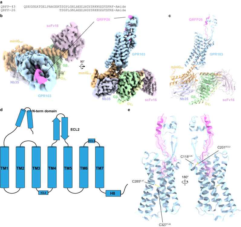
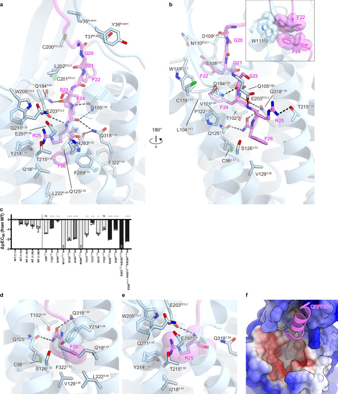
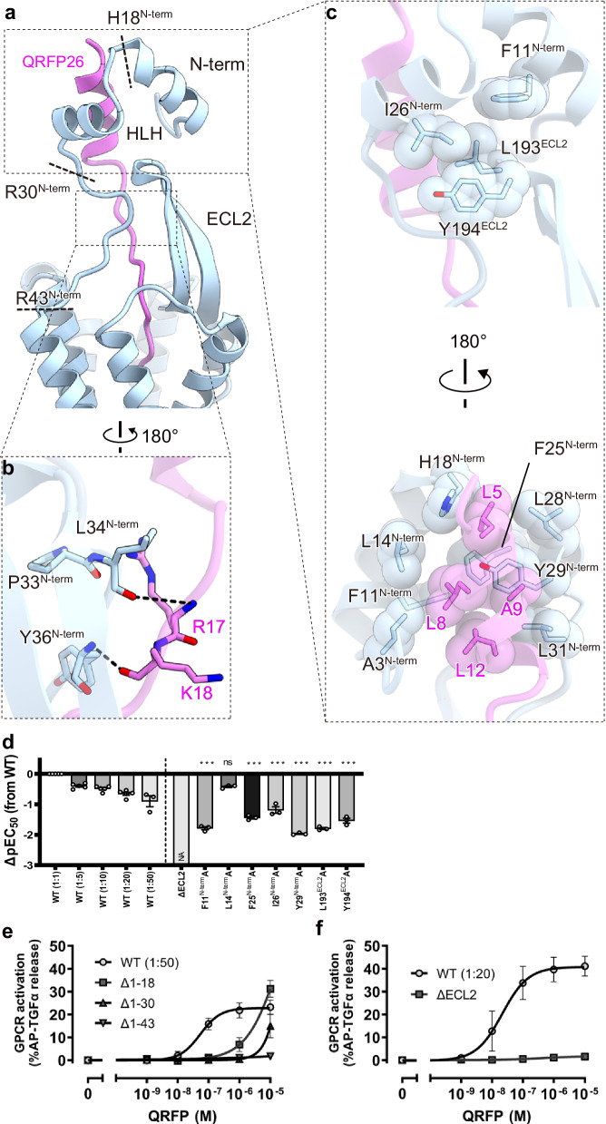
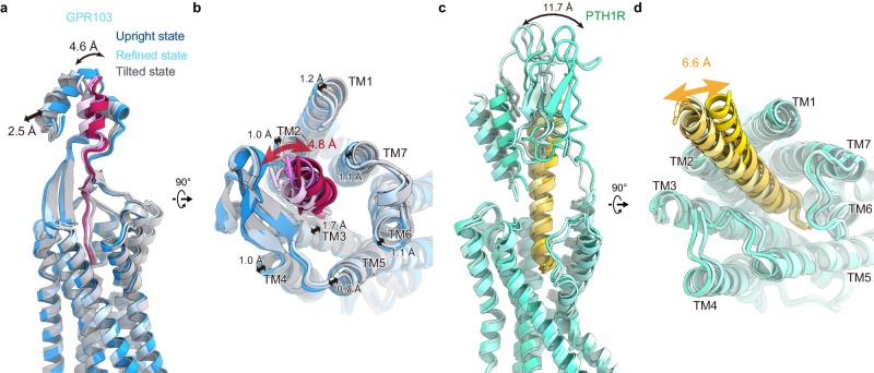
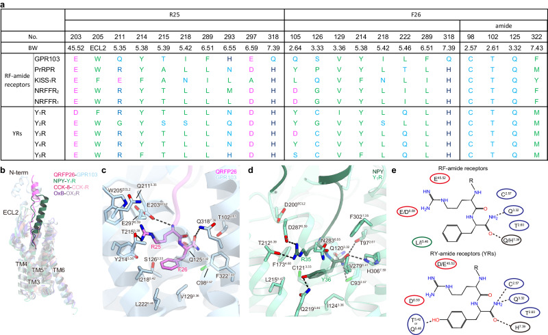
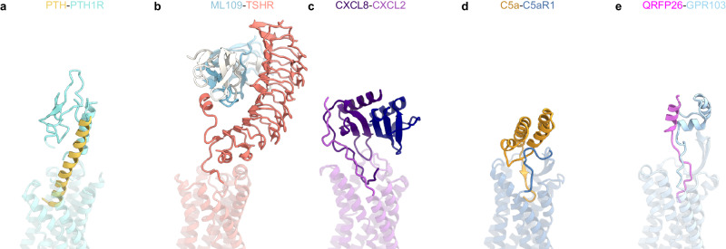
Pyroglutamylated RF-amide peptide (QRFP) is a peptide hormone with a C-terminal RF-amide motif. QRFP selectively activates a class A G-protein-coupled receptor (GPCR) GPR103 to exert various physiological functions such as energy metabolism and appetite regulation. Here, we report the cryo-electron microscopy structure of the QRFP26-GPR103-Gq complex at 3.19 Å resolution. QRFP26 adopts an extended structure bearing no secondary structure, with its N-terminal and C-terminal sides recognized by extracellular and transmembrane domains of GPR103 respectively. This movement, reminiscent of class B1 GPCRs except for orientation and structure of the ligand, is critical for the high-affinity binding and receptor specificity of QRFP26. Mutagenesis experiments validate the functional importance of the binding mode of QRFP26 by GPR103. Structural comparisons with closely related receptors, including RY-amide peptide-recognizing GPCRs, revealed conserved and diversified peptide recognition mechanisms, providing profound insights into the biological significance of RF-amide peptides. Collectively, this study not only advances our understanding of GPCR-ligand interactions, but also paves the way for the development of novel therapeutics targeting metabolic and appetite disorders and emergency medical care.
© 2024. The Author(s).
Conflict of interest statement
O.N. is a co-founder and scientific advisor for Curreio. All other authors declare no competing interests.
Figures

Similar articles
-
N-glycosylation of the human neuropeptide QRFP receptor (QRFPR) is essential for ligand binding and receptor activation.J Neurochem. 2021 Jul;158(2):138-152. doi: 10.1111/jnc.15337. Epub 2021 Mar 17. J Neurochem. 2021. PMID: 33655503
-
Structural basis for recognition of 26RFa by the pyroglutamylated RFamide peptide receptor.Cell Discov. 2024 Jun 4;10(1):58. doi: 10.1038/s41421-024-00670-3. Cell Discov. 2024. PMID: 38830850 Free PMC article.
-
Effects of systematic N-terminus deletions and benzoylations of endogenous RF-amide peptides on NPFF1R, NPFF2R, GPR10, GPR54 and GPR103.Peptides. 2015 Sep;71:156-61. doi: 10.1016/j.peptides.2015.07.016. Epub 2015 Jul 23. Peptides. 2015. PMID: 26211894
-
The Arg-Phe-amide peptide 26RFa/glutamine RF-amide peptide and its receptor: IUPHAR Review 24.Br J Pharmacol. 2017 Oct;174(20):3573-3607. doi: 10.1111/bph.13907. Epub 2017 Sep 8. Br J Pharmacol. 2017. PMID: 28613414 Free PMC article. Review.
-
Discovery of novel regulatory peptides by reverse pharmacology: spotlight on chemerin and the RF-amide peptides metastin and QRFP.Curr Protein Pept Sci. 2005 Jun;6(3):265-78. doi: 10.2174/1389203054065419. Curr Protein Pept Sci. 2005. PMID: 15974952 Review.
Cited by
-
Insights into G-protein coupling preference from cryo-EM structures of Gq-bound PTH1R.Nat Chem Biol. 2025 Jun 26. doi: 10.1038/s41589-025-01957-6. Online ahead of print. Nat Chem Biol. 2025. PMID: 40571720
-
Structural insights into the agonist selectivity of the adenosine A3 receptor.Nat Commun. 2024 Nov 7;15(1):9294. doi: 10.1038/s41467-024-53473-1. Nat Commun. 2024. PMID: 39511145 Free PMC article.
-
Promoter H3K4me3 and Gene Expression Involved in Systemic Metabolism Are Altered in Fetal Calf Liver of Nutrient-Restricted Dams.Int J Mol Sci. 2025 Aug 4;26(15):7540. doi: 10.3390/ijms26157540. Int J Mol Sci. 2025. PMID: 40806668 Free PMC article.
-
Structural insights into the selective recognition of RF-amide peptides by neuropeptide FF receptor 2.EMBO Rep. 2025 May;26(9):2413-2434. doi: 10.1038/s44319-025-00428-2. Epub 2025 Mar 24. EMBO Rep. 2025. PMID: 40128413 Free PMC article.
References
-
- Findeisen M, Rathmann D, Beck-Sickinger AG. RFamide peptides: structure, function, mechanisms and pharmaceutical potential. Pharmaceuticals. 2011;4:1248–1280. doi: 10.3390/ph4091248. - DOI
MeSH terms
Substances
Grants and funding
LinkOut - more resources
Full Text Sources
