. 2024 Jun 19;9(1):111.
doi: 10.1038/s41541-024-00899-9.

Mucosal bivalent live attenuated vaccine protects against human metapneumovirus and respiratory syncytial virus in mice

Affiliations

Mucosal bivalent live attenuated vaccine protects against human metapneumovirus and respiratory syncytial virus in mice

Daniela Ogonczyk-Makowska et al. NPJ Vaccines. .

Erratum in

Abstract

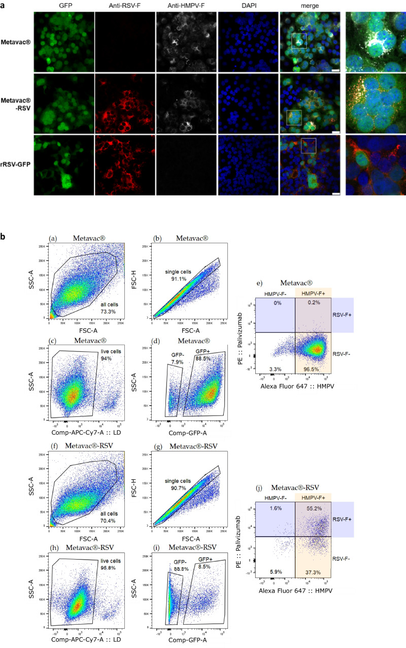
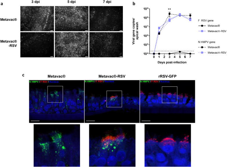
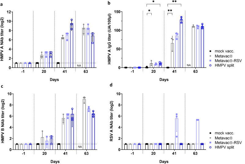
Live-Attenuated Vaccines (LAVs) stimulate robust mucosal and cellular responses and have the potential to protect against Respiratory Syncytial Virus (RSV) and Human Metapneumovirus (HMPV), the main etiologic agents of viral bronchiolitis and pneumonia in children. We inserted the RSV-F gene into an HMPV-based LAV (Metavac®) we previously validated for the protection of mice against HMPV challenge, and rescued a replicative recombinant virus (Metavac®-RSV), exposing both RSV- and HMPV-F proteins at the virion surface and expressing them in reconstructed human airway epithelium models. When administered to BALB/c mice by the intranasal route, bivalent Metavac®-RSV demonstrated its capacity to replicate with reduced lung inflammatory score and to protect against both RSV and lethal HMPV challenges in vaccinated mice while inducing strong IgG and broad RSV and HMPV neutralizing antibody responses. Altogether, our results showed the versatility of the Metavac® platform and suggested that Metavac®-RSV is a promising mucosal bivalent LAV candidate to prevent pneumovirus-induced diseases.


Conflict of interest statement

Manuel Rosa-Calatrava, Guy Boivin, Julia Dubois, and Marie-Eve Hamelin are co-founders and shareholders of Vaxxel SAS. Andrés Pizzorno and Olivier Terrier are shareholders of Vaxxel SAS. Julia Dubois was the R&D project manager of Vaxxel SAS. Caroline Chupin is an employee of Vaxxel SAS. The other authors declare no competing interests. The authors declare the following patent : EP22305240.2 – PCT/EP2023/055221 concerning Vaccine composition against two respiratory viruses (Inventors: Daniela Ogonczyk-Makowska, Jean-François Eléouët, Guy Boivin, Julia Dubois and Manuel Rosa-Calatrava ; Applicants : Institut National de la Santé et de la Recherche Médicale (INSERM), Centre National de la Recherche Scientifique (CNRS), Université Claude Bernard Lyon 1 (UCBL) Ecole Normale Supérieure de Lyon (ENS Lyon), Institut National de Recherche pour l’Agriculture, l’Alimentation et l’Environnement (INRAE) and Vaxxel SAS).

Figures

Fig. 1
Fig. 1. Rescue and characterization of recombinant Metavac®-RSV virus.
a Schematic genomic organization of the recombinant HMPV strain (rC-85473-GFP, rHMPV), monovalent Metavac® (ΔSH-rC-85473-GFP), and bivalent Metavac®-RSV (ΔSH-rC-85473-GFP/RSV-F) viruses is represented and the insertion site of the RSV-F ORF between HMPV-F and M2 genes in Metavac®-RSV genome is detailed. GS - Gene Start, GE - Gene End, IG - intergenic sequence. Sequences added to the ΔSH-rC-85473-GFP genome are underlined. Genomic sequence is presented from 3′ to 5′. b After the viral rescue, in vitro expression of the RSV-F protein at the surface of Metavac®-RSV viral particles was visualized by transmission electron microscopy after immunogold labeling with anti-HMPV serum (5 nm bead) and the Palivizumab (15 nm bead, black arrowhead) Scale bar = 100 nm. c Viral replication kinetics of the Metavac®-RSV virus were measured in LLC-MK2 cells and compared to the monovalent Metavac® counterpart. Over a 7-day period, culture supernatants were harvested and titrated in TCID50/ml. Results represent the mean of 3 experimental replicates for each time-point ± SD. *p < 0.05, **p < 0.01, ***p < 0.001 when comparing Metavac®-RSV to Metavac® virus using repeated measures two-way ANOVA.
Fig. 2
Fig. 2. Co-immunostaining of HMPV and RSV-F glycoproteins in infected LLC-MK2 cells.
a LLC-MK2 cells were infected with GFP-expressing Metavac®, Metavac®-RSV or RSV (rRSV-GFP) viruses, fixed and stained at 3 dpi with Palivizumab (red), HMPV24 mAb (white) and DAPI (blue). Merged fluorescent signals are represented (yellow). Images of representative cytopathic effects (CPEs) were taken using Zeiss880 confocal microscope (×40 magnification) and processed with ImageJ software. Scale bar = 25 µm. A numeric focus was made on CPEs (square) and presented in the right panel. b LLC-MK2 cells were infected with MOI 0.5 of either Metavac® (ae) or Metavac®-RSV (f, j), and antigens expression on the surface of the infected cells was measured by flow cytometry 48 h post-infection. HMPV-F protein was detected with HMPV24 mAb conjugated with Alexa Fluor™ 647 and RSV-F protein was detected with Palivizumab conjugated with R-Phycoerythrin. Cells were sorted and analyzed by LSR II Flow Cytometer (BD biosciences®). Approximately 30,000 single live cells were counted per each sample performed in triplicate. The figure shows representative gating of sub-populations on one of the three samples. a, f all cells in the sample; b, g single cells c, h single live cells d, i single live cells positive or negative for GFP expression e, j the percentage of GFP-expressing infected single live cells with HMPV-F expression revealed by HMPV24 mAb and RSV-F expression revealed by Palivizumab.
Fig. 3
Fig. 3. Viral replication and RSV-F expression in human airway epithelium (HAE) model.
Reconstituted HAEs were infected with Metavac® or Metavac®-RSV viruses at an MOI of 0.1 and monitored for 7 days. a Viral spread in HAE was monitored at 3, 5, and 7 dpi by GFP fluorescence observation (10× magnification). Scale bar = 100 µm. b Viral quantification from epithelium apical washes collected after 1, 3, 5, and 7 dpi was performed by RT-qPCR targeting the HMPV-N gene or the RSV-F gene. Data are shown as means ± SD and represent experimental triplicates. The dotted line represents the RT-qPCR quantification threshold. **p < 0.01 when comparing Metavac®-RSV N-HMPV gene expression to Metavac® virus using repeated measures two-way ANOVA. c Co-immunostaining of HMPV-N and RSV-F proteins was performed at 3 dpi. HAE infected by Metavac®, Metavac®-RSV, or rRSV-GFP viruses were fixed and cross-sections were stained with a mixture of mAbs specific to the HMPV-N protein (mAb hMPV123, green), RSV-F protein (Palivizumab, red) and with DAPI (blue) specific to the nucleus. Acquisition of images of representative infected areas was performed with confocal inverted microscope (Zeiss Confocal LSM 880) and processed with ImageJ software. Scale bar = 20 µm. A focus on the apical surface of ciliated infected cells was made (square) and is presented in the right panel.
Fig. 4
Fig. 4. Viral growth and attenuation of the Metavac®-RSV vaccine candidate in BALB/c mice.
BALB/c mice were infected by the IN route with 5 × 105 TCID50 of rHMPV virus, Metavac®, or Metavac®-RSV vaccine candidates. a Weight loss was monitored for 14 dpi (n = 16). Data are shown as means ± SEM. *p < 0.05, **p < 0.01, ***, p < 0.001 when compared to mock-infected mice using Repeated Measures Two-way ANOVA. b, c At 2 dpi, mice were euthanized, and BALs were harvested to measure HMPV-N gene (b) or RSV-F gene (c) copies by RT-qPCR (n = 2). d At 5 dpi, mean cumulative histopathological scores (peribronchial, intrabronchial, perivascular, interstitial, pleural, and intra-alveolar inflammation scores) of the lungs from infected mice were evaluated (n = 3). *p < 0.05, ***p < 0.001 when comparing mean global histopathological score to mock-infected mice using One-way ANOVA. e, f At 5 dpi, HMPV-N (e) or RSV-F (f) gene copies were measured by RT-qPCR from total RNA extracted from fixed lung tissues (n = 2–3). Data of viral gene quantification are shown as means ± SD.
Fig. 5
Fig. 5. Efficacy of Metavac®-RSV vaccine candidate against lethal challenge with HMPV.
BALB/c mice were immunized twice with a 21-day interval by the IN route with 5×10⁵ TCID50 of Metavac® or Metavac®-RSV LAV candidates or by the IM route with HMPV split preparation adjuvanted with AddaVax™. Three weeks after the last immunization, animals (n = 12/group) were inoculated with a lethal dose of the rHMPV virus. a Weight loss and b mortality rates were monitored for 14 days post-challenge (dpc, n = 8/group). Data are shown as means ± SEM. ***p < 0.001 when comparing to Metavac® vaccinated mice using Two-way ANOVA. c At 5 dpc, cumulative pulmonary histopathological scores (peribronchial, intrabronchial, perivascular, interstitial, pleural, and intra-alveolar inflammation scores) were also evaluated (n = 3/group). d At 2 dpc, mice were euthanized and nasal washes (NW) and bronchoalveolar lavages (BALs) were harvested to measure HMPV-N gene copies by RT-qPCR (n = 2/group). e At 5 dpc, RT-qPCR was performed on total RNA recovered from mouse lung homogenates (n = 4/group) to quantify HMPV-N gene copies. f, g Infectious TCID50 titers were measured from BAL samples collected at 2 dpc (f) or lung homogenates collected at 5 dpc (g). Data are shown as means ± SD. *p < 0.05, **p < 0.01, ***p < 0.001 when comparing mean global histopathological score to mock-vaccinated mice using one-way ANOVA.
Fig. 6
Fig. 6. Immunogenicity of Metavac®-RSV vaccine candidate before and after lethal challenge with HMPV.
BALB/c mice were immunized twice with a 21-day interval by the IN route with 5 × 105 TCID50 of Metavac® or Metavac® -RSV vaccine candidates or by the IM route with the adjuvanted HMPV split preparation. Three weeks after the last immunization, animals (n = 12/group) were inoculated with a lethal dose of rHMPV. Immunogenicity of vaccine candidates was measured at −1, 20, 41, or 63 days after the first immunization by microneutralization (a, c, d) or ELISA (b) assays from pools of sera (n = 3 pools/group). Neutralization titers were defined by an endpoint dilution assay based on fluorescent detection of (a) HMPV A, (c) HMPV B, or (d) RSV A and represented as mean log2 reciprocal neutralizing antibody (NAb) titers. b IgG titer specific to HMPV A virus was represented as an arbitrary unit based on endpoint absorbance. Naive status of mice was confirmed by processing the samples harvested one day before vaccination. Data are shown as means ± SD. *p < 0.05, **p < 0.01 when comparing each vaccinated group to the mock-vaccinated condition using Two-way ANOVA.
Fig. 7
Fig. 7. Efficacy and immunogenicity of Metavac®-RSV vaccine candidate following RSV challenge.
BALB/c mice were immunized twice with a 21-day interval by the IN route with 5 × 105 TCID50 of Metavac®-RSV vaccine candidate or rRSV-mCh (RSV WT) virus. Three weeks after the last immunization, animals (n = 12/group) were inoculated with 1 × 105 PFU of rRSV-Luc virus. a, b Bioluminescence was measured at 3 and 5 dpc by IN injection of 50 µl of D-Luciferin (200 mM). a Ventral views of 4 representative mice were taken using the IVIS system. The scale on the right indicates the average radiance (a sum of the photons per second from each pixel inside the region of interest, ps-1 cm-2 sr-1). b Luciferase activities were quantified using ‘Living Image’ software and were represented as mean ± SEM photons per second (p/s) (n = 8/group). c, d RT-qPCR was performed on total RNA recovered from mouse lung homogenates (n = 4/group) harvested at 4 dpc to quantify RSV-F (c) or residual HMPV-N gene copies (d). eg Immunogenicity of the Metavac®-RSV LAV candidate was measured by RSV A microneutralization assay, anti-total RSV or anti-preF RSV IgG ELISA assays from pools of sera (before each IN instillation at −1, 20, and 41 dpi) or individual sera (at the endpoint at 63 dpi, n = 6–8). e Neutralization of RSV A strain was represented as mean log2 reciprocal NAb titer. f, g IgG titer specific to RSV virus (f) or recombinant preF RSV protein (g) was represented as an arbitrary unit based on endpoint absorbance. Data are shown as means ± SD. *p < 0.05, **p < 0.01, ***p < 0.001 when comparing Metavac®-RSV or RSV WT vaccinated group to the mock-vaccinated condition using Two-way ANOVA.

Similar articles

Cited by

References

    1. Lozano R, et al. Global and regional mortality from 235 causes of death for 20 age groups in 1990 and 2010: a systematic analysis for the Global Burden of Disease Study 2010. Lancet. 2012;380:2095–2128. doi: 10.1016/S0140-6736(12)61728-0. - DOI - PMC - PubMed
    1. Li Y, et al. Global, regional, and national disease burden estimates of acute lower respiratory infections due to respiratory syncytial virus in children younger than 5 years in 2019: a systematic analysis. Lancet. 2022;399:2047–2064. doi: 10.1016/S0140-6736(22)00478-0. - DOI - PMC - PubMed
    1. Glezen WP, Taber LH, Frank AL, Kasel JA. Risk of primary infection and reinfection with respiratory syncytial virus. Am. J. Dis. Child. 1986;140:543–546. - PubMed
    1. Shi T, et al. Global disease burden estimates of respiratory syncytial virus–associated acute respiratory infection in older adults in 2015: a systematic review and meta-analysis. J. Infect. Dis. 2020;222:S577–S583. doi: 10.1093/infdis/jiz059. - DOI - PubMed
    1. Busack B, Shorr AF. Going viral—RSV as the neglected adult respiratory virus. Pathogens. 2022;11:1324. doi: 10.3390/pathogens11111324. - DOI - PMC - PubMed