. 2024 Jun 19;14(1):14146.
doi: 10.1038/s41598-024-64651-y.

Eribulin induces micronuclei and enhances the nuclear localization of cGAS in triple-negative breast cancer cells

Affiliations

Eribulin induces micronuclei and enhances the nuclear localization of cGAS in triple-negative breast cancer cells

Hideyuki Yamada et al. Sci Rep. .

Abstract

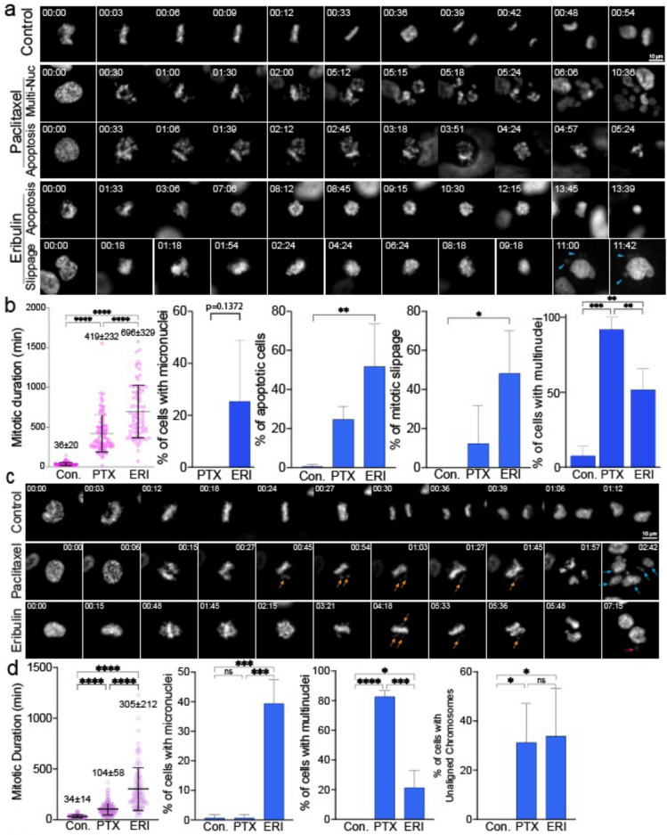
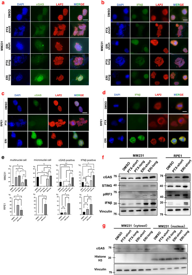
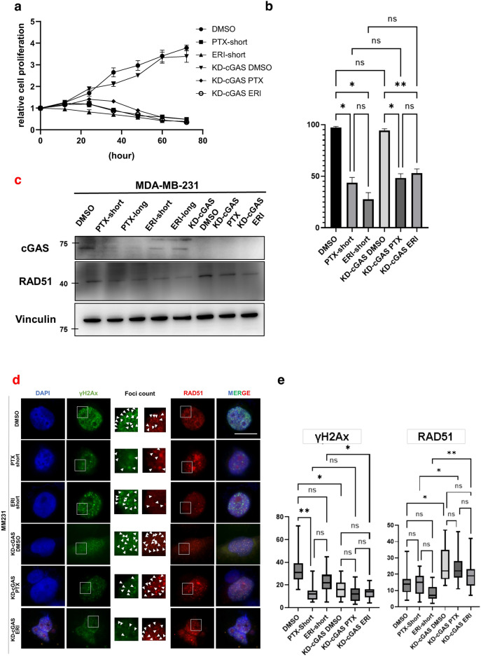
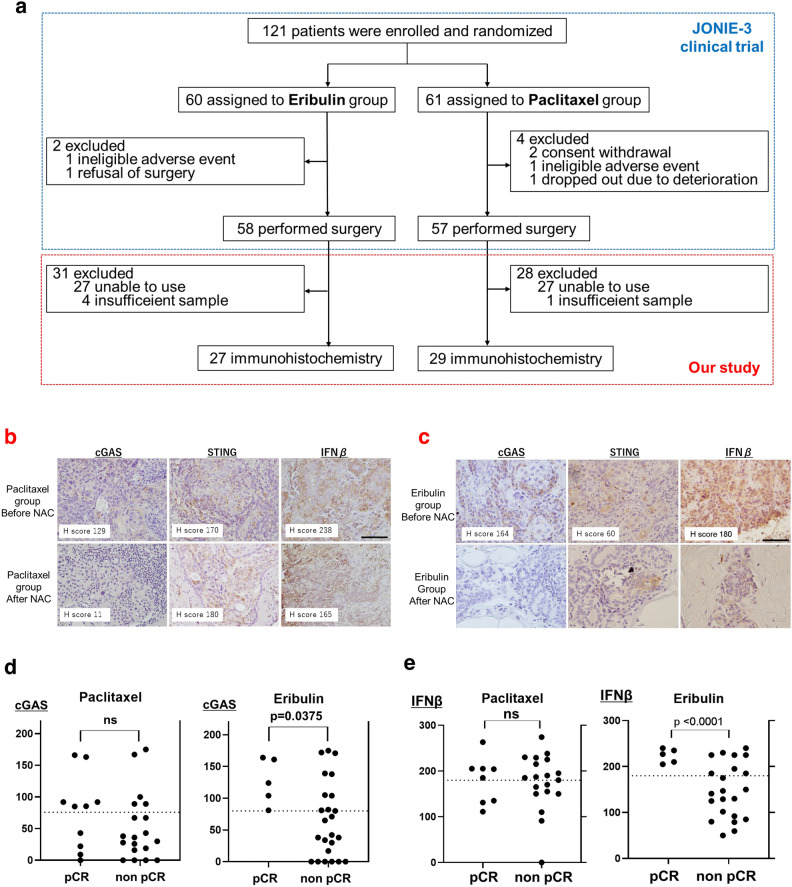
Eribulin (ERI), clinically utilized for locally advanced or metastatic breast tumors, has shown potential links to the immune system. Notably, the cGAS-STING pathway, a key component of innate immunity, has gained prominence. Yet, limited reports explore ERI's effects on the cGAS-STING pathway. Additionally, the nuclear presence of cGAS remains poorly understood. This study uniquely delves into ERI's impact on both the cytosolic cGAS-STING pathway and nuclear cGAS. ERI enhances nuclear localization of cGAS, resulting in hyper-activation of the cGAS-STING pathway in triple-negative breast cancer cells. Reduction of cGAS heightened both cell proliferation and ERI sensitivity. In clinical data using ERI in a neo-adjuvant setting, patients with low cGAS cases exhibited reduced likelihood of achieving pathological complete response after ERI treatment. These findings illuminate the potential of cGAS and IFNβ as predictive biomarkers for ERI sensitivity, providing valuable insights for personalized breast cancer treatment strategies.


Conflict of interest statement

The authors declare no competing interests.

Figures

Figure 1
Figure 1
Live cell imaging was used to show the effects of ERI on cell division. (a) MM231 cells and (c) RPE1 cells were treated with DMSO or PTX or ERI. Representative images of the mitotic morphology for the cells are shown. For (b) MM231 cells and (d) RPE1 cells, the length of mitotic duration and the ratio of cells with characteristic mitotic abnormalities are shown.
Figure 2
Figure 2
The effects of ERI on cGAS was evaluated. Immunofluorescence with (a) cGAS or (b) IFNβ was performed with MM231 cells treated with DMSO or PTX-short or PTX-long or ERI-short or ERI-long. Identically, Immunofluorescence with (c) cGAS or (d) IFNβ was performed with RPE1 cells treated with DMSO or PTX-short or PTX-long. DAPI and LAP2 were used for nuclear staining. The white lines are 10 μm. (e) The number of characteristic cells on average was counted and statistically compared. The meaning of the asterisks are as follows: *p < 0.001, **p < 0.01, ***p < 0.05 (f) MM231 cells and RPE1 cells were used and their protein expression of cGAS, STING, pIRF3 and IFNβ was evaluated by western blotting. Vinculin was used as loading control. (g) The cGAS expression of the cytoplasmic and nuclear fractions was evaluated by cell fractionation assay. Vinculin was used as cytoplasmic loading control and Histone H3 as nuclear loading control.
Figure 3
Figure 3
The effects of KD-cGAS on cell proliferation was evaluated. MM231 cells (DMSO, PTX-short, ERI-short, KD-cGAS-DMSO, KD-cGAS-PTX, KD-cGAS-ERI) were used (a) Proliferation assay was performed. 24 h after knockdown of cGAS was defined as 0 h, and cell proliferation was evaluated every 12 h. (b) Tripan blue stain was used to evaluate the percentage of live and dead cells 24 h after treatment. (c) We evaluated the effects of knocking down of cGAS on RAD51 expression. Vinculin was used as loading control. (d) Immunofluorescence with RAD51 and γH2Ax was performed. DAPI was used for nuclear staining. The white line is 10 μm. (e) We counted the number of nuclear foci in each cell stained with γH2Ax and RAD51 on average. The meaning of the asterisks are as follows: *p < 0.05, **p < 0.01.
Figure 4
Figure 4
Immunostaining was performed with clinical samples. (a) The method of the patients-extraction is shown. (b) In PTX group and (c) In ERI group, representative immunostaining images before and after chemotherapy are stained with cGAS, STING and IFNβ. The black line is 100 μm. (d) The H-score of cGAS was plotted for both PCR and non-PCR cases in both ERI and PTX group. (e) Similary, the H-score of IFNβ was plotted.

Update of

References

    1. Cortes J, et al. Eribulin monotherapy versus treatment of physician’s choice in patients with metastatic breast cancer (EMBRACE): A phase 3 open-label randomised study. Lancet. 2011;377:914–923. doi: 10.1016/S0140-6736(11)60070-6. - DOI - PubMed
    1. Kazutaka N, et al. A randomized controlled phase 2 study of neoadjuvant eribulin versus paclitaxel in women with operable breast cancer: The JONIE-3 Study. Clin. Breast Cancer. 2022;22:e881–e891. doi: 10.1016/j.clbc.2022.08.007. - DOI - PubMed
    1. Lengauer C, Kinzler KW, Vogelstein B. Genetic instabilities in human cancers. Nature. 1998;396:643–649. doi: 10.1038/25292. - DOI - PubMed
    1. Scribano CM, et al. Chromosomal instability sensitizes patient breast tumors to multipolar divisions induced by paclitaxel. Sci. Transl. Med. 2021;13:eabd4811. doi: 10.1126/scitranslmed.abd4811. - DOI - PMC - PubMed
    1. Harding SM, et al. Mitotic progression following DNA damage enables pattern recognition within micronuclei. Nature. 2017;548:466–470. doi: 10.1038/nature23470. - DOI - PMC - PubMed

MeSH terms