. 2024 Jul;631(8020):432-438.
doi: 10.1038/s41586-024-07576-w. Epub 2024 Jun 19.

dsRNA formation leads to preferential nuclear export and gene expression

Affiliations

dsRNA formation leads to preferential nuclear export and gene expression

Ivo Coban et al. Nature. 2024 Jul.

Abstract

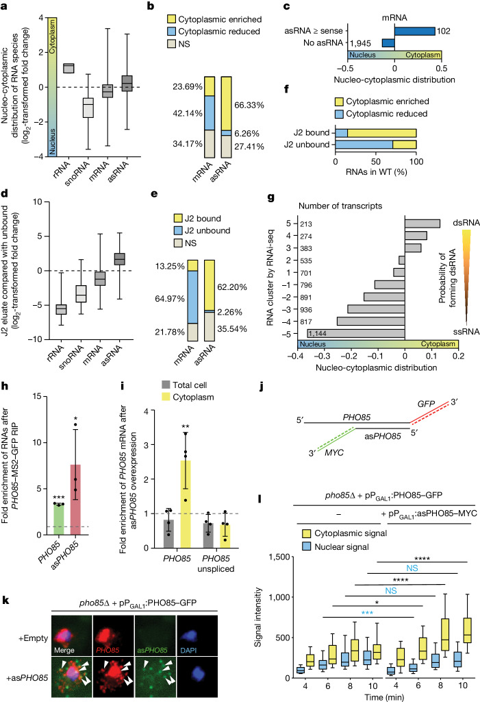
When mRNAs have been transcribed and processed in the nucleus, they are exported to the cytoplasm for translation. This export is mediated by the export receptor heterodimer Mex67-Mtr2 in the yeast Saccharomyces cerevisiae (TAP-p15 in humans)1,2. Interestingly, many long non-coding RNAs (lncRNAs) also leave the nucleus but it is currently unclear why they move to the cytoplasm3. Here we show that antisense RNAs (asRNAs) accelerate mRNA export by annealing with their sense counterparts through the helicase Dbp2. These double-stranded RNAs (dsRNAs) dominate export compared with single-stranded RNAs (ssRNAs) because they have a higher capacity and affinity for the export receptor Mex67. In this way, asRNAs boost gene expression, which is beneficial for cells. This is particularly important when the expression program changes. Consequently, the degradation of dsRNA, or the prevention of its formation, is toxic for cells. This mechanism illuminates the general cellular occurrence of asRNAs and explains their nuclear export.


Conflict of interest statement

The authors declare no competing interests.

Figures

Fig. 1
Fig. 1. dsRNAs are mainly localized in the cytoplasm.
a, Spatial RNA detection after fractionation RNA-seq experiment. The log2-transformed fold change of the cytoplasmic fraction compared with total lysate indicates the nucleo-cytoplasmic distribution. From n = 3 biologically independent samples. b, asRNAs are enriched in the cytoplasm. The percentage of unevenly distributed mRNAs and asRNAs from the fractionation RNA-seq is shown. NS, not significant. c, mRNAs localize to the cytoplasm when the asRNA is present in equal or higher amounts. Average nucleo-cytoplasmic distribution of mRNAs, based on their relative asRNA expression. d, J2 RIP experiments enriched asRNAs, as determined by the log2-transformed fold change of the J2 eluate relative to the unbound fraction. From n = 3 biologically independent samples. e, Percentage of significantly changed transcripts from the J2 RIP-seq. f, J2-enriched dsRNAs are cytoplasmic, as determined by their distribution in fractionation RNA-seq. WT, wild type. g, RNAs grouped by their enrichment in RNAi-seq analysed for their mean change in fractionation RNA-seq. h, PHO85 mRNA forms dsRNA with PHO85 asRNA (asPHO85) expressed from a plasmid. GFP pull-down with GFP-tagged MS2 loop-binding protein precipitates MS2-tagged PHO85 mRNA and co-precipitates PHO85 asRNA, determined by qPCR. From n = 4 biologically independent samples. i, Overexpression of PHO85 asRNA shifts PHO85 mRNA into the cytoplasm relative to no PHO85 asRNA (dotted line). The qPCR result after the fractionation experiment and RNA isolation of the total and unspliced PHO85 transcripts is shown. From n = 4 biologically independent samples. j, Hybridization with 12 Cy3-labelled probes detects a single GFP-tagged PHO85 mRNA and 15 Alexa647-labelled probes detects the MYC(15×)sequence of PHO85 asRNA. k, smFISH analysis shows dsRNA formation and a cytoplasmic shift of the PHO85 mRNA signal with PHO85 asRNA expression. The pho85∆ cells have a galactose-inducible PHO85–GFP and either an empty plasmid or the galactose-inducible asPHO85–MYC(15×) plasmid after 8 min galactose induction. The white arrowheads indicate colocalizing signal. From n = 4 biologically independent experiments with similar results. l, smFISH quantification of nuclear and cytoplasmic PHO85 signal at different time points after galactose induction with or without simultaneous asRNA induction; n > 52 cells over 3 biologically independent experiments. Source Data
Fig. 2
Fig. 2. Mex67 preferentially binds dsRNAs for faster nuclear export.
a, dsRNAs accumulate in the nucleus of mex67-5 xpo1-1, as determined by fractionation RNA-seq experiment. The average reduction of the cytoplasmic RNA relative to the wild type is based on groups defined by RNAi-seq. From n = 3 biologically independent samples. b, Exported dsRNAs reach the ribosome. J2 antibody IF with a Cy3-labelled secondary antibody (dsRNA) and FISH with a Cy3-labelled oligonucleotide d(T) probe were done for 1 h at 37 °C. From n = 3 biologically independent experiments. Scale bars, 3 μm. c, Fluorescence intensity quantification in J2 IF for 1  h at 37  °C for each condition shown in b; n > 20 cells over 3 biologically independent experiments. From left to right: P = 1.27 × 10−8, P = 3.03 × 10−25, P = 1.98 × 10−23 and P = 2.39 × 10−18. d, dsRNA levels increase when translation is inhibited. Dot-blots with RNA from the indicated strains and treatments after 1  h at 37  °C were detected with the J2 antibody. From n = 3 biologically independent experiments with similar results. e, dsRNAs contact ribosomes before ssRNAs. We shifted mex67-5 to 37 °C for 1 h to block RNA export. It was subsequently released by lowering the temperature to 25 °C. RIP experiments with Rps2–GFP from different time points were done. Three ssRNA and three dsRNA targets were analysed by qPCR. From n = 3 biologically independent experiments. f, Induction of PHO85 asRNA expression changes both protein and mRNA levels of PHO85, as shown by western blot quantification and qPCR. Hem15 served as loading control and for normalization. From n = 3 biologically independent experiments. g,h, More Mex67 molecules can bind to dsRNA than to ssRNA. EMSA with FAM-labelled ssRNAs (g) or dsRNAs (h) was carried out by adding increasing amounts of recombinant TAP-tagged Mex67–Mtr2. From n = 3 independent experiments with similar results. i,j, Competition assay detects preferential Mex67–Mtr2 binding to dsRNA. Cy5-labelled ssRNAs (red) were pre-incubated with Mex67–Mtr2. Subsequently, increasing amounts of a FAM-labelled dsRNA (green) were added as a competitor (i). Increasing amounts of Cy5-labelled ssRNA were added to pre-bound FAM-labelled dsRNA (j). From n = 3 independent experiments with similar results. Source Data
Fig. 3
Fig. 3. dsRNA formation is essential for cells changing their expression program.
a, Stress increases asRNA levels. A genome-wide RNA analysis of cells incubated with 0.6 M NaCl (ref. ) for 30 min was used to find changes in sense mRNA and asRNA expression compared with unstressed conditions, and is shown here by the log2-transformed fold change. From n = 2 biological independent samples. b, Stress-responsive mRNAs are accompanied by increased asRNA expression. Significantly changed asRNAs of significantly increased mRNAs from the RNA-seq are shown after 30 min exposure to 0.6 M NaCl. c, The amount of dsRNA increases under stress conditions. J2-IF is shown for the wild type exposed to the indicated stress conditions. From n = 3 biologically independent experiments. d, Quantification of the J2-IF displayed in e. From n = 30 cells over 3 independent experiments. Left to right: P = 6.91 × 10−9, P = 1.59 × 10−15, P = 7.57 × 10−4, P = 5.47 × 10−12. e, dsRNA degradation by the bacterial ribonuclease RNaseIII in the nucleus of yeast cells is lethal. We spotted 10-fold serial dilutions of the wild type containing the indicated plasmids onto glucose (no induction) or galactose (with induction) plates and incubated for 3 days. From n = 3 biologically independent experiments with similar results. f, J2-IF and localization of the GFP- and transport signal-tagged RNaseIII fusion proteins in yeast cells. Plasmid-containing wild-type cells were grown to the logarithmic phase before the RNaseIII expression was induced by adding galactose. From n = 3 biologically independent experiments with similar results. g, Cytoplasmic RNaseIII is not tolerated in cellular stress situations. We spotted 10-fold serial dilutions of wild-type cells containing either a constitutively expressed RNaseIII–NES from the ADH1 promoter or the RNAi system onto plates and incubated for 3 days at 25 °C. From n = 3 biologically independent experiments with similar results. h, Stress-induced dsRNA is degraded by cytoplasmic RNaseIII. J2-IF and oligonucleotide d(T) FISH are shown either without stress or after 30 min incubation with 0.7 M NaCl. From n = 3 biologically independent experiments. All scale bars, 3  μm.
Fig. 4
Fig. 4. Dbp2 induces dsRNA formation.
a, We found that dsRNAs accumulate in the nucleus of mtr4G677D at 37 °C. J2-IF and oligonucleotide d(T)-FISH are shown. b, Disturbed dsRNA formation in dbp2∆. Strains were changed to the non-permissive temperature for dbp2∆ of 25 °C. c, Signal quantification from a and b. From n > 40 cells over 3 biologically independent experiments. Left to right, P = 7.83 × 10−35, P = 8.56 × 10−34. d, J2 dot-blot of isolated RNA. From n = 3 biologically independent experiments with similar results. e, Dbp2 binds to dsRNA. Western blot of J2 Co-IP from cells expressing MYC-tagged DBP2 with or without the addition of recombinant RNaseIII. Grx4 is negative control. From n = 3 biologically independent replicates with similar results. f, dsRNA formation and cytoplasmic shift of PHO85 mRNA after PHO85 asRNA expression was lost in dbp2∆. Wild type and dbp2∆ carrying the galactose-inducible PHO85–GFP plasmid and either an empty vector or the galactose-inducible asPHO85–MYC(15×) plasmid were used for smFISH after 8 min galactose induction. From n = 3 biologically independent experiments. g, Quantification of smFISH for the cytoplasmic/total signal ratio of PHO85 mRNA either with or without (dotted line) simultaneous asRNA expression. From n > 23 cells examined over 3 biologically independent experiments. h, The increased presence of PHO85 mRNA in the cytoplasm after PHO85 asRNA overexpression was abolished in dbp2∆. After the induction of PHO85 asRNA, cells were shifted to 25 °C for 1 h before cytoplasmic fractionation, RNA isolation and qPCR. From n = 3 biologically independent samples. i, Model for the preferential export of dsRNAs. After transcription, ssRNAs are eventually bound by Mex67, leading to low-level export and translation in the cytoplasm. Gene expression is boosted by the transcription of asRNA and subsequent dsRNA formation of sense–antisense pairs by the helicase Dbp2 and its co-factor Yra1. The dsRNA preferentially binds to Mex67 for nuclear export and mRNA translation. Ribosomes recognize the non-coding property of the asRNAs and subsequent NMD-mediated degradation. This mechanism ensures preferential gene expression. All scale bars, 3 μm. Source Data
Extended Data Fig. 1
Extended Data Fig. 1. Supplement to Fig. 1.
(a) Cytoplasmic fractionation experiment eliminates the nuclear content of cells. RNA-seq was carried out from a total cell lysate and the cytoplasmic fraction. n = 3 biological independent samples. (b) Western blot analysis of nuclear and cytoplasmic proteins confirms the successful fractionation experiment. The cytosolic protein Zwf1, the nucleolar protein Nop1 and the nuclear protein Yra1 are shown before and after fractionation. (c) Fractionation-RNA-seq is highly reproducible. The read counts for each transcript across different replicates were compared against each other. (d) The calculated Spearman rank correlations between replicates are shown. (e, f) Vulcano plots of mRNAs (e) and asRNAs (f) from the fractionation-RNA-seq experiment, shown in Fig. 1a,b, are depicted. For null hypothesis testing the Wald test was used with multiple comparisons adjustments using the Benjamini and Hochberg method.
Extended Data Fig. 2
Extended Data Fig. 2. Supplement to Fig. 1.
(a) The J2 antibody recognizes ~40 bp long RNA double strands and was used to precipitate dsRNA for RNA-seq experiments. n = 3 biological independent samples. (b) The J2-RNA-seq is highly reproducible. The read counts for each transcript across different replicates were compared against each other. (c) The calculated Spearman rank correlations between replicates are shown. (d, e) Vulcano plot of mRNAs (d) and asRNA (e) of the J2-RNA-seq are shown. For null hypothesis testing the Wald test was used with multiple comparisons adjustments using the Benjamini and Hochberg method.
Extended Data Fig. 3
Extended Data Fig. 3. Supplement to Fig. 1.
(a) Most asRNAs are present in cells together with their sense transcripts. Single cell analysis of was analyzed for the simultaneously presence of sense and antisense transcript of a gene in a single cell. (b) The J2-RIP-seq and the RNAi-seq results strongly correlate. The log2-fold change of the J2-RIP-seq and RNAi-seq were plotted against each other and the spearman rank correlation was calculated (r = 0.72). (c) Groups of mRNAs based on their enrichment in RNAi-seq shown in Fig. 1g are depicted with their hit density of the degradation products along their gene bodies produced by the artificially expressed Dicer protein. (d) The probability of a cytoplasmic localization increases with the probability to be part of a dsRNA as determined by the RNAi-seq. Significantly cytoplasmic enriched and reduced targets of each group based on the RNAi-seq classification (Extended Data Fig. 3c) are depicted in percent. (e) XUTs are most likely to form dsRNA. lncRNAs were grouped by their classification into SUTs, CUTs and XUTs. The degradation products of the artificially expressed Dicer were then mapped onto the gene bodies of these separate groups. (f) The length overlaps in base pairs (bp) between mRNAs and their asRNA counterparts are shown in boxplots for SUTs, XUTs and CUTs. (g) asRNAs are on average more likely enriched in RNAi-seq and to localize in the cytoplasm (by fractionation-RNA-seq) than long intergenic non coding (linc)RNAs. asRNAs and lincRNAs were defined by whether they overlap a protein coding gene on the opposite strand, classified in XUTs, SUTs and CUTs and then analyzed for their log2 fold change in the RNAi-seq and the fractionation-RNA-seq.
Extended Data Fig. 4
Extended Data Fig. 4. Supplement to Fig. 1.
(a) Western blot of the cytoplasmic fractionation experiment shown in Fig. 1i and Extended Data Fig. 4b. The cytosolic protein Zwf1 and the nucleolar protein Nop1 are shown before and after fractionation. (b) Overexpression of asPHO85 results in similar amounts of sense and asRNA. Wild type cells either carrying an empty vector or a plasmid with asPHO85 under the GAL1 promoter were grown over night in glucose (uninduced) or 2% galactose (induced) to their log phase. Subsequently, the cells were lysed, the RNA isolated and quantified in qPCR. n = 4 independent biological experiments. (c) Quantification of smFISH at different time points after induction. The ratios of the PHO85 mRNA signal were calculated with or without simultaneous asRNA expression and subsequently related to the mean value of cells without asRNA (dotted line). n > 50 cells over 3 biological independent experiments. From left to right P = 6.79 × 10−06, P = 2.10 × 10⁻¹³, P = 4.79 × 10⁻¹⁸, P = 5.32 × 10⁻¹⁶. Source Data
Extended Data Fig. 5
Extended Data Fig. 5. Supplement to Fig. 1.
(a) dsRNAs do not show increased stability. mRNAs were divided in 4 groups (from left to right n = 3320, n = 1121, n = 322, n = 50) regarding their half-life determined by Chan et al.. The log2 fold change in RNAi-seq of the groups is represented. Only mRNAs that were present in both experiments were considered. The box plots are defined by the median as the center line, the 25th and 75th percentiles as the box boundaries and the Min and Max values as the whiskers. The presentation as a box plot was chosen for a clearer visualization of the respective comparison. The correlation was calculated independently of the grouping as the spearman correlation rank (r = −0.05). (b) The half-life of nuclear mRNAs is on average higher than that of cytoplasmic mRNAs. mRNAs were divided in 4 groups (from left to right n = 3489, n = 1214, n = 343, n = 53) regarding their half-life determined by Chan et al.. The groups were analyzed regarding their nucleo-cytoplasmic distribution as determined in the fractionation-RNA-seq. Only mRNAs that were present in both experiments were considered. The box plots are defined by the median as the center line, the 25th and 75th percentiles as the box boundaries and the Min and Max values as the whiskers. The presentation as a box plot was chosen for a clearer visualization of the respective comparison. The correlation was calculated independently of the grouping as the spearman correlation rank (r = −0.42). (c) Heatmap of spearman correlation ranks between different data sets.
Extended Data Fig. 6
Extended Data Fig. 6. Supplement to Fig. 2.
(a) Fractionation-RNA-seq in mex67-5 xpo1-1 is highly reproducible. The read counts for each transcript across different replicates were compared against each other. (b) The calculated Spearman rank correlations between replicates are shown.
Extended Data Fig. 7
Extended Data Fig. 7. Supplement to Fig. 2.
(a) mRNAs that belong to the top group in Fig. 2a encode for membrane proteins. GO-term analysis of this group uncovers mRNAs that possibly undergo intracellular transport. Overrepresentation was calculated with the Fishers exact test and corrected with the Bonferroni correction. (b) J2 precipitates dsRNA. J2-RIP was conducted followed by qPCR. Four highly confident dsRNA targets were chosen to verify the dsRNA pulldown by J2 in RIP experiments compared to a control with no added antibody and subsequent qPCRs. n = 5 biological independent experiment (c) dsRNA accumulates in the export mutant. Same experiment as shown in (b) in the indicated strains and related to J2 pulldown in wild type. n = 4 biological independent experiments (d) dsRNAs are exported to reach the ribosome. IF experiment with the J2 antibody (primary) and a Cy3-labelled secondary antibody and a FISH with a Cy3-labelled oligo d(T) probe are shown in the indicated strains and treatments. n = 3 biological independent experiments (e) dsRNA contacts ribosomes before ssRNAs. Rps3-GFP was precipitated at different timepoints before and after export release. The successful pull-down and isolation of Rps3-GFP is shown in an example Western blot. The bound RNA was purified and subsequently analyzed in Fig. 2e. n = 3 biological independent experiments (f) RNA substrates run differently in agarose gel electrophoresis depending on their tag and whether they are single or double stranded. RNA substrates tagged with Cy5 or FAM and their complementary untagged oligos for subsequent EMSA were loaded onto a 0.5 % TAE agarose gel supplemented with HD-green. HD-green intercalates with double strand nucleic acids and thus stains the provided RNA only when a double strand was formed. In this case, HD-green staining was only visible when the tagged RNA substrate and the complementary oligo were combined, proving the formation of the double strand in vitro. n = 1 (g, h) Competition assay reveals a preferential binding of Mex67 to dsRNA. The EMSA shown in Fig. 2i and j were repeated with changed labels. FAM-labelled (green) ssRNA (g) or Cy5-labeled (red) dsRNA (h) were pre-incubated with low concentrations of Mex67 for complex formation. Subsequently, increasing amounts of a Cy5-labeled (red) dsRNA (left) or FAM-labeled (green) ssRNA (right) was added as a competitor. n = 3 independent experiments with similar results. Source Data
Extended Data Fig. 8
Extended Data Fig. 8. Supplement to Fig. 3.
(a) Absence of the transcription factor Set2 leads to increased dsRNA production. IF with J2-antibody and FISH with a Cy3-labelled oligo d(T) probe in indicated strains are shown. (b) Quantification of the signal intensities depicted in (a). Signal intensity was determined via Fiji. n = 20 cells examined over 3 biological independent samples. P = 1.37 × 10−08. (c) set2∆ contains elevated amounts of dsRNA. J2-dot blot of 1 µg RNA is shown. n = 3 biological independent experiments with similar results. (d) The level of the Set2-responsive antisense RNA of SEG2 is significantly increased in set2∆. SEG2 mRNA and asRNA amounts in wild type and set2∆ were measured by strand specific qPCR. n = 5 biological independent experiments. (e) The SEG2 mRNA and its asRNA form dsRNA in set2∆. J2-RIPs were carried out in wild type and set2∆ followed by strand specific qPCR of SEG2 and asSEG2. n = 3 biological independent experiments. (f) Western blot of the cytoplasmic fractionation experiment shown in Extended Data Fig. 8g. (g) Increased presence of SEG2 in the cytosol of set2∆ cells. Cytoplasmic fractionation experiment in wild type and set2∆ was carried out, followed by strand specific qPCR of indicated targets. n = 5 biological independent experiments. (h) RNaseIII degrades dsRNA from yeast. Recombinant RNaseIII was added to 1 µg of total RNA isolated from wild type cells. After 20 min incubation at 37 °C the RNA was dropped onto a membrane together with a control of untreated 1 µg total RNA. Subsequently a J2 dot-blot was conducted. n = 1. (i) dsRNA amounts increase under stress conditions. J2-IF with a secondary Cy3 labeled anti-mouse antibody is shown in wild type cells, exposed to the indicated stress conditions for the indicated time. n = 3 biological independent experiments. (j) Directing the dsRNA degrading bacterial RNaseIII into the nucleus of yeast cells is lethal. Growth analyses of wild type cells expressing the indicated constructs on glucose (repressing) and galactose (inducing conditions) containing plates at the indicated temperatures. n = 3 biological independent experiments with similar results. (k) Localization of the GFP- and transport signal-tagged RNaseIII fusion proteins in yeast cells. Plasmid containing wild type cells were grown to log phase before the RNaseIII expression was induced through the addition of 2 % galactose for 6 h. The GFP signal was detected using fluorescence microscopy. n = 3 biological independent experiments. (l) J2-IF in cells expressing GFP- and transport signal-tagged RNaseIII fusion proteins. Cells were treated as in Extended Data Fig. 8k. n = 3 biological independent experiments. (m) Stress-induced dsRNA is degraded by cytoplasmic RNaseIII. J2-IF and oligo d(T) FISH are shown for the indicated strains either without stress or after a 30 min incubation with 0.7 M NaCl. n = 3 biological independent experiments. (n) Cytoplasmic RNaseIII-NES degrades accumulated dsRNA in the translation mutant rpl10(G161D). rpl10(G161D) either containing an empty vector or a plasmid encoding the galactose inducible RNaseIII-NES was grown in galactose containing media at 25 °C until log phase. Half of the cells were shifted to 37 °C for 1 h before the RNA was isolated and 1 µg spotted onto a nylon membrane. The dsRNA level was detected with the J2-antibody. n = 3 biological independent experiments with similar results. Source Data
Extended Data Fig. 9
Extended Data Fig. 9. Supplement to Fig. 4.
(a) Deletion of DBP2 leads to severe cold sensitivity. 10-fold serial dilutions of wild type and dbp2∆ after 3 days of growth on full medium plates. n = 3 independent biological replicates with similar results. (b,c) J2-IF and FISH with an oligo d(T) probe is shown. Strains were shifted for 2.5 h to the non-permissive temperature of mtr4(G677D) at 37 °C (b) or for 1 h to the non-permissive temperature of dbp2∆ at 25 °C (c). (d) On average, dsRNAs are less structured in dbp2∆. The measured DMS reactivity in Structure-seq data from dbp2∆ was compared to wild type and analyzed for every group based on the RNAi-seq. n = 2 independent biological replicates. (e) Dbp2 binds to ssRNA and dsRNA targets in similar amounts. mRNAs with hits in Dbp2 iCLIP were counted for each group based by RNAi-Seq and are given in percentage related to the total amount of mRNAs of each group. (f) Western blot of the cytoplasmic fractionation experiment shown in Fig. 4h for the cytoplasmic protein Zwf1 and the nuclear protein Nop1 is shown. n = 3 biological independent experiments with similar results. (g) Overexpression of the asPHO85 results in similar amounts of sense and asRNA in wild type and dbp2∆. Wild type and dbp2∆ strains were either transformed with an empty vector or with a plasmid carrying asPHO85 under the GAL1 promoter. The strains were grown over night under inducing conditions (2% galactose) to log phase. Subsequently, cells were lysed, the RNA was isolated and used in qPCR. n = 3 biological independent experiments. Source Data

References

    1. Segref A, et al. Mex67p, a novel factor for nuclear mRNA export, binds to both poly(A)+ RNA and nuclear pores. EMBO J. 1997;16:3256–3271. doi: 10.1093/emboj/16.11.3256. - DOI - PMC - PubMed
    1. Zenklusen D, Stutz F. Nuclear export of mRNA. FEBS Lett. 2001;498:150–156. doi: 10.1016/S0014-5793(01)02482-6. - DOI - PubMed
    1. van Dijk EL, et al. XUTs are a class of Xrn1-sensitive antisense regulatory non-coding RNA in yeast. Nature. 2011;475:114–117. doi: 10.1038/nature10118. - DOI - PubMed
    1. Berretta J, Morillon A. Pervasive transcription constitutes a new level of eukaryotic genome regulation. EMBO Rep. 2009;10:973–982. doi: 10.1038/embor.2009.181. - DOI - PMC - PubMed
    1. Djebali S, et al. Landscape of transcription in human cells. Nature. 2012;489:101–108. doi: 10.1038/nature11233. - DOI - PMC - PubMed

MeSH terms