

Arteriovenous fistula of the mandible: a case report
- PMID: 38898353
- PMCID: PMC11379768
- DOI: 10.1007/s11282-024-00762-6
Arteriovenous fistula of the mandible: a case report
Abstract
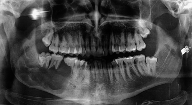
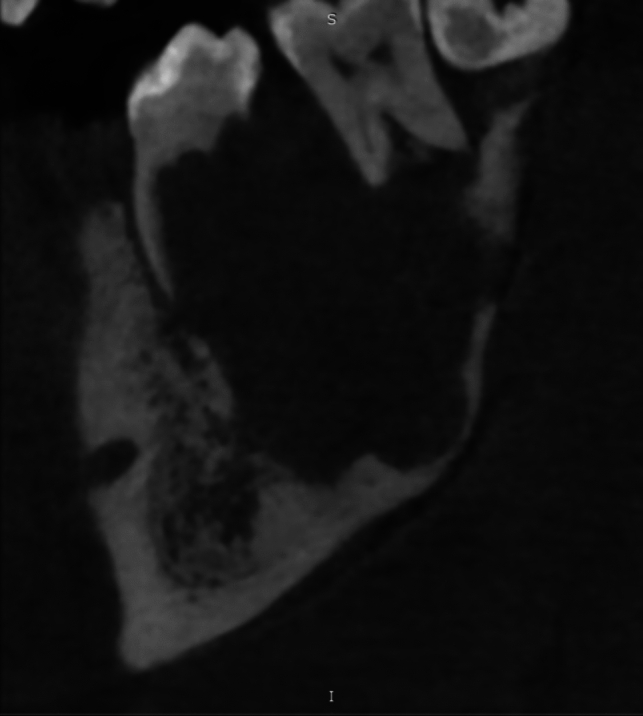
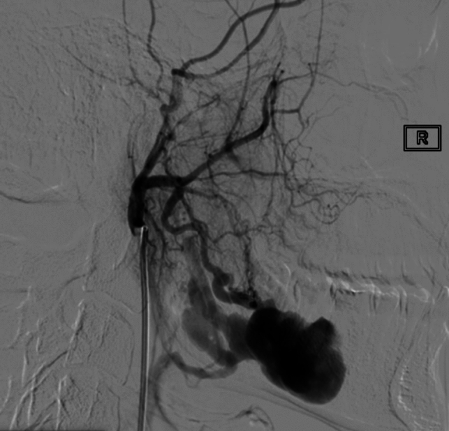
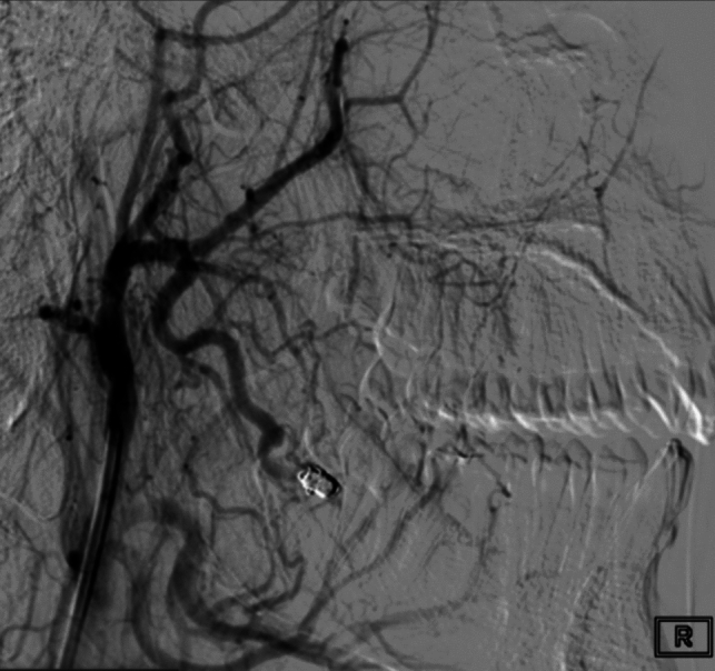
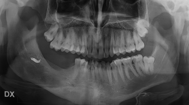
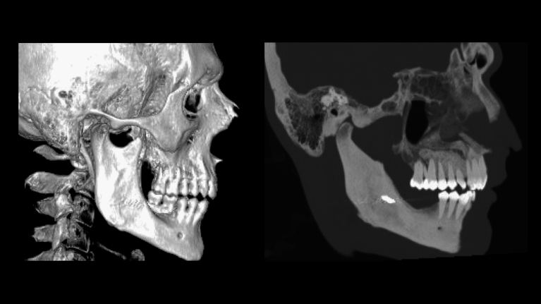
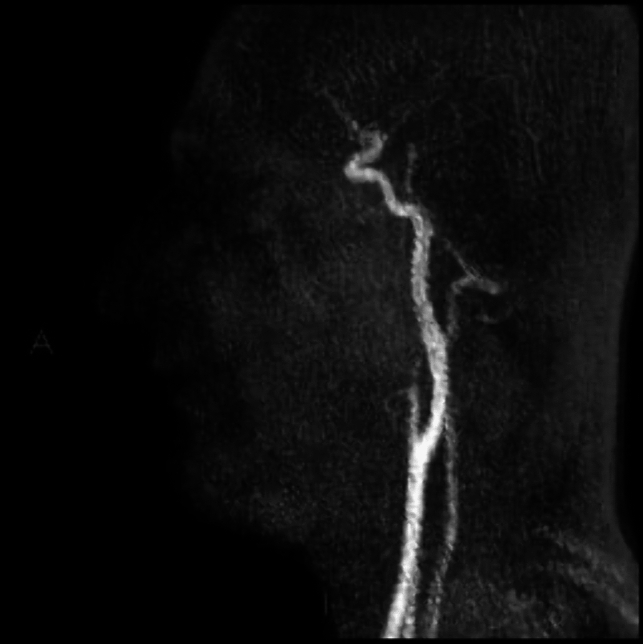
Intraosseous arteriovenous malformations (AVM) are uncommon high-flow vascular malformation that can affect the maxilla or mandible. AVM may present with aspecific and misleading signs and symptoms. The diagnosis is often accidental and bleeding may represent the first symptom. Radiographically, there are few characteristic features and misdiagnosis is easy. Here we report the case of a young male affected by arteriovenous fistula on the right side of the mandible initially misdiagnosed as a cystic lesion. The patient underwent transarterial embolization of the vascular malformation and subsequently the lesion was surgically removed. 1-year follow-up showed complete healing of the mandibular bone and absence of recurrence. Intraosseous arteriovenous malformations are rare entities. However, due to their harmfulness, both clinicians and radiologists must be aware of this type of lesion and should always consider them in the differential diagnosis of osteolytic lesions.
Keywords: Arteriovenous; Dentigerous cyst; Embolization; Fistula; Oral and maxillofacial pathology; Vascular malformation.
© 2024. The Author(s).
Conflict of interest statement
The authors have no conflicts of interest to declare that are relevant to the content of this article.
Figures
References
-
- Mulliken JB, Young AE. Vascular birthmarks: hemangiomas and malformations. Philadelphia: WB Saunders; 1988.
-
- Lasjaunias P, Berenstein A. Craniofacial hemangioma, vascular malformations and angiomatosis: specific aspects (chapter 10) pp: 341–397 in: Surgical Neuroangiography (vol 2). Springer-Verlag Berlin Heidelberg 1987.
Publication types
MeSH terms
LinkOut - more resources
Full Text Sources