

Association of disease severity and genetic variation during primary Respiratory Syncytial Virus infections
- PMID: 38898440
- PMCID: PMC11188216
- DOI: 10.1186/s12920-024-01930-7
Association of disease severity and genetic variation during primary Respiratory Syncytial Virus infections
Abstract
Background: Respiratory Syncytial Virus (RSV) disease in young children ranges from mild cold symptoms to severe symptoms that require hospitalization and sometimes result in death. Studies have shown a statistical association between RSV subtype or phylogenic lineage and RSV disease severity, although these results have been inconsistent. Associations between variation within RSV gene coding regions or residues and RSV disease severity has been largely unexplored.
Methods: Nasal swabs from children (< 8 months-old) infected with RSV in Rochester, NY between 1977-1998 clinically presenting with either mild or severe disease during their first cold-season were used. Whole-genome RSV sequences were obtained using overlapping PCR and next-generation sequencing. Both whole-genome phylogenetic and non-phylogenetic statistical approaches were performed to associate RSV genotype with disease severity.
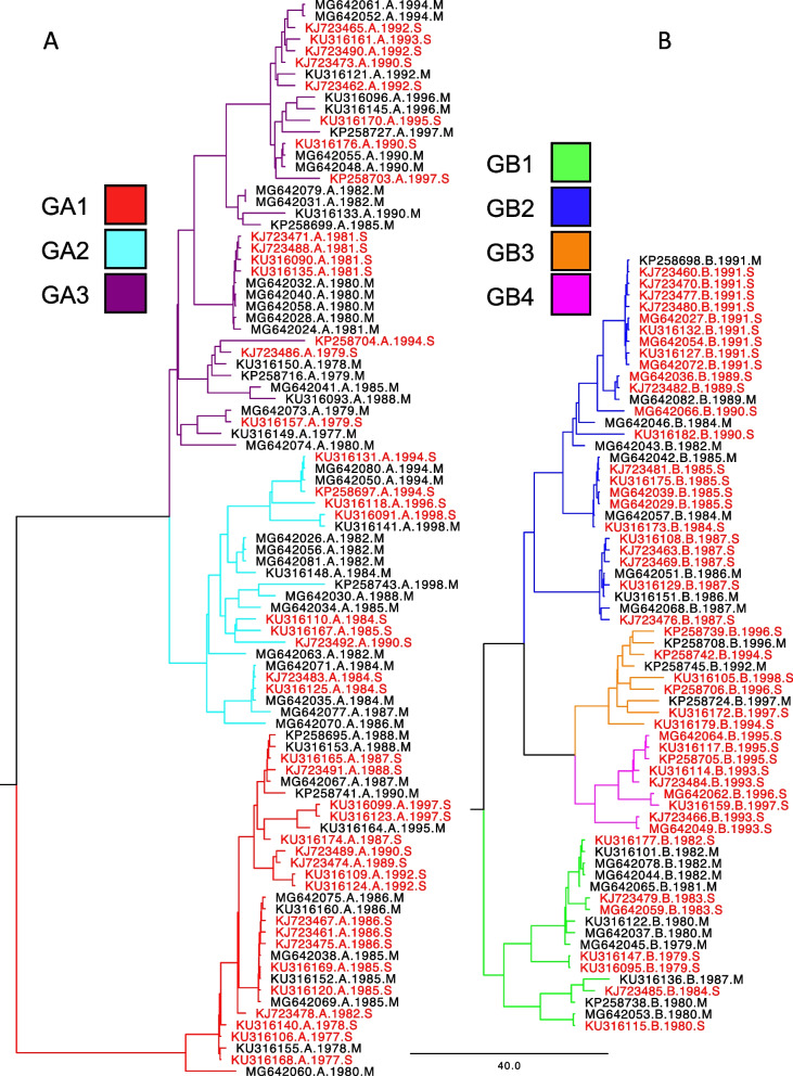
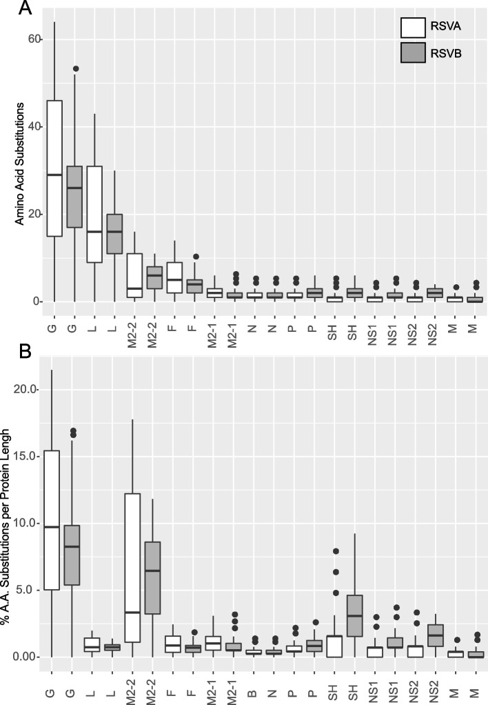
Results: The RSVB subtype was statistically associated with disease severity. A significant association between phylogenetic clustering of mild/severe traits and disease severity was also found. GA1 clade sequences were associated with severe disease while GB1 was significantly associated with mild disease. Both G and M2-2 gene variation was significantly associated with disease severity. We identified 16 residues in the G gene and 3 in the M2-2 RSV gene associated with disease severity.
Conclusion: These results suggest that phylogenetic lineage and the genetic variability in G or M2-2 genes of RSV may contribute to disease severity in young children undergoing their first infection.
Keywords: Genetic variation; RSV; Respiratory infection; Severe disease; Whole-genome.
© 2024. The Author(s).
Conflict of interest statement
The authors declare no competing interests.
Figures

Similar articles
-
Genomic characterization of circulating human respiratory syncytial viruses A and B in Kuwait using whole-genome sequencing.Microbiol Spectr. 2024 Jul 2;12(7):e0015924. doi: 10.1128/spectrum.00159-24. Epub 2024 May 29. Microbiol Spectr. 2024. PMID: 38808977 Free PMC article.
-
Epidemic Outbreak of Respiratory Syncytial Virus Infection After the end of the Zero-COVID-19 Policy in China: Molecular Characterization and Disease Severity Associated With a Novel RSV-B Clade.J Med Virol. 2025 Apr;97(4):e70343. doi: 10.1002/jmv.70343. J Med Virol. 2025. PMID: 40207902
-
[Epidemiologic characteristics and the relationship with disease severity of respiratory syncytial virus genotypes from children with lower respiratory tract infection in the southern Zhejiang province].Zhonghua Er Ke Za Zhi. 2015 Jul;53(7):537-41. Zhonghua Er Ke Za Zhi. 2015. PMID: 26310648 Chinese.
-
Phylogenetic and phylodynamic analysis of respiratory syncytial virus strains circulating in children less than five years of age in Karachi-Pakistan.Infect Genet Evol. 2024 Dec;126:105694. doi: 10.1016/j.meegid.2024.105694. Epub 2024 Nov 26. Infect Genet Evol. 2024. PMID: 39608424 Free PMC article.
-
Respiratory Syncytial Virus: The Influence of Serotype and Genotype Variability on Clinical Course of Infection.Int J Mol Sci. 2017 Aug 6;18(8):1717. doi: 10.3390/ijms18081717. Int J Mol Sci. 2017. PMID: 28783078 Free PMC article. Review.
Cited by
-
Human respiratory syncytial virus genetic diversity and lineage replacement in Ireland pre- and post-COVID-19 pandemic.Microb Genom. 2025 Mar;11(3):001379. doi: 10.1099/mgen.0.001379. Microb Genom. 2025. PMID: 40096248 Free PMC article.
-
Genetic diversity of respiratory syncytial virus in children with community-acquired pneumonia in Guangzhou: an epidemiological update.Pediatr Res. 2025 Jul 1. doi: 10.1038/s41390-025-04214-7. Online ahead of print. Pediatr Res. 2025. PMID: 40593188
-
Epidemiology of respiratory syncytial virus and its subtypes among cases of influenza like illness and severe acute respiratory infection: findings from nationwide sentinel surveillance in Ethiopia.BMC Infect Dis. 2025 Jul 24;25(1):941. doi: 10.1186/s12879-025-11330-6. BMC Infect Dis. 2025. PMID: 40707913 Free PMC article.
References
-
- Shi T, McAllister DA, O'Brien KL, Simoes EAF, Madhi SA, Gessner BD, Polack FP, Balsells E, Acacio S, Aguayo C, et al. Global, regional, and national disease burden estimates of acute lower respiratory infections due to respiratory syncytial virus in young children in 2015: a systematic review and modelling study. Lancet. 2017;390(10098):946–958. doi: 10.1016/S0140-6736(17)30938-8. - DOI - PMC - PubMed
-
- Fields BN, Knipe DM, Howley PM, Griffin DE. Fields virology. 4. Philadelphia: Lippincott Williams & Wilkins; 2001.
MeSH terms
Grants and funding
LinkOut - more resources
Full Text Sources
Medical
