. 2024 Jun 19;22(1):347.
doi: 10.1186/s12951-024-02638-7.

Advancing fluorescence imaging: enhanced control of cyanine dye-doped silica nanoparticles

Affiliations

Advancing fluorescence imaging: enhanced control of cyanine dye-doped silica nanoparticles

Taewoong Son et al. J Nanobiotechnology. .

Abstract

Background: Silica nanoparticles (SNPs) have immense potential in biomedical research, particularly in drug delivery and imaging applications, owing to their stability and minimal interactions with biological entities such as tissues or cells.

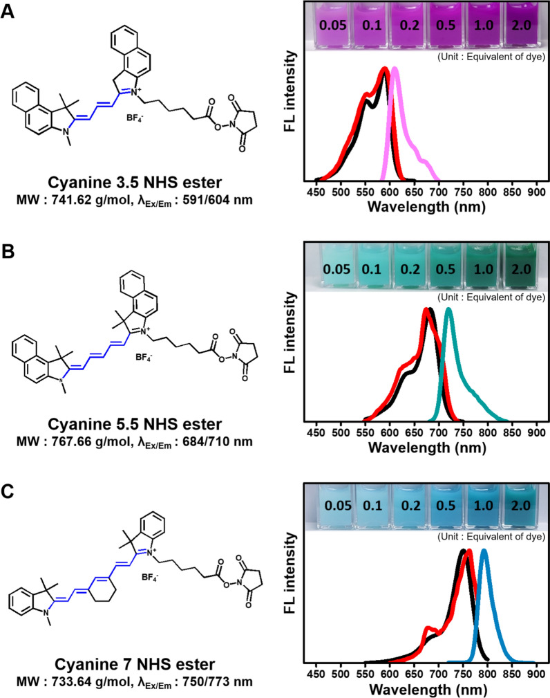
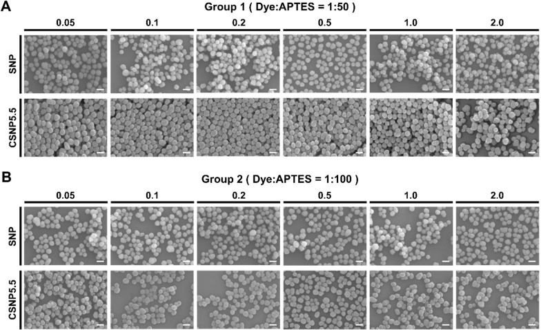
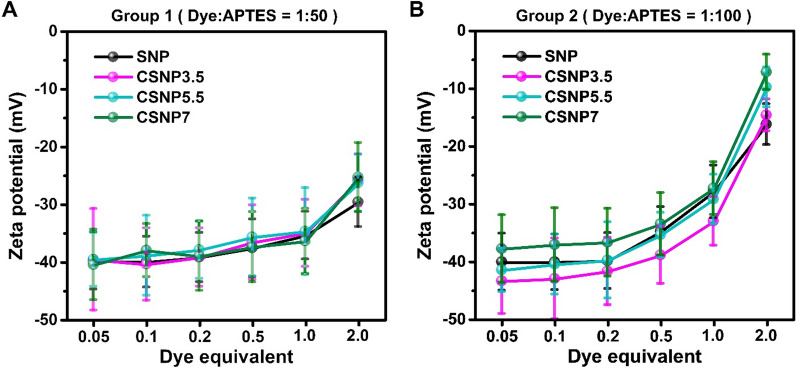
Results: With synthesized and characterized cyanine-dye-doped fluorescent SNPs (CSNPs) using cyanine 3.5, 5.5, and 7 (Cy3.5, Cy5.5, and Cy7). Through systematic analysis, we discerned variations in the surface charge and fluorescence properties of the nanoparticles contingent on the encapsulated dye-(3-aminopropyl)triethoxysilane conjugate, while their size and shape remained constant. The fluorescence emission spectra exhibited a redshift correlated with increasing dye concentration, which was attributed to cascade energy transfer and self-quenching effects. Additionally, the fluorescence signal intensity showed a linear relationship with the particle concentration, particularly at lower dye equivalents, indicating a robust performance suitable for imaging applications. In vitro assessments revealed negligible cytotoxicity and efficient cellular uptake of the nanoparticles, enabling long-term tracking and imaging. Validation through in vivo imaging in mice underscored the versatility and efficacy of CSNPs, showing single-switching imaging capabilities and linear signal enhancement within subcutaneous tissue environment.

Conclusions: This study provides valuable insights for designing fluorescence imaging and optimizing nanoparticle-based applications in biomedical research, with potential implications for targeted drug delivery and in vivo imaging of tissue structures and organs.

Keywords: Characterization; Cyanine N-hydroxysuccinimide ester; Fluorescence in vitro and in vivo image; Imaging optimization; Silica nanoparticle.


Conflict of interest statement

The authors declare no competing financial interests.

Figures

Fig. 1
Fig. 1
Structures and material properties of amine-reactive cyanine NHS ester dyes. The material properties of dye include molecular weights, fluorescence excitation/emission and experimental spectra of Cy3.5 (A), Cy5.5 (B), and Cy7 (C). The black spectrum indicates absorbance, the red spectrum indicates excitation scan, and the colored spectrum indicates emission scan. The numbers shown in the inset image represent the equivalents of dye. The molecular weights and fluorescence excitation/emission are referred from Lumiprobe
Fig. 2
Fig. 2
Synthetic reaction formula of silica nanoparticle (SNP)-based on Stöber method. A Ammonium catalyst based hydrolysis and condensation of silica. B Coupling reaction between the ester group on the cyanine dye and the amine group on APTES. C Fluorescent SNP formation with cyanine NHS ester dye. Scale bar = 100 nm
Fig. 3
Fig. 3
Morphological characteristics analyses. Representative scanning electron microscopy images of SNPs and CSNP5.5 synthesized at varying APTES ratios and equivalents. A Group synthesized with a 50-fold molar ratio excess of APTES to dye. B Group with a 100-fold. Within each group, nanoparticles were synthesized under various conditions depending on the equivalents. Scale bars = 100 nm
Fig. 4
Fig. 4
Surface charge of CSNPs. The zeta potentials of CSNPs synthesized under various APTES ratios and equivalents were measured using a DLS. A Group synthesized with a 50-fold molar ratio excess of APTES to dye. B Group with a 100-fold. The samples were suspended in distilled water with 2.5 mg/ml concentration and titrated to pH 7.4
Fig. 5
Fig. 5
Encapsulation of cyanine dyes in CSNP synthesized under various APTES ratios and equivalents. A Group synthesized with a 50-fold molar ratio excess of APTES to dye. B Group with a 100-fold
Fig. 6
Fig. 6
Emission spectra of CSNP5.5 synthesized under various APTES ratios and equivalents were measured using a fluorescence spectrophotometer. A Group synthesized with a 50-fold molar ratio excess of APTES to dye. B Group with a 100-fold. The black spectrum indicates excitation scan, and the cyanine series colored spectra indicate emission scan of each equivalent
Fig. 7
Fig. 7
Comparison of fluorescent signal increase depending on the concentration of CSNP5.5 synthesized at varying APTES ratios and equivalents. AC Group synthesized with a 50-fold molar ratio excess of APTES to dye. DF Group with a 100-fold. A, D Increase of fluorescence signal with concentrations ranging from 0 to 50 mg/ml. B, E A range where the signal increases linearly (R2 > 0.99) in proportion to concentration. F, D Comparison of fluorescent signal efficiency at low, middle, and high concentrations. All signal intensities were obtained using a microplate reader
Fig. 8
Fig. 8
In vitro application of CSNPs. A Cytotoxicity of CSNPs to in vitro. The HeLa cells were incubated with CSNPs for 24 h and the cell viability were analyzed by MTS assay. B Wide field microscope images of CSNPs loaded to HeLa cell (scale bars = 30 μm), MRC-5 cell (20 μm), and Raw264.7 cell (10 μm). C Single particle microscope images of CSNPs after spin-coating (scale bars = 10 μm). The red, blue, and green colors indicate CSNP3.5, CSNP5.5, and CSNP7, respectively
Fig. 9
Fig. 9
In vivo application of CSNPs. All imaging was conducted using an IVIS spectrum, with CSNPs being imaged at excitation/emission wavelengths of 570/620 nm, 675/720 nm, and 745/800 nm for CSNP3.5, CSNP5.5, and CSNP7, respectively. A Representative fluorescence image of CSNP5.5 in a black 96-well plate before injection into the mice. B Switchable imaging of CSNPs by their own wavelength. C In vivo fluorescence imaging of CSNPs. Red, blue, and green indicate CSNP3.5, CSNP5.5, and CSNP7, respectively. D Fluorescence intensities of CSNPs depending on concentration and their linearity

Similar articles

Cited by

References

    1. Xiao D, Qi H, Teng Y, Pierre D, Kutoka PT, Liu D. Advances and challenges of fluorescent nanomaterials for synthesis and biomedical applications. Nanoscale Res Lett. 2021;16(167):1–23. - PMC - PubMed
    1. Anselmo AC, Mitragotri S. Nanoparticles in the clinic: an update post COVID-19 vaccines. Bioeng Transl Med. 2021;6:e10246. doi: 10.1002/btm2.10246. - DOI - PMC - PubMed
    1. Ma D, Kell AJ, Tan S, Jakubek ZJ, Simard B. Photophysical properties of dye-doped silica nanoparticles bearing different types of dye-silica interactions. J Phys Chem C. 2009;113:15974–15981. doi: 10.1021/jp905812f. - DOI
    1. Janjua TI, Cao Y, Yu C, Popat A. Clinical translation of silica nanoparticles. Nat Rev Mater. 2021;6(12):1072–1074. doi: 10.1038/s41578-021-00385-x. - DOI - PMC - PubMed
    1. Kharlamov AN, Tyurnina AE, Veselova VS, Kovtun OP, Shur VY, Gabinsky JL. Silica–gold nanoparticles for atheroprotective management of plaques: results of the NANOM-FIM trial. Nanoscale. 2015;7:8003–8015. doi: 10.1039/C5NR01050K. - DOI - PubMed

LinkOut - more resources