. 2024 Oct;23(10):e14256.
doi: 10.1111/acel.14256. Epub 2024 Jun 19.

Aging-induced short-chain acyl-CoA dehydrogenase promotes age-related hepatic steatosis by suppressing lipophagy

Affiliations

Aging-induced short-chain acyl-CoA dehydrogenase promotes age-related hepatic steatosis by suppressing lipophagy

Dan Deng et al. Aging Cell. 2024 Oct.

Abstract

Hepatic steatosis, the first step in the development of nonalcoholic fatty liver disease (NAFLD), is frequently observed in the aging population. However, the underlying molecular mechanism remains largely unknown. In this study, we first employed GSEA enrichment analysis to identify short-chain acyl-CoA dehydrogenase (SCAD), which participates in the mitochondrial β-oxidation of fatty acids and may be associated with hepatic steatosis in elderly individuals. Subsequently, we examined SCAD expression and hepatic triglyceride content in various aged humans and mice and found that triglycerides were markedly increased and that SCAD was upregulated in aged livers. Our further evidence in SCAD-ablated mice suggested that SCAD deletion was able to slow liver aging and ameliorate aging-associated fatty liver. Examination of the molecular pathways by which the deletion of SCAD attenuates steatosis revealed that the autophagic degradation of lipid droplets, which was not detected in elderly wild-type mice, was maintained in SCAD-deficient old mice. This was due to the decrease in the production of acetyl-coenzyme A (acetyl-CoA), which is abundant in the livers of old wild-type mice. In conclusion, our findings demonstrate that the suppression of SCAD may prevent age-associated hepatic steatosis by promoting lipophagy and that SCAD could be a promising therapeutic target for liver aging and associated steatosis.

Keywords: acetyl‐coenzyme A; aging; autophagy; lipid droplet; lipophagy; liver steatosis; short‐chain acyl‐CoA dehydrogenase.


Conflict of interest statement

The authors declare that there is no conflict of interest.

Figures

FIGURE 1
FIGURE 1
The levels of SCAD in the liver and serum increase during aging. (a, b) GSEA plots of FATTY_ACID_METABLISM and BUTANOATE_METABLISM (a) and hepatic ACADS mRNA expression (b) using the GSE133815 gene expression profile between 11 young (21–45 years old, male% = 54.5%) and 12 old (>69 years of age, male% = 58.3%) individuals. FDR, false discovery rate; NES, normalized enrichment score. (c) Correlation analysis of serum SCAD levels and age was conducted using the Pearson correlation test. The serum SCAD concentrations of 51 individuals with normal liver tissue were quantified via ELISA. (d) SCAD protein levels in PBMCs from normal‐liver individuals were assessed by Western blotting. (e, f) mRNA expression levels of acyl‐CoA dehydrogenase family members (VLCAD, LCAD, MCAD and SCAD) (e) and the SCAD protein levels (f) in liver tissues from wild‐type mice at the ages of 3, 10, and 20 months were quantified by real‐time PCR and Western blotting, respectively (n = 5 for each group). (g) The protein levels of p21 and SCAD in H2O2‐induced senescent HepG2 cells were detected by Western blotting. The values are presented as the means ± SDs of 3 independent experiments. *p < 0.05, **p < 0.01, and ***p < 0.001.
FIGURE 2
FIGURE 2
SCAD levels are positively associated with age‐related liver steatosis. (a) Hepatic ACADS mRNA expression in normal controls and NAFLD patients from the GEO datasets GSE66676 (control: n = 34, male% = 17.6%; NAFLD: n = 26, male% = 23.1%) (left) and GSE48452 (control: n = 27, male% = 7.4%; NAFLD: n = 14, male% = 41.7%) (right). (b) Representative H&E staining (upper) and SCAD IHC staining (lower) of liver sections from NAFLD patients and healthy individuals. (c) SCAD protein levels in PBMCs from NAFLD patients (n = 4) and age‐matched healthy controls (n = 4) were assessed via Western blotting. (d) Correlation analysis of serum SCAD levels and age was conducted using the Pearson correlation test. Serum SCAD concentrations were measured by ELISA in 68 NAFLD patients at various ages. *p < 0.05, **p < 0.01, and ***p < 0.001.
FIGURE 3
FIGURE 3
SCAD depletion protects mice against age‐related liver pathological damage. Acads+/+ mice and Acads−/− mice were fed a chow diet for 3 months (3 M) or 24 months (24 M). (a) Liver/body weight (LW/BW) ratio. (b) Hepatic triglyceride and fatty acid (FA) concentrations. (c) Representative images of liver sections stained with H&E, Oil Red O, Masson's trichrome, senescence‐associated (SA)‐β‐galactosidase, or p21 antibody (scale bar = 50 μm). (d) Quantitative analysis of the number of lipid vacuoles in the liver and the areas of Oil Red O‐, Masson's trichrome‐, β‐galactosidase‐, or p21‐positive staining. Three micrographs of each mouse were used for quantification (n = 5). (e) Serum ALT and AST concentrations (n = 3). The data are presented as the mean ± SD. *p < 0.05, **p < 0.01, and ***p < 0.001.
FIGURE 4
FIGURE 4
SCAD inhibits autophagy in the liver during aging. (a) GSEA was conducted by examining the transcriptome (GSE128608) of mice with an Acads+/+ or Acads−/− genetic background, and enrichment plots showing the regulation of autophagy and lysosomal pathways were generated. NES, normalized enrichment score; FDR, false discovery rate. (b–d) Hepatic protein levels of autophagy markers (p62, LC3 and p21) were measured by Western blotting (b) and autolysosome ultrastructural features were assessed by TEM (c) in young (3 M) and old (24 M) mice with an Acads+/+ or Acads−/− genetic background. The overall average number of autophagosomes in both genotypes was determined from 8 randomly selected fields (d). (e–h) HepG2 cells were transfected with Acads siRNA for 2 days and then treated with 300 μM H2O2 for 4 days. Autophagic flux was assessed with a dual tandem‐tagged GFP‐RFP‐LC3 adenovirus. (e) Western blot analysis of the effect of Acads siRNA on SCAD protein levels. (f) Images of representative HepG2‐LC3 cells (scale bar: 10 μm). (g) Quantification of LC3 puncta in HepG2‐LC3 cells in at least three independent experiments. (h) HepG2‐LC3 cells were analyzed by flow cytometry. *p < 0.05, **p < 0.01, and ***p < 0.001.
FIGURE 5
FIGURE 5
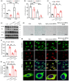
SCAD depletion alleviates senescence‐associated liver steatosis through the acetyl‐coenzyme A‐dependent lipophagy pathway. (a, b) Hepatic acetyl‐CoA concentrations (a) and ATP production (b) were quantified via ELISA in 3‐month‐old (young) and 24‐month‐old (old) Acads+/+ or Acads−/− mice (n = 3 for each group). (c–h) Senescence was induced in HepG2 cells with or without Acads knockdown by Acads siRNA via treatment with 300 μM H2O2 for 4 days; thereafter, the cells were treated with 12 μM dichloroacetic acid (DCA) for 24 h. (c) Cellular acetyl‐CoA production, (d) SA‐β‐galactosidase staining (bar scale: 100 μm), (e) immunoblots of SCAD, p21, LC3I/LC3II and p62, (f) cellular triglyceride content, (g) representative immunofluorescence images of colocalization between lysosomes labeled with LysoTracker Red and lipid droplets labeled with the green lipid dye BODIPY (bar scale: 20 μm), (h) bar graphs showing the percentage of colocalization between lysosomes and lipid droplets. *p < 0.05, **p < 0.01, and ***p < 0.001.
FIGURE 6
FIGURE 6
SCAD ablation protects cells against replicative senescence. Primary MEFs with an Acads+/+ or Acads−/− genetic background were prepared from mouse embryos on embryonic day 14 and cultured in DMEM for 2 days; these cells were labeled passage 0 (P0). Thereafter, the MEFs were serially cultured to passage 10 (P10) to establish replicative senescent cells. (a) SCAD protein levels were assessed in primary MEFs at P0 and P10 by Western blotting. (b) Cellular morphology was monitored with a light microscope (upper, scale bar: 100 μm), and lipid accumulation in cells was assessed by Oil Red O staining (lower, scale bar: 50 μm). (c) γ‐H2AX content in the P10 MEFs was examined via immunofluorescence staining. (d) Representative immunofluorescence images showing the colocalization of lysosomes labeled with LysoTracker Red and lipid droplets labeled with the green lipid dye BODIPY (scale bar: 50 μm). (e) Bar graphs showing the percentage of colocalization between lysosomes and lipid droplets. ***p < 0.001.

Similar articles

References

    1. Ahrens, M. , Ammerpohl, O. , von Schonfels, W. , Kolarova, J. , Bens, S. , Itzel, T. , Teufel, A. , Herrmann, A. , Brosch, M. , Hinrichsen, H. , Erhart, W. , Egberts, J. , Sipos, B. , Schreiber, S. , Häsler, R. , Stickel, F. , Becker, T. , Krawczak, M. , Röcken, C. , … Hampe, J. (2013). DNA methylation analysis in nonalcoholic fatty liver disease suggests distinct disease‐specific and remodeling signatures after bariatric surgery. Cell Metabolism, 18, 296–302. - PubMed
    1. Bertolotti, M. , Lonardo, A. , Mussi, C. , Baldelli, E. , Pellegrini, E. , Ballestri, S. , Romagnoli, D. , & Loria, P. (2014). Nonalcoholic fatty liver disease and aging: Epidemiology to management. World Journal of Gastroenterology, 20, 14185–14204. - PMC - PubMed
    1. Bradshaw, P. C. (2021). Acetyl‐CoA metabolism and histone acetylation in the regulation of aging and lifespan. Antioxidants, 10, 572. - PMC - PubMed
    1. Carrer, A. , Parris, J. L. D. , Trefely, S. , Henry, R. A. , Montgomery, D. C. , Torres, A. , Viola, J. M. , Kuo, Y. M. , Blair, I. A. , Meier, J. L. , Andrews, A. J. , Snyder, N. W. , & Wellen, K. E. (2017). Impact of a high‐fat diet on tissue acyl‐CoA and histone acetylation levels. The Journal of Biological Chemistry, 292, 3312–3322. - PMC - PubMed
    1. Chalasani, N. , Younossi, Z. , Lavine, J. E. , Diehl, A. M. , Brunt, E. M. , Cusi, K. , Charlton, M. , & Sanyal, A. J. (2012). The diagnosis and management of non‐alcoholic fatty liver disease: Practice guideline by the American Association for the Study of Liver Diseases, American College of Gastroenterology, and the American Gastroenterological Association. Hepatology, 55, 2005–2023. - PubMed

Substances

LinkOut - more resources