. 2024 Jun 19;14(28):19806-19822.
doi: 10.1039/d4ra01459f. eCollection 2024 Jun 18.

Direction-oriented fiber guiding with a tunable tri-layer-3D scaffold for periodontal regeneration

Affiliations

Direction-oriented fiber guiding with a tunable tri-layer-3D scaffold for periodontal regeneration

Sarin Abraham et al. RSC Adv. .

Abstract

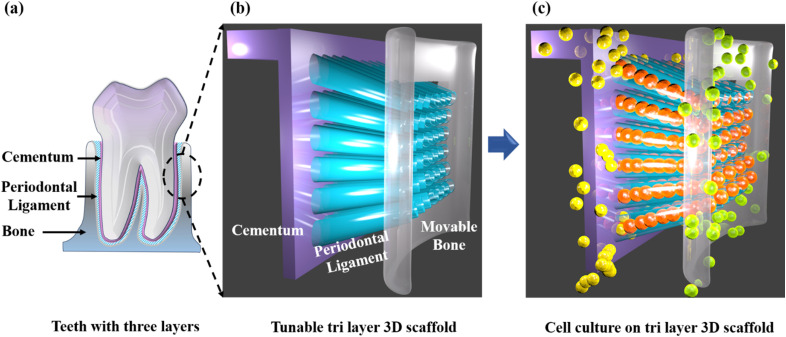
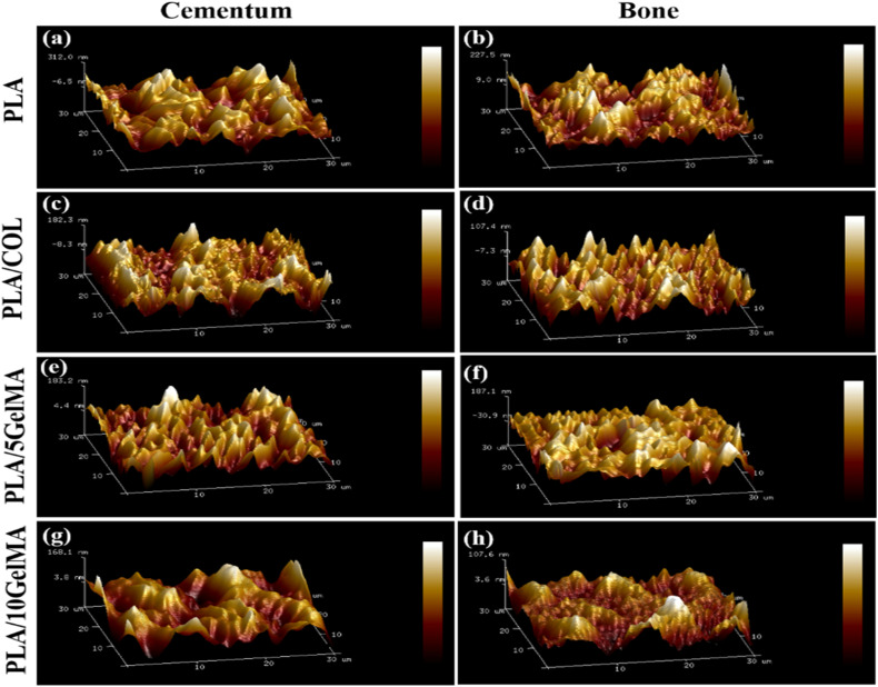
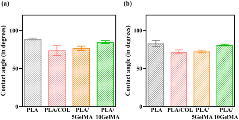
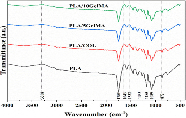
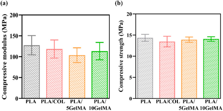
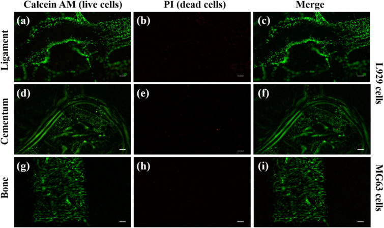
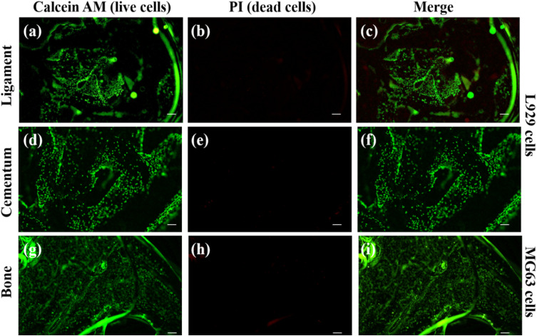
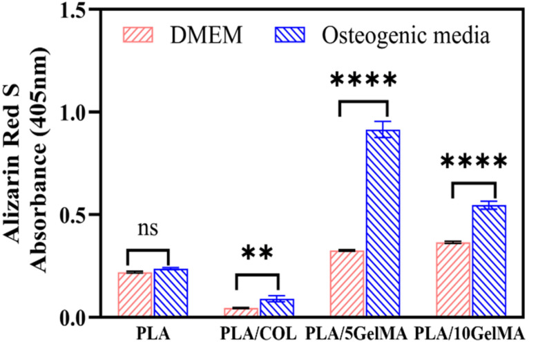
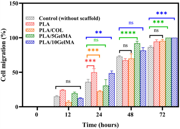
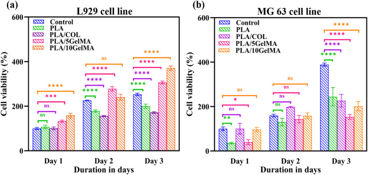
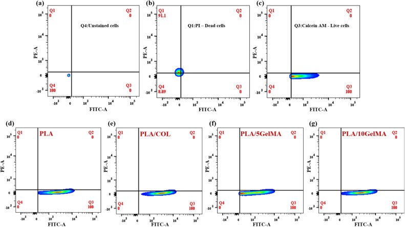
Multilayered scaffolds mimicking mechanical and biological host tissue architectures are the current prerequisites for successful tissue regeneration. We propose our tunable tri-layered scaffold, designed to represent the native periodontium for potential regenerative applications. The fused deposition modeling platform is used to fabricate the novel movable three-layered polylactic acid scaffold mimicking in vivo cementum, periodontal ligament, and alveolar bone layers. The scaffold is further provided with multiple angulated fibers, offering directional guidance and facilitating the anchorage dependence on cell adhesion. Additionally, surface modifications of the scaffold were made by incorporating coatings like collagen and different concentrations of gelatin methacryloyl to enrich the cell adhesion and proliferation. The surface characterization of our designed scaffolds was performed using tribological studies, atomic force microscopy, contact angle measurement, scanning electron microscopy, and micro-computed tomography. Furthermore, the material characterization of this scaffold was investigated by attenuated total reflectance-Fourier transformed infrared spectroscopy. The scaffold's mechanical characterization, such as strength and compression modulus, was demonstrated by compression testing. The L929 mouse fibroblast cells and MG63 human osteosarcoma cells have been cultured on the scaffold. The scaffold's superior biocompatibility was evaluated using fluorescence dye with fluorescence microscopy, scanning electron microscopy, in vitro wound healing assay, MTT assay, and flow cytometry. The mineralization capability of the scaffolds was also studied. In conclusion, our study demonstrated the construction of a multilayered movable scaffold, which is highly biocompatible and most suitable for various downstream applications such as periodontium and in situ tissue regeneration of complex, multilayered tissues.


Conflict of interest statement

There are no conflicts to declare.

Figures

Fig. 1
Fig. 1. Schematic of direction-oriented fiber guiding with tunable tri-layer-3D scaffold (a) teeth with three layers (b) fabricated direction-oriented fiber guiding with tunable tri layer -3D scaffold (c) cell culture on direction-oriented fiber guiding tri layer 3D scaffold.
Fig. 2
Fig. 2. (a) Schematic depicting the scaffold fabrication steps (b) optical images of the fabricated direction oriented tunable scaffold, where angulated ligament fiber is anchored between cementum and bone (c) optical image of the surface of cementum layer anchored with ligament.
Fig. 3
Fig. 3. 3D model of the tri-layered scaffold (a) isometric view of the tri-layered scaffold (b) top view of the scaffold design showing the three distinct layers (c) optical image of the fabricated top surface of the ligaments which are attached on the cementum scaffold (d) side view of the scaffold model (e) side view of fabricated scaffold using micro-CT imaging (f) micro-CT image of cementum layer and ligaments interconnection (g) micro-CT image of the sliced periodontal ligament.
Fig. 4
Fig. 4. Plot of coefficient of friction for PLA, PLA/COL, PLA/5 GelMA and PLA/10 GelMA scaffolds for time period from 0 seconds to 1800 seconds.
Fig. 5
Fig. 5. AFM images of the scaffold. Roughness variation in (a) PLA cementum layer (b) PLA bone layer (c) PLA/COL cementum layer (d) PLA/COL bone layer (e) PLA/5 GelMA cementum layer (f) PLA/5 GelMA bone layer (g) PLA/10 GelMA cementum layer and (h) PLA/10 GelMA bone layer.
Fig. 6
Fig. 6. Contact angle measurement (a) cementum layer for PLA, PLA/COL, PLA/5 GelMA and PLA/10 GelMA (b) bone layer for PLA, PLA/COL, PLA/5 GelMA and PLA/10 GelMA.
Fig. 7
Fig. 7. The ATR-FTIR spectra indicating the functional groups present in PLA, PLA/COL, PLA/5 GelMA and PLA/10 GelMA. The significance bands for PLA, collagen and GelMA are indicated in the figure.
Fig. 8
Fig. 8. Compression testing (a) compressive modulus for PLA, PLA/COL, PLA/5 GelMA and PLA/10 GelMA (b) compressive strength for PLA, PLA/COL, PLA/5 GelMA and PLA/10 GelMA.
Fig. 9
Fig. 9. Cell culture on PLA scaffold (a) live L929 cell staining on ligament layer using calcein AM (b) dead L929 cells staining on ligament layer using PI dye (c) merged image of live and dead cells (d) live L929 cell staining on cementum layer using calcein AM (e) dead L929 cells staining on cementum layer using PI dye (f) merged image of live and dead cells (g) live MG63 cell staining on bone layer using calcein AM (h) dead MG63 cells staining on bone layer using PI dye (i) merged image of live and dead cells. Scale for all images is 200 μm.
Fig. 10
Fig. 10. Cell culture on PLA/10 GelMA scaffold (a) live L929 cell staining on ligament layer using calcein AM (b) dead L929 cells staining on ligament layer using PI dye (c) merged image of live and dead cells (d) live L929 cell staining on cementum layer using calcein AM (e) dead L929 cells staining on cementum layer using PI dye (f) merged image of live and dead cells (g) live MG63 cell staining on bone layer using calcein AM (h) dead MG63 cells staining on bone layer using PI dye (i) merged image of live and dead cells. Scale for all images is 200 μm.
Fig. 11
Fig. 11. SEM images of cell morphologies on scaffolds when the L929 cells had been cultured for 48 hours. (a,b,c) Shows cells on the control PLA scaffold. (d,e,f) Illustrate the relatively interconnected cells proliferating on PLA/COL cementum layers. (g,h,i) Indicate the elongated and highly interconnected cells on the surface of PLA/5 GelMA cementum layer. (j,k,l) Shows L929 cells elongated and thriving on the surface of PLA/10 GelMA cementum layers.
Fig. 12
Fig. 12. Mineralization on scaffolds cultured in DMEM and osteogenic media. Statistical analysis is shown on the bar graphs. Data are presented as the mean and SD of the three independent experiments. *P < 0.0332, **P < 0.0021, ***P < 0.0002, and ****P < 0.0001.
Fig. 13
Fig. 13. Quantitative analysis of in vitro wound closure expressed as the area covered by the L929 cells. Data are presented as the mean and SD of the three independent experiments. *P < 0.0332, **P < 0.0021, ***P < 0.0002, and ****P < 0.0001.
Fig. 14
Fig. 14. The MTT assay test results showing the viability of (a) L929 cells on control, PLA scaffold, PLA/COL scaffold, PLA/5 GelMA scaffold and PLA/10 GelMA scaffold (b) MG63 cells in control, PLA scaffold, PLA/COL scaffold, PLA/5 GelMA scaffold and PLA/10 GelMA scaffold after 1, 2, and 3 days of incubation. Statistical analysis was performed using two-way ANOVA test. Statistical significance was assumed for p-values (*p < 0.0332, **p < 0.0021, ***p < 0.0002, and ****p < 0.0001).
Fig. 15
Fig. 15. Flow cytometry-based quantification of live and dead L929 cells on scaffolds. (a) Unstained L929 cell population (b) cell stained with PI dye (dead cells) (c) cells stained with calcein AM (live cells) (d) the percentage of live and dead cells on PLA scaffold (e) the percentage of live and dead cells on PLA/COL scaffold (f) the percentage of live and dead cells on 5 GelMA scaffold (g) the percentage of live and dead cells on PLA/10 GelMA scaffold after 1 day incubation.

Similar articles

Cited by

References

    1. de Jong T. Bakker A. D. Everts V. Smit T. H. J. Periodontal Res. 2017;52:965–974. doi: 10.1111/jre.12477. - DOI - PubMed
    1. Larsen T. Fiehn N. E. Apmis. 2017;125:376–384. doi: 10.1111/apm.12688. - DOI - PubMed
    1. Nazir M. Al-Ansari A. Al-Khalifa K. Alhareky M. Gaffar B. Almas K. Sci. World J. 2020;2020:2146160. - PMC - PubMed
    1. Kim J. Amar S. Odontology. 2006;94:10–21. doi: 10.1007/s10266-006-0060-6. - DOI - PMC - PubMed
    1. Liang Y. Luan X. Liu X. Bioact. Mater. 2020;5:297–308. - PMC - PubMed