Multispectral Imaging-Based System for Detecting Tissue Oxygen Saturation With Wound Segmentation for Monitoring Wound Healing
- PMID: 38899145
- PMCID: PMC11186648
- DOI: 10.1109/JTEHM.2024.3399232
Multispectral Imaging-Based System for Detecting Tissue Oxygen Saturation With Wound Segmentation for Monitoring Wound Healing
Abstract
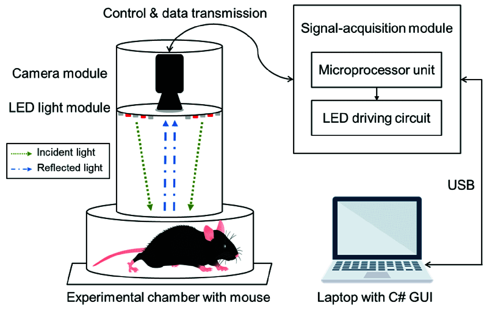
Objective: Blood circulation is an important indicator of wound healing. In this study, a tissue oxygen saturation detecting (TOSD) system that is based on multispectral imaging (MSI) is proposed to quantify the degree of tissue oxygen saturation (StO2) in cutaneous tissues.
Methods: A wound segmentation algorithm is used to segment automatically wound and skin areas, eliminating the need for manual labeling and applying adaptive tissue optics. Animal experiments were conducted on six mice in which they were observed seven times, once every two days. The TOSD system illuminated cutaneous tissues with two wavelengths of light - red ([Formula: see text] nm) and near-infrared ([Formula: see text] nm), and StO2 levels were calculated using images that were captured using a monochrome camera. The wound segmentation algorithm using ResNet34-based U-Net was integrated with computer vision techniques to improve its performance.
Results: Animal experiments revealed that the wound segmentation algorithm achieved a Dice score of 93.49%. The StO2 levels that were determined using the TOSD system varied significantly among the phases of wound healing. Changes in StO2 levels were detected before laser speckle contrast imaging (LSCI) detected changes in blood flux. Moreover, statistical features that were extracted from the TOSD system and LSCI were utilized in principal component analysis (PCA) to visualize different wound healing phases. The average silhouette coefficients of the TOSD system with segmentation (ResNet34-based U-Net) and LSCI were 0.2890 and 0.0194, respectively.
Conclusion: By detecting the StO2 levels of cutaneous tissues using the TOSD system with segmentation, the phases of wound healing were accurately distinguished. This method can support medical personnel in conducting precise wound assessments. Clinical and Translational Impact Statement-This study supports efforts in monitoring StO2 levels, wound segmentation, and wound healing phase classification to improve the efficiency and accuracy of preclinical research in the field.
Keywords: Multispectral imaging; principal component analysis; tissue oxygen saturation; wound healing; wound segmentation.
© 2024 The Authors.
Figures


References
-
- Thomas Hess C., “Checklist for factors affecting wound healing,” Adv. Skin Wound Care, vol. 24, no. 4, p. 192, Apr. 2011. - PubMed
-
- Strauss M. B., Bryant B. J., and Hart G. B., “Transcutaneous oxygen measurements under hyperbaric oxygen conditions as a predictor for healing of problem wounds,” Foot Ankle Int., vol. 23, no. 10, pp. 933–937, Oct. 2002. - PubMed
MeSH terms
Substances
LinkOut - more resources
Full Text Sources
Other Literature Sources

