. 2024 Mar 8;1(2):272-282.
doi: 10.1039/d3pm00060e. eCollection 2024 Jun 18.

Insight into the liposomal encapsulation of mono and bis-naphthalimides

Affiliations

Insight into the liposomal encapsulation of mono and bis-naphthalimides

Abdullahi Magaji Dauda et al. RSC Pharm. .

Abstract

Mitonafide-loaded liposomes are a promising strategy to overcome the neurotoxicity observed in clinical trials for this drug. This study investigates the influence of loaded mitonafide or a dimer analogue on different liposomal formulations and their therapeutic efficacy in vitro. Physicochemical properties of the liposomes were manipulated using different loading methods (namely bilayer or core loading) and varying the rigidity of the bilayer using distinct phospholipid compositions. Our results demonstrated that the mitonafide dimer analogue had a comparable encapsulation efficiency (EE%) into the liposomes when loaded into rigid or flexible bilayers in contrast to the low mitonafide monomer EE%. A pH gradient core loading method resulted in a more efficient mechanism to load the monomer into the liposomes. DOSY NMR and spectrofluorometric studies revealed key differences in the structure of the vesicles and the arrangement of the monomer or the dimer in the bilayer or the core of the liposomes. The in vitro assessment of the formulations using MDA-MB-231 and RT-112 cells revealed that a flexible lipid bilayer allows a faster drug release, which correlated well with the spectroscopy studies. This study investigated for the first time that the characteristics of the lipid bilayer and the loading method influence the encapsulation efficacy, colloidal properties, photoactivity and stability of mono and bis-naphthalimides loaded in a liposomal carrier, essential factors that will impact the performance of the formulation in a biological scenario.


Conflict of interest statement

There are no conflicts to declare.

Figures

Fig. 1
Fig. 1. (a) Gas optimised structure predictions of mitonafide monomer (left) and analogue dimer (right) used for UV/Vis absorption calculations. (b) Calculated absorption spectrum of monomer and dimer electronic structures (220–400 nm range) compared to experimentally measured values of samples.
Fig. 2
Fig. 2. Schematic representation of the liposomal bilayer of two formulations in the study: (left) DSPC or (right) SoyPC/Chol/DSPE-PEG2000 (molar ratio: 95/50/5).
Fig. 3
Fig. 3. Colloidal properties and encapsulation efficiency of DSPC or SoyPC/Chol/DSPE-PEG2000 liposomes loaded with the monomer (M) or the dimer (D) in the bilayer of the vesicle. (a) Hydrodynamic size. (b) Polydispersity index, (c) Z-potential, (d) encapsulation efficiency. Data represent the mean ± SD of at least three independent measurements.
Fig. 4
Fig. 4. Colloidal properties and encapsulation efficiency of DSPC or SoyPC/Chol/DSPE-PEG2000 liposomes loaded with the monomer in the core of the vesicle using different methods (passive loading with mannitol, active loading via pH gradient using ammonium sulphate). (a) Hydrodynamic size. (b) Polydispersity index, (c) Z-potential, (d) encapsulation efficiency. (e) Solubility of the monomer in different buffers: NH4-EDTA (300 mM ammonium EDTA), DHE (300 mM dextrose, 20 mM HEPES and 15 mM EDTA, pH 7.4), mannitol (5% mannitol), (NH4)2SO4 (240 mM ammonium sulphate pH 5.4). Data represent the mean ± SD of at least three independent measurements. eL: empty liposomes, M: liposomes loaded via passive loading, AS: liposomes loaded via pH gradient.
Fig. 5
Fig. 5. 1H PEG (3.5–3.6 ppm) NMR analysis across storage stability test. (a) The solvent-suppressed NMR indicates the line shapes of empty liposome particles on Day 0 (solid line) and Day 50 (dashed line). (b) Solvent suppressed 1H NMR indicating line shapes of loaded liposome particles on Day 0 (solid line) and Day 50 (dashed line) of Dimer (left and middle) and monomer (right) systems from 3.5–3.7 ppm. (c) Relative 1H Intensity of PEG over sample storage (average of 3 integration comparisons). (d) Apparent hydrodynamic radii of PEG protons. Liposomes are listed as eL (empty liposomes), M (monomer loaded via pH gradient) and D (dimer loaded in the bilayer) systems.
Fig. 6
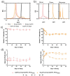
Fig. 6. Fluorescence spectroscopy analysis of monomer (M) and dimer (D) in aqueous solution. Photoluminescence spectra following excitation at 340 nm were carried out for monomer (A) and dimer (B), to determine peak intensity (C). TCSPC measurements were also carried out on monomer (D) and dimer (E) solutions to determine the average fluorescent excited state lifetime (F). Data shows the resultant excited state lifetime determined from a dual exponential fit of the luminescent decay.
Fig. 7
Fig. 7. Cytotoxicity studies of mitonafide monomer and dimer in two cell lines: MDA-MB-231 breast adenocarcinoma cells and RT-112 urinary bladder carcinoma. Cells were seeded in 96-well plates (1.7 × 10⁴ cells per well), and the next day, they were incubated with both compounds over 24 (coloured bars) and 48 (white bars) hours. Cell viability was assessed using MTT assay. The results are expressed as average ± SD (n = 6).
Fig. 8
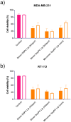
Fig. 8. Cytotoxicity studies of different formulations loaded with the monomer or the dimer in 2 different cell lines: MDA-MB-231 breast adenocarcinoma cells (a) and RT-112 urinary bladder carcinoma (b). Cells were seeded in 96-well plates (1.7 × 10⁴ cells per well), and the next day they were incubated with different formulations over 24 (coloured bars) and 48 (white bars) hours. Cell viability was assessed using resazurin assay. The results are expressed as average ± SD (n = 6).

References

    1. Berlanga J. M. C., Brana M. F. and Roldan C. M., Patent, DE2318136A1, 1973
    1. Banerjee S. Veale E. B. Phelan C. M. Murphy S. A. Tocci G. M. Gillespie L. J. Frimannsson D. O. Kelly J. M. Gunnlaugsson T. Recent advances in the development of 1,8-naphthalimide based DNA targeting binders, anticancer and fluorescent cellular imaging agents. Chem. Soc. Rev. 2013;42:1601. doi: 10.1039/C2CS35467E. - DOI - PubMed
    1. Braña M. F. Sanz A. M. Castellano J. M. Roldan C. M. Roldan C. Synthesis and cytostatic activity of benz de isoquinoline 1 3 diones structure activity relationships. Eur. J. Med. Chem. 1981;16:207–212.
    1. Allen S. L. Kolitz J. E. Lundberg A. S. Bennett J. M. Capizzi R. L. Budman D. R. Phase I trials of amonafide as monotherapy and in combination with cytarabine in patients with poor-risk acute myeloid leukemia. Leuk. Res. 2010;34:487–491. doi: 10.1016/j.leukres.2009.07.038. - DOI - PubMed
    1. U.S. National, Library of Medicine, Clinicaltrials.gov, https://clinicaltrials.gov/ct2/home

LinkOut - more resources