. 2024 May 30:18:1382036.
doi: 10.3389/fnana.2024.1382036. eCollection 2024.

The amygdaloid body of the family Delphinidae: a morphological study of its central nucleus through calbindin-D28k

Affiliations

The amygdaloid body of the family Delphinidae: a morphological study of its central nucleus through calbindin-D28k

Simona Sacchini et al. Front Neuroanat. .

Abstract

Introduction: The amygdala is a noticeable bilateral structure in the medial temporal lobe and it is composed of at least 13 different nuclei and cortical areas, subdivided into the deep nuclei, the superficial nuclei, and the remaining nuclei which contain the central nucleus (CeA). CeA mediates the behavioral and physiological responses associated with fear and anxiety through pituitary-adrenal responses by modulating the liberation of the hypothalamic Corticotropin Releasing Factor/Hormone.

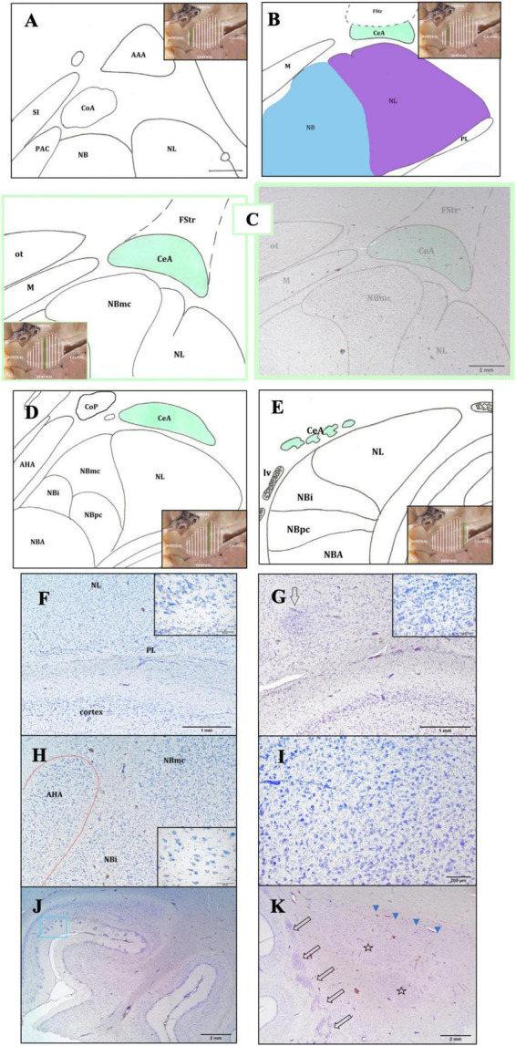
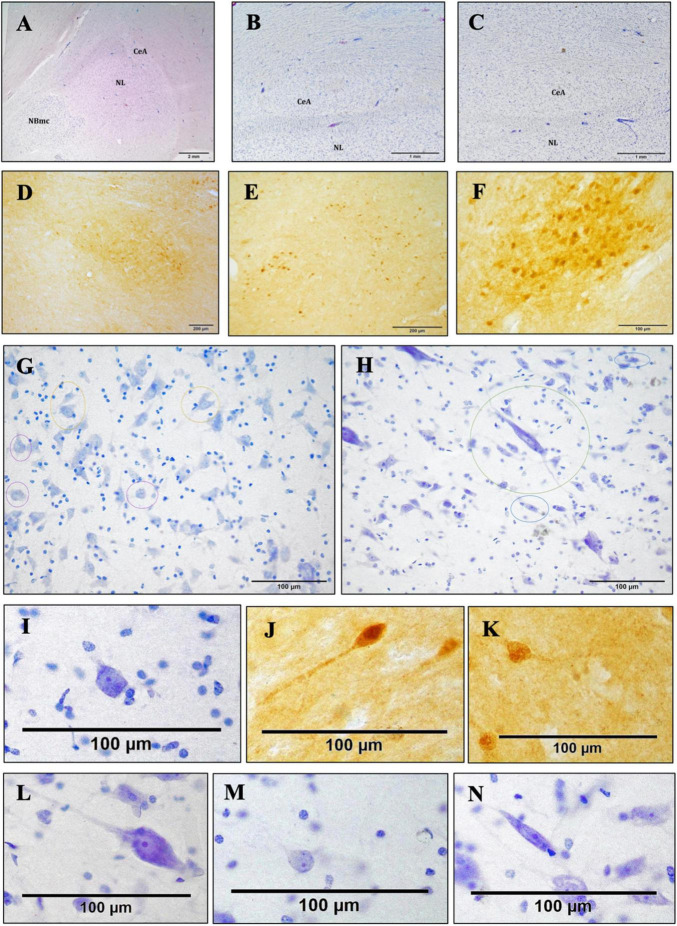
Methods: Five dolphins of three different species, belonging to the family Delphinidae (three striped dolphins, one common dolphin, and one Atlantic spotted dolphin), were used for this study. For a precise overview of the CeA's structure, thionine staining and the immunoperoxidase method using calbindin D-28k were employed.

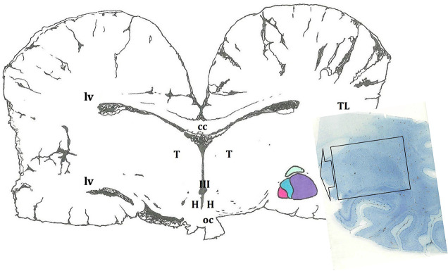
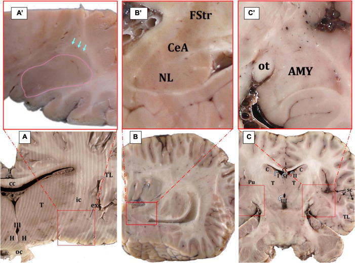
Results: CeA extended mainly dorsal to the lateral nucleus and ventral to the striatum. It was medial to the internal capsule and lateral to the optic tract and the medial nucleus of the amygdala.

Discussion: The dolphin amygdaloid complex resembles that of primates, including the subdivision, volume, and location of the CeA.

Keywords: amygdaloid body; calbindin-D28k; central nucleus of the amygdala; dolphins; toothed whales.


Conflict of interest statement

The authors declare that the research was conducted in the absence of any commercial or financial relationships that could be construed as a potential conflict of interest.

Figures

FIGURE 1
FIGURE 1
Schematic representation of the amygdaloid complex (in colors), in a cross section of the brain obtained at the level of the hypothalamus (H). In this diagram, as in the rest of the cross sections, a certain asymmetry of the two hemispheres predominates. The microscopic image depicts a histological preparation of the amygdala (enclosed by a black box). III, third ventricle; cc, corpus callosum; oc, optic chiasm; T, thalamus; TL, temporal lobe; lv, lateral ventricle.
FIGURE 2
FIGURE 2
Gross anatomy of the AMY (surrounded by red squares) and the adjoining structures (A–C). The edges of the central nucleus are indicated by arrows while the basolateral AMY in surrounded by a pink line; Cuvier’s beaked whale, transverse plane (A,A’). The central nucleus is located ventral to the fundus striati and dorsal to the lateral nucleus; Atlantic spotted dolphin, parasagittal plane (B,B’). In the horizontal plane, the AMY is also clearly evident; Atlantic spotted dolphin (C,C’). AMY, amygdaloid body; cc, corpus callosum; C, caudate nucleus; CeA, central nucleus of the amygdala; exc, extreme capsule; FStr, fundus striati; H, hypothalamus; ic, internal capsule; oc, optic chiasm; ot, optic tract; Pu, putamen; sc, sylvian cleft; T, thalamus; TL, temporal lobe; I, II, III, first, second, and third ventricle.
FIGURE 3
FIGURE 3
Rostro-caudal description of the CeA through schematic representations. The purpose of the insets is to aid the reader in comprehending the precise position and evolution of the AMY and CeA (A–E). In the image, the following structures are observed: the lateral nucleus (NL), the basal nucleus (NB); the periamygdaloid cortex (PAC); the anterior cortical area (CoA), dorsal to the PAC; the anterior amygdaloid area (AAA), dorsal to the abovementioned nuclei; the substantia innominata (SI), medial to the amygdaloid complex (A). Caudally, the CeA is relatively elongated in shape, dorsal to the NL (in purple) and ventral to the fundus striati (FStr); the medial nucleus (M) is medial to the CeA; the NL is ventral to the CeA and lateral to the NB (in blue). The NB and the paralaminar nucleus (PL) are also represented (B). Maximum size of the CeA. The magnocellular division of the NB (NBmc) has a more rounded shape, reflected by its great extension and its large neurons. The optic tract (ot) which is situated medial to the CeA, dorsal to the M, and dorsal-medial to the NBmc, is clearly visible (C). Caudally, both the CeA and the NBmc are diminished. The whole NB has a significant reduction in all three of its subdivisions, included the intermediate (NBi) and parvicellular (NBpc) divisions, but NBmc is the most affected. The NL shows the disappearance of its small accessory portion. Two additional superficial nuclei are also present: the posterior cortical nucleus (CoP) and the anterior amygdalohippocampal area (AHA) (D). Finally, only isolated islets of CeA neurons are visible at this stage, and its perimeter and area cannot be ascertained. Only the deep nuclei—the NB, NL, and NBA—and the beginning of the lateral ventricle (lv) are visible, aside from the CeA (E). Bright-field photomicrograph of Nissl-stained sections of the lower part of the AMY (F,G). Histological images of the PL with its neuronal morphology (inset) (F) and the intercalated cell masses (arrow) and their neuronal morphology (inset) (G); common dolphin, rostral right AMY, thionine. Histological image of the anterior amygdalohippocampal area (AHA), located medially to the NB, and its large neurons, striped dolphin, caudal right AMY, thionine (H). The histological image displays the magnocellular division of the NB (NBmc), rostral right AMY, striped dolphin, thionine (I). Entorhinal cortex histology, ventral to the AMY. Neurons with polygonal cell bodies and dark staining are seen in layer II; these neurons are typically grouped into islands (blue box), common dolphin, thionine (J). Histological images of the caudal left claustrum, whose segmented appearance (arrows) and non-stratified neuronal arrangement define its caudalmost portion. The NL mostly represents the first part of the AMY (stars) and is accompanied by the substantia innominata (arrowheads); Atlantic spotted dolphin, thionine (K).
FIGURE 4
FIGURE 4
Bright-field photomicrograph of Nissl-stained and c-D28k immunoreactive sections showing the cytoarchitectonics of the CeA. Histological image of the right AMY and CeA of an Atlantic spotted dolphin. Comparing this species to the others, the CeA showed a lower dimension, despite having reached its highest size. In fact, the CeA does not extend to the NBmc; rather, it is solely dorsal to the NL, thionine (A). Histological image of the CeA, which is characterized by the presence of nerve fiber bundles and a very reduced size. Common dolphin, caudal right AMY, thionine (B); striped dolphin, caudal right AMY, thionine (C). The CeA exhibits immunoreactivity against c-D28k. Atlantic spotted dolphin, rostral right AMY (D); striped dolphin, caudal left AMY (E); common dolphin, rostral right AMY (F). Neurons of different morphologies; thionine (G,H). Poligonal (yellow circles), round (purple circles) Atlantic spotted dolphin (G). Fusiform (blue circles), LP (green circle), striped dolphin (H). Poligonal neurons, thionine (I) and c-D28k free-floating immunohistochemistry (J,K). LP (L). Round (M). Fusiform (N).

Similar articles

References

    1. Amunts K., Kedo O., Kindler M., Pieperhoff P., Mohlberg H., Shah N. J., et al. (2005). Cytoarchitectonic mapping of the human amygdala, hippocampal region and entorhinal cortex: intersubject variability and probability maps. Anat. Embryol. 210 343–352. 10.1007/s00429-005-0025-5 - DOI - PubMed
    1. Bickart K. C., Wright C. I., Dautoff R. J., Dickerson B. C., Barrett L. F. (2011). Amygdala volume and social network size in humans. Nat. Neurosci. 14 163–164. 10.1038/nn.2724 - DOI - PMC - PubMed
    1. Boccardi M., Frisoni G. B., Hare R. D., Cavedo E., Najt P., Pievani M., et al. (2011). Cortex and amygdala morphology in psychopathy. Psychiatry Res. 193 85–92. 10.1016/j.pscychresns.2010.12.013 - DOI - PubMed
    1. Bombardi C. (2014). Neuronal localization of the 5-HT2 receptor family in the amygdaloid complex. Front. Pharmacol. 5:68. 10.3389/fphar.2014.00068 - DOI - PMC - PubMed
    1. Bombardi C., Grandis A., Chiocchetti R., Lucchi M. L. (2006). Distribution of calbindin-D28k, neuronal nitric oxide synthase, and nicotinamide adenine dinucleotide phosphate diaphorase (NADPH-d) in the lateral nucleus of the sheep amygdaloid complex. Anat. Embryol. 211 707–720. 10.1007/s00429-006-0133-x - DOI - PubMed

LinkOut - more resources