

Cerebellar Hemangioblastoma Mimicking Arteriovenous Malformation: A Case Report
- PMID: 38899263
- PMCID: PMC11186394
- DOI: 10.7759/cureus.60671
Cerebellar Hemangioblastoma Mimicking Arteriovenous Malformation: A Case Report
Abstract
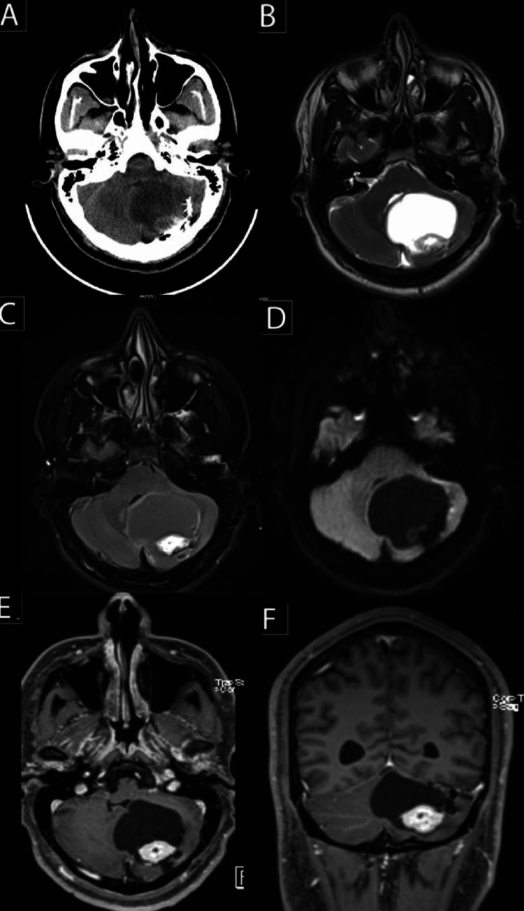
Hemangioblastoma (HBM) is a tumor distinguished by the presence of stromal cells and small vessels. These stromal cells represent stem cells, which, due to the influence of the neoplasm, proliferate and differentiate into "vasoformative elements" that create new blood vessels. Hemangioblastomas resemble arteriovenous malformation (AVM) in imaging features, characterized by an apparent vascular blush, the presence of multiple feeding vessels, and evident draining veins observed on digital subtraction angiography (DSA). Our study presents a case of HBM in the right cerebellar hemisphere mimicking AVM. The patient had been diagnosed with AVM in the same location two years ago and managed with endovascular embolization. One month prior, the patient experienced severe headaches, imbalance, nausea, left ear fullness, blurry vision, and high blood pressure. The imaging feature suggests HBM rather than AVM. The patient next underwent sub-occipital craniotomy and tumor resection with external ventricular drainage (EVD) insertion. The histopathological report of the excised mass confirmed HBM. In conclusion, AVM and HBM rarely occur together. Recent research indicates that HBM and AVM have exact embryologic origins and need later genetic alterations to develop into symptomatic lesions. Further research is required to clarify the uncommon combination of these lesions.
Keywords: avm; case report; hemangioblastoma; imaging; neuroradiology.
Copyright © 2024, Al-Mutairi et al.
Conflict of interest statement
The authors have declared that no competing interests exist.
Figures


References
-
- Central nervous system vascular malformations: a clinical review. Sabayan B, Lineback C, Viswanathan A, Leslie-Mazwi TM, Shaibani A. https://pubmed.ncbi.nlm.nih.gov/33434339/ Ann Clin Transl Neurol. 2021;8:504–522. - PMC - PubMed
-
- Central nervous system capillary haemangioblastoma: the pathologist's viewpoint. Hussein MR. https://pubmed.ncbi.nlm.nih.gov/17877533/ Int J Exp Pathol. 2007;88:311–324. - PMC - PubMed
-
- Hemangioblastomas of the central nervous system in Von Hippel-Lindau syndrome and sporadic disease. Conway JE, Chou D, Clatterbuck RE, Brem H, Long DM, Rigamonti D. https://pubmed.ncbi.nlm.nih.gov/11152361/ Neurosurgery. 2001;48:55–62. - PubMed
-
- A case report of cerebellar hemangioblastoma simulated brain metastasis shown by magnetic resonance imaging. Xue J, Mo C. https://pubmed.ncbi.nlm.nih.gov/38335432/ Medicine (Baltimore) 2024;103:0. - PMC - PubMed
-
- Case report of a hemangioblastoma with large blood vessels and rare vascular anomalies: is it fibromuscular dysplasia or arteriovenous malformation association? Rodríguez-Hernández LA, Sangrador-Deitos MV, Montano-Tello H, Mondragon-Soto M, Tena Suck ML. https://www.ncbi.nlm.nih.gov/pmc/articles/PMC9138196/ Cureus. 2022;14:0. - PMC - PubMed
Publication types
LinkOut - more resources
Full Text Sources