. 2024 Jul 19;135(3):453-469.
doi: 10.1161/CIRCRESAHA.123.323360. Epub 2024 Jun 20.

Fibroblast Smad7 Induction Protects the Remodeling Pressure-Overloaded Heart

Affiliations

Fibroblast Smad7 Induction Protects the Remodeling Pressure-Overloaded Heart

Claudio Humeres et al. Circ Res. .

Erratum in

Abstract

Background: Cardiac fibroblast activation contributes to adverse remodeling, fibrosis, and dysfunction in the pressure-overloaded heart. Although early fibroblast TGF-β (transforming growth factor-β)/Smad (small mother against decapentaplegic)-3 activation protects the pressure-overloaded heart by preserving the matrix, sustained TGF-β activation is deleterious, accentuating fibrosis and dysfunction. Thus, endogenous mechanisms that negatively regulate the TGF-β response in fibroblasts may be required to protect from progressive fibrosis and adverse remodeling. We hypothesized that Smad7, an inhibitory Smad that restrains TGF-β signaling, may be induced in the pressure-overloaded myocardium and may regulate fibrosis, remodeling, and dysfunction.

Methods: The effects of myofibroblast-specific Smad7 loss were studied in a mouse model of transverse aortic constriction, using echocardiography, histological analysis, and molecular analysis. Proteomic studies in S7KO (Smad7 knockout) and overexpressing cells were used to identify fibroblast-derived mediators modulated by Smad7. In vitro experiments using cultured cardiac fibroblasts, fibroblasts populating collagen lattices, and isolated macrophages were used to dissect the molecular signals responsible for the effects of Smad7.

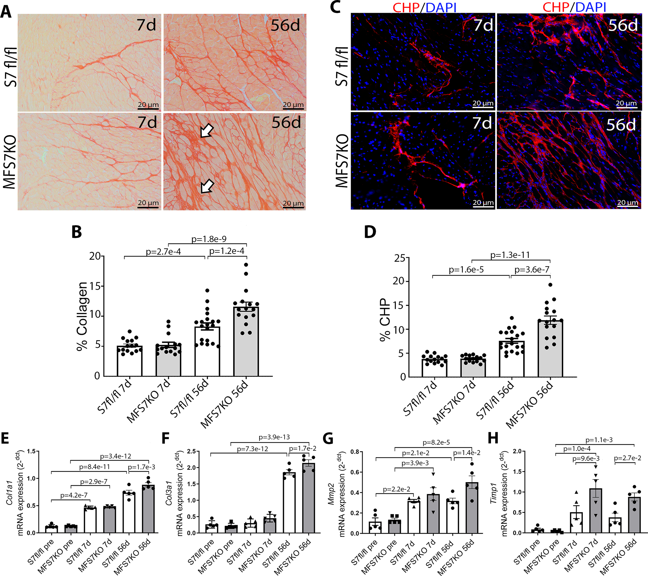
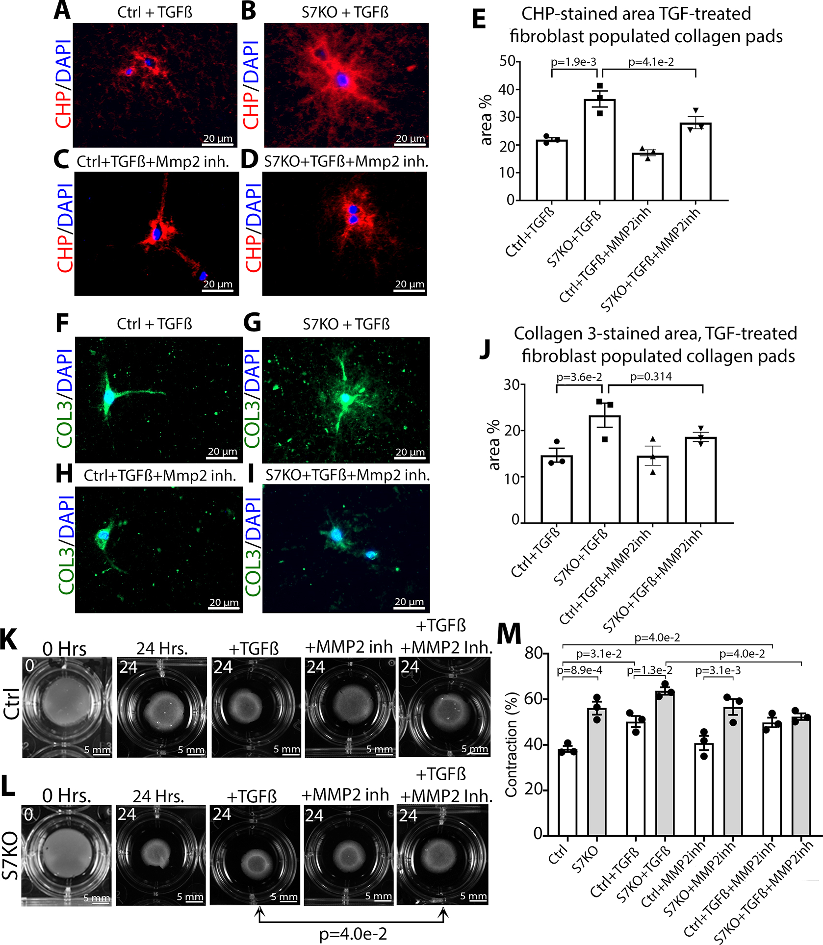
Results: Following pressure overload, Smad7 was upregulated in cardiac myofibroblasts. TGF-β and angiotensin II stimulated fibroblast Smad7 upregulation via Smad3, whereas GDF15 (growth differentiation factor 15) induced Smad7 through GFRAL (glial cell line-derived neurotrophic factor family receptor α-like). MFS7KO (myofibroblast-specific S7KO) mice had increased mortality, accentuated systolic dysfunction and dilative remodeling, and accelerated diastolic dysfunction in response to transverse aortic constriction. Increased dysfunction in MFS7KO hearts was associated with accentuated fibrosis and increased MMP (matrix metalloproteinase)-2 activity and collagen denaturation. Secretomic analysis showed that Smad7 loss accentuates secretion of structural collagens and matricellular proteins and markedly increases MMP2 secretion. In contrast, Smad7 overexpression reduced MMP2 levels. In fibroblasts populating collagen lattices, the effects of Smad7 on fibroblast-induced collagen denaturation and pad contraction were partly mediated via MMP2 downregulation. Surprisingly, MFS7KO mice also exhibited significant macrophage expansion caused by paracrine actions of Smad7 null fibroblasts that stimulate macrophage proliferation and fibrogenic activation. Macrophage activation involved the combined effects of the fibroblast-derived matricellular proteins CD5L (CD5 antigen-like), SPARC (secreted protein acidic and rich in cysteine), CTGF (connective tissue growth factor), ECM1 (extracellular matrix protein 1), and TGFBI (TGFB induced).

Conclusions: The antifibrotic effects of Smad7 in the pressure-overloaded heart protect from dysfunction and involve not only reduction in collagen deposition but also suppression of MMP2-mediated matrix denaturation and paracrine effects that suppress macrophage activation through inhibition of matricellular proteins.

Keywords: collagen; echocardiography; fibroblasts; fibrosis; growth factors; heart failure; macrophages.


Conflict of interest statement

None.

Figures

Figure 1.
Figure 1.. Fibroblast Smad 7 upregulation in fibroblasts in the pressure-overloaded myocardium.
(A) Myocardial Smad7 is markedly increased after 3, 7 and 28 days of TAC. (B) To examine whether Smad7-expressing interstitial cells are fibroblasts, we used PDGFRαEGFP fibroblast reporter mice, in which the fibroblast nucleus is labeled. Dual immunofluorescence shows abundant PDGFRα+/Smad7+ cardiac fibroblasts (white arrows) 7–56 days after TAC (upper row). Representative Smad7-expressing PDGFRα+ fibroblasts (yellow squares) are shown in high magnification images in the lower row. (C) Representative images of dual immunofluorescent staining for Smad7 and α-SMA shows upregulation of Smad7 expression in α-SMA+ myofibroblasts after 7–56 days of TAC (white arrows). (D-E) TGF-β1 and GDF15 induce Smad7 upregulation in mouse cardiac fibroblasts after 4 and 24h of stimulation. TGFβ2 increased Smad7 levels only at the 4h timepoint (D), whereas TGFβ3, BMP4 and Angiotensin II induce late Smad7 upregulation (E). BMP7, IL-6, GDF11, activin-A and IL-1β had no effects on Smad7 expression. Statistical analysis was performed using one-way ANOVA followed by Sidak multiple comparison test (n=3/timepoint). Each data point represents an independent biological experiment. Values are shown as fold change over the mean of the shams (A) and fold change over the mean of the controls (Ctrl) (D-E). For histology experiments: Sham Control n=8, 7 days TAC n=9, 28 days TAC n= 9, 56 days TAC n=9. Scale bar=20μm. Scale bar=10μm for zoomed images
Figure 2.
Figure 2.. Myofibroblast-specific Smad7 protects the pressure-overloaded heart from systolic dysfunction and dilative remodeling.
(A) Myofibroblast-specific Smad7 knockout (MFS7KO) mice had significantly increased mortality after 56 days of TAC, in comparison to Smad7fl/fl (S7fl/fl) (p=0.021, n=27–28/group). (B-C) MFS7KO mice had worse systolic dysfunction, suggested by a lower ejection fraction (LVEF) and a higher reduction in left-ventricular ejection fraction (ΔLVEF) after 56 days of TAC. (D-E) MSF7KO had accentuated chamber dilation, evidenced by a significant increase in left ventricular end-diastolic volume (LVEDV) at 28 days and by increased ΔLVEDV at all timepoints. (F) MFS7KO mice had a transient increase in the E/E’ ratio (indicator of diastolic dysfunction) after 7 days of TAC, consistent with delayed diastolic dysfunction. Although myofibroblast-specific loss of Smad7 was associated with worse dysfunction and increased dilation in response to pressure overload in both male (H-K) and female mice (N-Q), only male MFS7KO animals had increased mortality after TAC (G, M). Male but not female MFS7KO mice had increased E/E’ (suggesting worse diastolic dysfunction) after 7 days of TAC (L, R). Survival analysis was performed using the Kaplan-Meier method. Mortality was compared using the log-rank test. Statistical analysis of echocardiographic endpoints (B-F, H-L, N-R) was performed using 2-way repeated measures ANOVA followed by Sidak post-hoc test. Each data point represents an independent experiment (S7fl/fl pre, n =24 (male=9, female=15); MFS7KO pre, n=24 (male=16, female=9); S7fl/fl 7d, n=23 (male=8, female=15); MFS7KO 7d, n=22 (male=13, female=9); S7fl/fl 28d, n=23 (male=8 Male, female=15); MFS7KO 28d, n=17 (male=8 Male, female=9); S7fl/fl 56d, n=23 (male=8, female=15); MFS7KO 56d, n=17 (male=13 Male, female=9).
Figure 3.
Figure 3.. Myofibroblast-specific Smad7 protects the pressure-overloaded myocardium from excessive collagen deposition and from collagen denaturation.
(A) Picrosirius red staining was used to label collagen Smad7fl/fl (S7fl/fl) and myofibroblast-specific Smad7-knockouts (MFS7KO) after 7–56 days of TAC. Arrows show increased deposition of interstitial collagen in pressure-overloaded MFS7KO hearts. (B) MFS7KO mice had significantly higher collagen content than S7fl/fl animals after 56 days of TAC. (C) Staining with collagen hybridizing peptide (CHP), which specifically binds to denatured unfolded collagen, was used to assess collagen denaturation. (D) MFS7KO mice had increased collagen denaturation after 56 days of TAC. (E-H) MFS7KO TAC hearts showed increased expression of Col1a1, Col3a1 and Mmp2 56 days after TAC, and higher Timp1 levels at both 7d and 56d timepoints. Statistical comparison was performed using 2-way ANOVA followed by Sidak multiple comparison test. Each data point represents an independent experiment (S7 fl/fl 7d, n=15; MFS7KO 7d, n=15, S7 fl/fl 56d, n=20; MFS7KO 56d, n=16). For qPCR experiments, n=5/timepoint. Scale bars=20μm.
Figure 4.
Figure 4.. Myofibroblast-specific Smad7 protects the pressure-overloaded myocardium from excessive macrophage infiltration.
(A) Macrophages were identified in the pressure overloaded myocardium of Smad7fl/fl (S7fl/fl) and myofibroblast-specific Smad7 knockout (MFS7KO) mice using Mac2. (B-D) Both male and female MFS7KO mice exhibited increased macrophage density after 7 days of TAC. No significant differences were noted at the 56-day timepoint. (E-F) MFS7KO hearts had significantly increased expression of the macrophage-specific gene Adgre1 (F4/80) and of Ccl2/Mcp1 after 7 days of TAC. Statistical comparisons were performed using 2-way ANOVA followed by Sidak multiple comparison test. Each data point represents an independent experiment (S7fl/fl 7d, n=16 (male=9, female=7); MFS7KO 7d, n=14 (male=7, female=7), S7fl/fl 56d, n=19 (male=9, female=10); MFS7KO 56d, n=16 (male=6, female 10)). For qPCR, n=4–5/timepoint. Scale bar=50μm.
Figure 5.
Figure 5.. Smad7 restrains fibroblast secretion of structural and matricellular ECM proteins.
Heatmap of significantly upregulated secreted proteins in the secretome of Smad7KO (S7) fibroblasts compared to control (WT) fibroblasts, in the presence (A) and absence (B) of TGF-β1 stimulation (24 h). (A) TGF-β-treated Smad7KO fibroblasts (TS7) have accentuated secretion of MMP2, COL3A1, COL1A1 and of the matricellular glycoproteins TGFBI (TGF-β-induced protein), CD5L (CD5 antigen Like), FN1 (Fibronectin), ECM1 (Extracellular matrix protein 1) and PSAP (Precursor of sphingolipid activators), when compared with TGF-β-treated control fibroblasts (TWT). (B) In the absence of TGFβ stimulation, in addition to increased MMP2, TGFBI, ECM1, CD5L, COL1A1 and COL3A1, Smad7 KO fibroblasts (CS7) also secreted higher amounts of EFEMP1 (associated EGF-containing fibulin-like extracellular matrix protein 1), PCOLCE (Procollagen C-endopeptidase enhancer), FBLN2 (Fibulin-2), SPARC (Secreted protein acidic and rich in cysteine), IGF-1 (insulin-like growth factor), and CCN2/CTGF. (C) Secretomic analysis of TGF-β-treated Smad7 overexpressing (TS7OE) and corresponding control fibroblasts (TWT) was performed to compare the differentially regulated proteins secreted in gain- and loss-of function Smad7 experiments. Out of the proteins identified to be upregulated in Smad7 KO cells (red), MMP2 and COL3A1 (blue) were significantly downregulated in the secretome of TGF-β-stimulated Smad7-overexpressing cells (TS7OE). COL1A1 levels were reduced in Smad7 OE cells; however, this did not reach statistical significance. Significantly upregulated (p<0.05) secreted proteins in each heatmap are ranked based on their protein fold increase in S7KO supernatant compared to WT. Comparisons between two groups for each protein were performed by unpaired two-tailed Student’s t test. Each data point (n=3/group) represents an independent experiment. It should be noted that multiple testing correction was not performed. This may increase the probability of false associations.
Figure 6.
Figure 6.. The effects of Smad7 on fibroblast-mediated collagen denaturation and pad contraction involve suppression of MMP2 activity.
Representative images of CHP staining (labeling denatured collagen) (A-D) and Collagen 3 staining (F-I) in collagen I lattices populated with TGFβ-treated control (Ctrl) or TGFβ-treated Smad7KO (S7KO) fibroblasts, in the presence or absence of MMP2 inhibitor (ARP-100, 5μM). (E) Smad7 loss increases collagen denaturation; this effect is attenuated upon MMP2 inhibition. (J) In contrast, the increased collagen deposition in pads populated with TGFβ-treated Smad7KO fibroblasts was unaffected by MMP2 inhibition. (K-L) Representative images of control (Ctrl) and S7KO fibroblasts-populated collagen pads in presence or absence of TGFβ and MMP2 inhibitor. (M) MMP2 inhibition abrogated the increased contraction induced by Smad7 loss. Statistical analysis using 1-way ANOVA followed by Sidak post-hoc test. Each data point (n=3) represents an independent biological experiment. Scalebar=20μm (A-I), scalebar=5mm (K-L).
Figure 7.
Figure 7.. Smad7 overexpression attenuates collagen deposition and denaturation and inhibits fibroblast-induced pad contraction.
Representative images of Collagen 3 staining (A) and CHP staining (which labels denatured collagen) (B) in collagen I lattices populated with control (Ctrl) and Smad7 overexpressing (S7OE) fibroblasts, in the presence and absence of TGF-β1. Quantification shows that overexpression of Smad7 decreased both collagen deposition and denaturation in both unstimulated and TGF-β1 treated conditions. (C) Representative images and quantification of contraction of Ctrl and S7OE fibroblasts-populated collagen pads with/without TGF-β1 stimulation (24 hours) demonstrate that Smad7 overexpression significantly reduced lattice contraction. Statistical comparison was performed using 1-way ANOVA followed by Sidak multiple comparison test. Each data point represents an independent biological experiment. n=4/group for A, B; n=3 for C. Scale bar=20 μm for A-B, scalebar=5mm for C.
Figure 8.
Figure 8.. Fibroblast Smad7 restrains macrophage proliferation and fibrogenic activation through paracrine actions.
(A) Proliferation assay of bone marrow macrophages (BMMs) stimulated without (control) or with the supernatant of control (WT FB Sup) or Smad7KO (S7KO FB Sup) fibroblasts. Macrophages treated with S7KO fibroblast supernatant had significantly increased proliferation when compared to untreated macrophages. (B-F) The supernatant of S7KO fibroblasts stimulated expression of M2 macrophage markers and fibrogenic mediators, such as Tgfb1, Cd206, Pparg and Vegfa and downmodulated Cxcl1 gene expression. (I-M) To study paracrine mechanisms that may mediate the effects of the secretome of Smad7 KO fibroblasts on macrophages, we stimulated BMMs with candidate mediators identified through our secretomic analysis (CTGF/CCN2, SPARC, TGFBI, ECM1 and CD5L, Figure 5). Of the 5 mediators tested, only CTGF/CCN2 and TGFBI induced expression of Tgfb1 by macrophages, whereas CTGF/CCN2 and SPARC stimulated expression of the M2 marker Cd206. Only SPARC stimulated expression of Pparg. Moreover, CTGF/CCN2, CD5L and ECM1 increased Vegfa synthesis, whereas CTGF/CCN2 and SPARC recapitulated the suppressive effects of the S7KO fibroblast supernatant on Cxcl1 expression. These results suggest that the paracrine activating effects of myofibroblast-specific Smad7 loss on macrophages are not due to induction of a single mediator but may rather involve the combined actions of CTGF/CCN2, SPARC, TGFBI, CD5L and ECM1. Statistical comparison using Student’s t test with Welch’s correction for unequal variances (B-F). Multiple groups analysis was done by 1-way ANOVA followed by Sidak post-hoc test (A, I-M). Each data point (n=3/4 group) represents an independent biological replicate.

Similar articles

Cited by

References

    1. Tallquist MD, Molkentin JD. Redefining the identity of cardiac fibroblasts. Nat Rev Cardiol. 2017; 14:484–491. - PMC - PubMed
    1. Souders CA, Bowers SL, Baudino TA. Cardiac fibroblast: the renaissance cell. Circ Res. 2009; 105:1164–1176. - PMC - PubMed
    1. Liu X, Burke RM, Lighthouse JK, Baker CD, Dirkx RA Jr., Kang B, Chakraborty Y, Mickelsen DM, Twardowski J, Mello SS, Ashton JM, Small EM. P53 Regulates the Extent of Fibroblast Proliferation and Fibrosis in Left Ventricle Pressure Overload. Circ Res. 2023. - PMC - PubMed
    1. Moore-Morris T, Guimaraes-Camboa N, Banerjee I, Zambon AC, Kisseleva T, Velayoudon A, Stallcup WB, Gu Y, Dalton ND, Cedenilla M, Gomez-Amaro R, Zhou B, et al. Resident fibroblast lineages mediate pressure overload-induced cardiac fibrosis. J Clin Invest. 2014; 124:2921–2934. - PMC - PubMed
    1. Ali SR, Ranjbarvaziri S, Talkhabi M, Zhao P, Subat A, Hojjat A, Kamran P, Muller AM, Volz KS, Tang Z, Red-Horse K, Ardehali R. Developmental heterogeneity of cardiac fibroblasts does not predict pathological proliferation and activation. Circ Res. 2014; 115:625–635. - PubMed