. 2024 Aug;11(32):e2402740.
doi: 10.1002/advs.202402740. Epub 2024 Jun 20.

Hierarchical Protofilament Intertwining Rules the Formation of Mixed-Curvature Amyloid Polymorphs

Affiliations

Hierarchical Protofilament Intertwining Rules the Formation of Mixed-Curvature Amyloid Polymorphs

Jiangtao Zhou et al. Adv Sci (Weinh). 2024 Aug.

Abstract

Amyloid polymorphism is a hallmark of almost all amyloid species, yet the mechanisms underlying the formation of amyloid polymorphs and their complex architectures remain elusive. Commonly, two main mesoscopic topologies are found in amyloid polymorphs characterized by non-zero Gaussian and mean curvatures: twisted ribbons and helical fibrils, respectively. Here, a rich heterogeneity of configurations is demonstrated on insulin amyloid fibrils, where protofilament packing can occur, besides the common polymorphs, also in a combined mode forming mixed-curvature polymorphs. Through AFM statistical analysis, an extended array of heterogeneous architectures that are rationalized by mesoscopic theoretical arguments are identified. Notably, an unusual fibrillization pathway is also unraveled toward mixed-curvature polymorphs via the widespread recruitment and intertwining of protofilaments and protofibrils. The results present an original view of amyloid polymorphism and advance the fundamental understanding of the fibrillization mechanism from single protofilaments into mature amyloid fibrils.

Keywords: amyloid polymorphism; atomic force microscopy; filament intertwining mechanism; mixed‐curvature amyloid.


Conflict of interest statement

The authors declare no conflict of interest.

Figures

Figure 1
Figure 1
The evolution of insulin amyloid fibrils during incubation. a) Aggregation kinetics monitored by ThT fluorescence intensity as a function of time. b) AFM images of insulin aggregation, from monomers to protofilaments, protofibrils, and then mature fibrils at different time points (0, 1, 2, 4, 6, and 8 h) of incubation. Noticeable periodic fluctuations in fibril morphologies are evident. c) Relative abundance of fluctuating chiral fibrils within the whole insulin amyloid population. d) Evolution of average height of amyloid fibrils over incubation time showing the tendency for forming thicker fibrils with prolonged incubation.
Figure 2
Figure 2
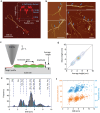
Fibril–fibril interaction and morphological investigation on insulin chiral fibrils. a) AFM snapshot of a self‐folded protofilament with a nanoracket‐type shape. The inset shows the height profile along the direction of the arrows. b) Snapshots of intertwining protofilaments and protofibrils at the early stage of fibrillization, indicated by the arrows. Scale bars are 500 nm. c) Schematic representation of a chiral fibril consisting of two protofilaments under AFM investigation. The extracted height profile (red) along the fibril ridge is used for calculating the average height, crossover pitch (crossover distance), and amplitude of the chiral fibril. d) Density map of the average height distribution computed from the full profile against the mean height calculated as the arithmetic mean between minimum and maximum height. e) IMH distribution of chiral fibrils obtained in the experiment, featuring seven distinct families peaked at 1.2 ± 0.2, 2.7 ± 0.4, 3.4±0.3, 5.1±0.7,6.0±0.3, 7.4±0.6 and 9.4±0.4 nm, corresponding to n  =  1,  2,  …,  7. An extra IMH peak located at 4 nm unveils the presence of a richer morphological complexity and possible packing arrangements are reported in Figure S8 (Supporting Information). f) Scatter plot of crossover pitch and amplitude against IMH of the chiral fibril. The clustered distribution in the plot indicates a correlation between the amplitude and the crossover pitch of chiral fibrils with their IMH height.
Figure 3
Figure 3
Classification of hierarchical configurations of chiral fibrils. a) Histogram of the amplitude distribution of all chiral fibrils with IMH height of more than 2 nm i) and chiral fibrils from IMH family 2–6 ii–vi). Regions shaded in grey refer to the IMH of fibrils with 1, 2, and 3 protofilaments. The labels on the peaks of the multimodal distributions refer to distinct fibril configurations. b) Schematic models of possible configurations of chiral fibrils consisting of up to 6 protofilaments with the cross‐section of each fibril model at the indicated position. The corresponding fibrils are illustrated in the AFM images and the insets show the height profiles of the respective fibril along the dashed lines, in which the IMH, crossover pitch, and amplitude are noted.
Figure 4
Figure 4
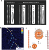
Cryo‐EM observation of fibril polymorphs and the intertwining of early protofibrils. a) The reconstituted fibrils (stitched 2D class averages) on the left side of the panels illustrate the heterogeneous nature of individual fibrils for each polymorph, which includes a twisted ribbon and three mixed‐curvature polymorphs. In each case, a 3D model of each polymorph displays the 3D averaged cross‐sections and side views, further highlighting the differences between polymorphs. Scale bars represent 8 nm. b) A snapshot of the intertwining of two identical early protofibrils 2[1] into the higher‐ordered protofibril 4[2]. These protofibrils are identified by their height fingerprints in Figure S11 (Supporting Information). c) The protofilament‐based schemes of the protofibrils from panel b, as a representative model demonstrating the transition from twisted ribbons into mixed‐curvature polymorphs. Twisted ribbon 2[1] involves the torsion of protofilaments, while the merged 4[2] protofibril, formed by the helical intertwining of two twisted ribbons, has non‐zero torsional and bending energy.
Figure 5
Figure 5
Visualization of protofibril intertwining and the consensus model for fibril formation. a, b) AFM snapshots of the a) intertwining of 2[1]and 4[2] protofibrils into higher‐ordered fibrils in family 6 and b) of the entangling between two identical 3[2] protofibrils that are both formed by 2[1] protofibrils, recruiting another protofilament and generating a higher‐ordered family‐7 fibril. The inset shows the AFM‐profile fingerprint of chiral fibrils before and after intertwining along the dashed lines and arrows in the images. c) Representative case of the combination of protofibrils into mature fibrils, showing a protofibril 2[1] and a protofilament 1[0] combine into a three‐stranded fibril, either with k  =  1 (2[1]1/21[0] →  3[1]) or with k  =  2 (2[1]11[0] →  3[2]). The right sections corresponding to the blue plane are depicted, and the corresponding height is indicated. d) Structures and AFM profiles for the four‐stranded fibrils 4[1] (left) and 4[2] (right). In the AFM profiles, the real height is depicted as a black dashed line, while red continuous lines give the height obtained after convolution with a tip with an effective radius R  =  90 nm.

References

    1. a) Chiti F., Dobson C. M., Annu. Rev. Biochem. 2006, 75, 333; - PubMed
    2. b) Dubay K. F., Pawar A. P., Chiti F., Zurdo J., Dobson C. M., Vendruscolo M., J. Mol. Biol. 2004, 341, 1317. - PubMed
    1. a) Close W., Neumann M., Schmidt A., Hora M., Annamalai K., Schmidt M., Reif B., Schmidt V., Grigorieff N., Fändrich M., Nat. Commun. 2018, 9, 699; - PMC - PubMed
    2. b) Adamcik J., Mezzenga R., Angew. Chem. Int. Ed. 2018, 57, 8370. - PubMed
    1. a) Annamalai K., Gührs K.‐H., Koehler R., Schmidt M., Michel H., Loos C., Gaffney P. M., Sigurdson C. J., Hegenbart U., Schönland S., Fändrich M., Angew. Chem. Int. Ed. 2016, 55, 4822; - PMC - PubMed
    2. b) Bansal A., Schmidt M., Rennegarbe M., Haupt C., Liberta F., Stecher S., Puscalau‐Girtu I., Biedermann A., Fändrich M., Nat. Commun. 2021, 12, 1013. - PMC - PubMed
    1. Jiménez J. L., Nettleton E. J., Bouchard M., Robinson C. V., Dobson C. M., Saibil H. R., Proc. Natl. Acad. Sci. USA 2002, 99, 9196. - PMC - PubMed
    1. a) Congdon E. E., Wu J. W., Myeku N., Figueroa Y. H., Herman M., Marinec P. S., Gestwicki J. E., Dickey C. A., Yu W. H., Duff K. E., Autophagy 2012, 8, 609; - PMC - PubMed
    2. b) Fitzpatrick A. W. P., Falcon B., He S., Murzin A. G., Murshudov G., Garringer H. J., Crowther R. A., Ghetti B., Goedert M., Scheres S. H. W., Nature 2017, 547, 185; - PMC - PubMed
    3. c) Lutter L., Aubrey L. D., Xue W., JMB 2021, 483, 167124. - PubMed

LinkOut - more resources