. 2024 Jul 23;98(7):e0050424.
doi: 10.1128/jvi.00504-24. Epub 2024 Jun 20.

A BSL-2 compliant mouse model of SARS-CoV-2 infection for efficient and convenient antiviral evaluation

Affiliations

A BSL-2 compliant mouse model of SARS-CoV-2 infection for efficient and convenient antiviral evaluation

Zinuo Chen et al. J Virol. .

Abstract

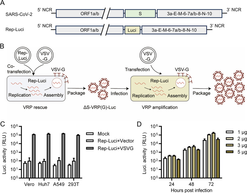
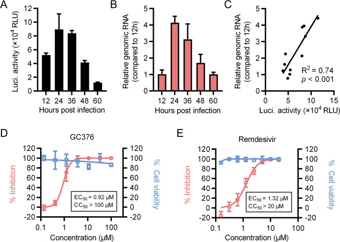
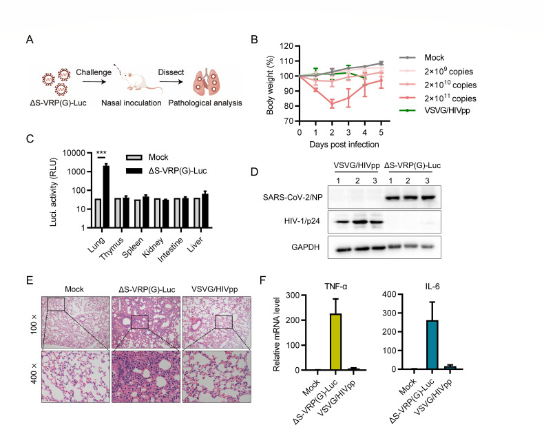
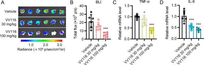
Animal models of authentic severe acute respiratory syndrome coronavirus 2 (SARS-CoV-2) infection require operation in biosafety level 3 (BSL-3) containment. In the present study, we established a mouse model employing a single-cycle infectious virus replicon particle (VRP) system of SARS-CoV-2 that can be safely handled in BSL-2 laboratories. The VRP [ΔS-VRP(G)-Luc] contains a SARS-CoV-2 genome in which the spike gene was replaced by a firefly luciferase (Fluc) reporter gene (Rep-Luci), and incorporates the vesicular stomatitis virus glycoprotein on the surface. Intranasal inoculation of ΔS-VRP(G)-Luc can successfully transduce the Rep-Luci genome into mouse lungs, initiating self-replication of Rep-Luci and, accordingly, inducing acute lung injury mimicking the authentic SARS-CoV-2 pathology. In addition, the reporter Fluc expression can be monitored using a bioluminescence imaging approach, allowing a rapid and convenient determination of viral replication in ΔS-VRP(G)-Luc-infected mouse lungs. Upon treatment with an approved anti-SARS-CoV-2 drug, VV116, the viral replication in infected mouse lungs was significantly reduced, suggesting that the animal model is feasible for antiviral evaluation. In summary, we have developed a BSL-2-compliant mouse model of SARS-CoV-2 infection, providing an advanced approach to study aspects of the viral pathogenesis, viral-host interactions, as well as the efficacy of antiviral therapeutics in the future.IMPORTANCESevere acute respiratory syndrome coronavirus 2 (SARS-CoV-2) is highly contagious and pathogenic in humans; thus, research on authentic SARS-CoV-2 has been restricted to biosafety level 3 (BSL-3) laboratories. However, due to the scarcity of BSL-3 facilities and trained personnel, the participation of a broad scientific community in SARS-CoV-2 research had been greatly limited, hindering the advancement of our understanding on the basic virology as well as the urgently necessitated drug development. Previously, our colleagues Jin et al. had generated a SARS-CoV-2 replicon by replacing the essential spike gene in the viral genome with a Fluc reporter (Rep-Luci), which can be safely operated under BSL-2 conditions. By incorporating the Rep-Luci into viral replicon particles carrying vesicular stomatitis virus glycoprotein on their surface, and via intranasal inoculation, we successfully transduced the Rep-Luci into mouse lungs, developing a mouse model mimicking SARS-CoV-2 infection. Our model can serve as a useful platform for SARS-CoV-2 pathological studies and antiviral evaluation under BSL2 containment.

Keywords: SARS-CoV-2; animal model; replicon.


Conflict of interest statement

The authors declare no conflict of interest.

Figures

Fig 1
Fig 1
Generation of single-cycle infectious VRPs of SARS-CoV-2 carrying a Fluc reporter. (A) Schematic representation of SARS-CoV-2 and viral replicon (Rep-Luci) genomes. The S gene in the SARS-CoV-2 genome was replaced with the Fluc reporter. (B) Generation and amplification of SARS-CoV-2 VRPs. Initially, 293T cells were transfected with Rep-Luci bacmid and VSV-G plasmid, generating VRPs termed ΔS-VRP(G)-Luc. Supernatants from rescue transfections were used as seed to amplify ΔS-VRP(G)-Luc in 293T cells transfected with VSV-G plasmid. (C) Infectivity determination of ΔS-VRP(G)-Luc. 293T cells were co-transfected with Rep-Luci bacmid and VSV-G plasmid, or with Rep-Luci bacmid and empty plasmid as control. At 72 h (H) post-transfection (p.t.), the supernatants were harvested and used to infect indicated cells lines. Mock infected cells were used as blank control. At 24 h post-infection (p.i.), the cells were subjected to luciferase assay. (D) Optimization of ΔS-VRP(G)-Luc amplification. 293T cells grown in six-well plates were transfected with various doses of VSV-G plasmid. At 24 h p.t., the Vero-E6 cells were inoculated with ΔS-VRP(G)-Luc at a multiplicity of infection of 0.01 relative light unit (RLU) per cell. At different time points p.i., a portion of the supernatants were removed for infectivity determination.
Fig 2
Fig 2
In vitro characterization of ΔS-VRP(G)-Luc. (A and B) The Vero-E6 cells were infected with ΔS-VRP(G)-Luc at an MOI of 1 RLU/cell. At indicated time points, the cells were harvested for luciferase assay (A) or genomic RNA quantification by RT-qPCR (B). (C) Linear correlation analysis between the Fluc expression and relative viral genome level in cells from individual wells. (D and E) Antiviral evaluation. Vero-E6 cells infected with ΔS-VRP(G)-Luc were treated with increasing concentrations of GC376 (D) and remdesivir (E), separately. The percent reduction of luciferase values compared to dimethyl sulfoxide (DMSO) control were used to indicate the inhibition of viral replication.
Fig 3
Fig 3
Pathological analysis of ΔS-VRP(G)-Luc infection in mice. (A) The diagram to establish the mouse model of ΔS-VRP(G)-Luc infection. Intranasal inoculation was used to transduce the Rep-Luci into mouse lungs. (B) Body weight changes. Female BALB/c mice (4–6 weeks old, n = 5) were nasally challenged with increasing doses of ΔS-VRP(G)-Luc and the body weight of mice were monitored daily. Mock (n = 3) or mice infected with the VSV-G-based pseudotyped lentivirus (VSVG/HIVpp, n = 3) were used as controls. (C) Mice infected with ΔS-VRP(G)-Luc (2 × 10¹⁰ copies/mouse) were euthanized at 24 h p.i. and indicated tissues were dissected for luciferase assay. Data are shown as mean ± SD for each group. ***, P < 0.001, Student’s t-test. (D) Western blot analysis of nucleoprotein and p24 protein expression at 24 h p.i. in individual lungs of ΔS-VRP(G)-Luc- and VSVG/HIVpp-infected mice, respectively. Glyceraldehyde-3-phosphate dehydrogenase (GAPDH) levels are shown as loading controls. (E) Histopathologic analysis of mouse lung tissues. The mice were sacrificed at 24 h p.i. and the lung tissues were subjected to hematoxylin-and-eosin (H&E) staining. Representative microscopic pictures of H&E staining in each group are shown. (F) The expression level of pro-inflammatory cytokines tumor necrosis factor-α (TNF)-α and interleukin (IL)-6 in the indicated lung tissues collected at 24 h p.i. were determined by RT-qPCR. Data are shown as mean ± SD for each group.
Fig 4
Fig 4
Time course of ΔS-VRP(G)-Luc infection in mouse lungs. (A–D) Female BALB/c mice (4–6 weeks old) were nasally inoculated with ΔS-VRP(G)-Luc at 2 × 10¹⁰ copies/mouse. At various time points, the mouse lungs (n = 3 for each time point) were dissected. The lung index (A), luciferase expression (B), viral RNA (C), as well as the expression level of pro-inflammatory cytokines TNF-α (D) and IL-6 (E) in the lung tissues were determined. Data are shown as mean ± SD for each time point. *, P < 0.05; **, P < 0.01; and ***, P < 0.001, Student’s t-test. (E) Correlation analysis of the expression level of luciferase, TNF-α, or IL-6 with the viral RNA in ΔS-VRP(G)-Luc-infected mouse lungs.
Fig 5
Fig 5
Bioluminescence imaging of ΔS-VRP(G)-Luc-infected mouse lungs. Female BALB/c mice (4–6 weeks old, n = 3) were nasally inoculated with increasing doses of ΔS-VRP(G)-Luc. At 24 h p.i., the lungs were dissected for further analysis. (A) Ex vivo bioluminescence imaging of ΔS-VRP(G)-Luc virus-infected mouse lungs. (B) Analysis of the total bioluminescence signals from ex vivo imaging of mouse lungs. (C) Viral RNA determination. The viral RNA in ΔS-VRP(G)-Luc virus-infected lungs were determined by RT‐qPCR. (D) Correlation analysis between the bioluminescence density and viral RNA of individual mouse lungs. (E and F) Expression levels of pro-inflammatory cytokines TNF-α (E) and IL-6 (F) in mouse lungs infected by various doses of ΔS-VRP(G)-Luc. Data are shown as mean ± SD for each group. *, P < 0.05; **, P < 0.01; ***, P < 0.001; and ****, P < 0.0001, Student’s t-test.
Fig 6
Fig 6
Antiviral evaluation using the mouse models of ΔS-VRP(G)-Luc infection. Female BALB/c mice (4–6 weeks old, n = 6) were inoculated with ΔS-VRP(G)-Luc virus at a dose of 3 × 10¹⁰ copies/mouse, and VV116 was orally administered at 30 to 100 mg/kg/day, starting 2 h before infection. At 24 h p.i., the lung tissues were excised and subjected to ex vivo imaging (A), and the bioluminescence signals were analyzed (B). Subsequently, the expression levels of TNF-α (C) and IL-6 (D) in the lung tissues were measured by RT-qPCR. Data are shown as mean ± SD for each group. *, P < 0.05; ***, P < 0.001, Student’s t-test.

Similar articles

Cited by

References

    1. Qi F, Qin C. 2022. Characteristics of animal models for COVID-19. Animal Model Exp Med 5:401–409. doi:10.1002/ame2.12278 - DOI - PMC - PubMed
    1. Kim MI, Lee C. 2023. Human coronavirus OC43 as a low-risk model to study COVID-19. Viruses 15:578. doi:10.3390/v15020578 - DOI - PMC - PubMed
    1. Andrade AC dos SP, Campolina-Silva GH, Queiroz-Junior CM, de Oliveira LC, Lacerda L de SB, Pimenta JC, de Souza FRO, de Meira Chaves I, Passos IB, Teixeira DC, et al. . 2021. A biosafety level 2 mouse model for studying betacoronavirus-induced acute lung damage and systemic manifestations. J Virol 95:e0127621. doi:10.1128/jvi.01276-21 - DOI - PMC - PubMed
    1. Zhu Y, Scholle F, Kisthardt SC, Xie DY. 2022. Flavonols and dihydroflavonols inhibit the main protease activity of SARS-CoV-2 and the replication of human coronavirus 229E. Virology 571:21–33. doi:10.1016/j.virol.2022.04.005 - DOI - PMC - PubMed
    1. Yamada H, Sasaki SI, Tani H, Somekawa M, Kawasuji H, Saga Y, Yoshida Y, Yamamoto Y, Hayakawa Y, Morinaga Y. 2022. A novel hamster model of SARS-CoV-2 respiratory infection using a pseudotyped virus. Sci Rep 12:11125. doi:10.1038/s41598-022-15258-8 - DOI - PMC - PubMed

MeSH terms

LinkOut - more resources