Review
. 2024 Jun 20;15(1):157.
doi: 10.1186/s13244-024-01729-1.

CT imaging post-TAVI: Murphy's first law in action-preparing to recognize the unexpected

Affiliations
Review

CT imaging post-TAVI: Murphy's first law in action-preparing to recognize the unexpected

Costanza Lisi et al. Insights Imaging. .

Abstract

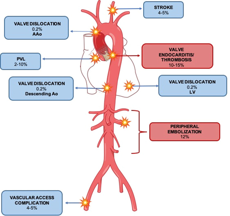
Transfemoral aortic valve implantation (TAVI) has been long considered the standard of therapy for high-risk patients with severe aortic-stenosis and is now effectively employed in place of surgical aortic valve replacement also in intermediate-risk patients. The potential lasting consequences of minor complications, which might have limited impact on elderly patients, could be more noteworthy in the longer term when occurring in younger individuals. That's why a greater focus on early diagnosis, correct management, and prevention of post-procedural complications is key to achieve satisfactory results. ECG-triggered multidetector computed tomography angiography (CTA) is the mainstay imaging modality for pre-procedural planning of TAVI and is also used for post-interventional early detection of both acute and long-term complications. CTA allows detailed morphological analysis of the valve and its movement throughout the entire cardiac cycle. Moreover, stent position, coronary artery branches, and integrity of the aortic root can be precisely evaluated. Imaging reliability implies the correct technical setting of the computed tomography scan, knowledge of valve type, normal post-interventional findings, and awareness of classic and life-threatening complications after a TAVI procedure. This educational review discusses the main post-procedural complications of TAVI with a specific imaging focus, trying to clearly describe the technical aspects of CTA Imaging in post-TAVI and its clinical applications and challenges, with a final focus on future perspectives and emerging technologies. CRITICAL RELEVANCE STATEMENT: This review undertakes an analysis of the role computed tomography angiography (CTA) plays in the assessment of post-TAVI complications. Highlighting the educational issues related to the topic, empowers radiologists to refine their clinical approach, contributing to enhanced patient care. KEY POINTS: Prompt recognition of TAVI complications, ranging from value issues to death, is crucial. Adherence to recommended scanning protocols, and the optimization of tailored protocols, is essential. CTA is central in the diagnosis of TAVI complications and functions as a gatekeeper to treatment.

Keywords: Cardiac imaging; Complications; Computed tomography angiography (CTA); Transfemoral aortic valve implantation (TAVI).


Conflict of interest statement

The authors declare that they have no competing interests.

Figures

Fig. 1
Fig. 1
Schematic representation of main post-TAVI complications identified by CTA and their incidence. Blue square for peri-procedural complications, red square for late complications
Fig. 2
Fig. 2
MPR of a infaranular valve dislocation (a, b, c). Transcatheter sel-expandable aortic valve (Portico) dislocated to the left ventricular outflow tract (a, c, blue arrow) with anterior mitral valve leaflet impairment (a). After a ViV procedure, a second prosthetic module was correctly positioned (c, yellow arrow)
Fig. 3
Fig. 3
A 55-year-old patient CTA control after TAVI positioning for prosthesis dislocation. CTA images show, aortic prosthesis dislocation in the descending aorta (a, b, c blue arrow), causing focal ascending aorta dissection (blue asterisk). Sagittal MPR also shows the presence of a vascular catheter inside the thoracoabdominal aorta, trapped in the dislocated prosthesis (c, yellow arrow)
Fig. 4
Fig. 4
An 83-year-old patient presenting to the emergency room with dysarthria, difficulty in walking, and dropping lip 7 days after TAVI for severely calcific aortic-stenosis (a). A brain CTA was performed, demonstrating a calcific embolus in the right middle cerebral artery (b, c arrow), determining incomplete occlusion of the vessel (d, arrow), as a result of distant calcific embolization during TAVI. Perfusion-CT shows an area of ischemic penumbra in the right cerebral hemisphere, with reduced cerebral blood flow (CBF) (e). The patient was treated with a thrombectomy and got a resolution of clinical symptoms
Fig. 5
Fig. 5
An 85-year-old woman, subjected to TAVI, developing fever and abdominal pain with elevated white blood cell count and C-reactive protein. CCTA is performed in the suspicion of endocarditis and also extended to the abdomen to rule out peripheral embolization. CCTA para-axial, coronal, and sagittal vie (a, b, c, d) show thick ipodense thrombotic apposition over prosthesis leaflets (b, c blue arrow). Abdominal CT also shows splenic infarction due to septic embolization (e, f asterisk). Blood culture turns positive for Enterococcus faecium
Fig. 6
Fig. 6
A 68-year-old male patient with severe aortic-stenosis, treated with Edwards SAPIENS aortic bioprosthesis, undergoing ECG-gated CTA for a TTE suspicion of the periprosthetic leak. CTA confirms a thin (6 mm thickness, arrow) PVL located just below the ostium of the main trunk in the axial oblique (a, arrow) and coronal plane (b, arrow)
Fig. 7
Fig. 7
An 85-year-old patient candidate for TAVI for severe symptomatic aortic-stenosis. Intraprocedural death for cardiac arrest was likely due to annular iatrogenic rupture as documented by angiographic images: Iodinated contrast extravasations outside the aorta are demonstrated (arrow, a), as well as contrast extravasation into the pericardial sac (asterisks, b)
Fig. 8
Fig. 8
A 70-year-old patient subjected to TAVI 2 days before developed hypotension, anemia (Hb: 9 g/dL) and altered mental status. Contrast-enhanced CTA was performed to rule out acute bleeding, showing a massive lower abdominal spontaneously hyperdense collection (a) (asterisk, mean HU = 52), with active bleeding inside (b, arrow, arterial phase) (c, arrow, venous phase), as a result of an iatrogenic rupture of an arterial femoral-iliac vessel on the left (d, e asterisks) of 6 × 20 × 8.5 cm (a, f)
Fig. 9
Fig. 9
A 55-year-old female patient undergoing post-TAVI CTA for anemia onset (Hb: 9.5 g/dL) after the procedure showing focal blood leakage anteriorly to the left ventricle (a, b, c, arrow) due to an iatrogenic ventricular wall lesion

References

    1. Vahanian A, Beyersdorf F, Praz F, et al. 2021 ESC/EACTS guidelines for the management of valvular heart disease: developed by the task force for the management of valvular heart disease of the European Society of Cardiology (ESC) and the European Association for Cardio-Thoracic Surgery (EACTS) Rev Esp Cardiol (Engl Ed) 2022;75:524. doi: 10.1016/j.rec.2022.05.006. - DOI - PubMed
    1. Van Mieghem NM, Deeb GM, Søndergaard L, et al. Self-expanding transcatheter vs surgical aortic valve replacement in intermediate-risk Patients: 5-year Outcomes of the SURTAVI randomized clinical trial. JAMA Cardiol. 2022;7:1000–1008. doi: 10.1001/jamacardio.2022.2695. - DOI - PMC - PubMed
    1. Mack MJ, Leon MB, Thourani VH, et al. Transcatheter aortic-valve replacement in low-risk patients at five years. N Engl J Med. 2023;389:1949–1960. doi: 10.1056/NEJMoa2307447. - DOI - PubMed
    1. Mack MJ, Leon MB, Thourani VH, et al. Transcatheter aortic-valve replacement with a balloon-expandable valve in low-risk patients. N Engl J Med. 2019;380:1695–1705. doi: 10.1056/NEJMoa1814052. - DOI - PubMed
    1. Tully A, Tom S, Xie J, et al. Evolving computed tomography angiography for aortic valve replacement: optimizing transcatheter and surgical therapies. J Card Surg. 2022;37:4124–4132. doi: 10.1111/jocs.16977. - DOI - PubMed

LinkOut - more resources