. 2024 Jun 20;20(6):e1012300.
doi: 10.1371/journal.ppat.1012300. eCollection 2024 Jun.

Human cytomegalovirus deploys molecular mimicry to recruit VPS4A to sites of virus assembly

Affiliations

Human cytomegalovirus deploys molecular mimicry to recruit VPS4A to sites of virus assembly

Benjamin G Butt et al. PLoS Pathog. .

Abstract

The AAA-type ATPase VPS4 is recruited by proteins of the endosomal sorting complex required for transport III (ESCRT-III) to catalyse membrane constriction and membrane fission. VPS4A accumulates at the cytoplasmic viral assembly complex (cVAC) of cells infected with human cytomegalovirus (HCMV), the site where nascent virus particles obtain their membrane envelope. Here we show that VPS4A is recruited to the cVAC via interaction with pUL71. Sequence analysis, deep-learning structure prediction, molecular dynamics and mutagenic analysis identify a short peptide motif in the C-terminal region of pUL71 that is necessary and sufficient for the interaction with VPS4A. This motif is predicted to bind the same groove of the N-terminal VPS4A Microtubule-Interacting and Trafficking (MIT) domain as the Type 2 MIT-Interacting Motif (MIM2) of cellular ESCRT-III components, and this viral MIM2-like motif (vMIM2) is conserved across β-herpesvirus pUL71 homologues. However, recruitment of VPS4A by pUL71 is dispensable for HCMV morphogenesis or replication and the function of the conserved vMIM2 during infection remains enigmatic. VPS4-recruitment via a vMIM2 represents a previously unknown mechanism of molecular mimicry in viruses, extending previous observations that herpesviruses encode proteins with structural and functional homology to cellular ESCRT-III components.


Conflict of interest statement

The authors have declared that no competing interests exist.

Figures

Fig 1
Fig 1. HCMV pUL71 binds the VPS4A MIT domain via a potential MIM2 motif.
(A) Normalised sequence conservation of pUL71 homologues across Betaherpesvirinae. Inset shows an alignment of the canonical MIM2 sequence of human CHMP6, the potential MIM2 of pUL71, and the MIM2 consensus sequence where x denotes any residue,–an acidic residue and ϕ a hydrophobic residue [46]. Vertical lines denote hydrophobic (black) and backbone hydrogen bond (red) interactions between residues of the CHMP6 MIM2 and the MIT domain of VPS4A [46]. Selected MIM2 residues important for the interaction with VPS4A are highlighted in pink. (B) Co-transfection of pUL71 and pp28-EGFP with VPS4A-FLAG. Nuclei are outlined in single channel images and shown (DAPI, blue) in merge. Scale bars = 10 μm. (C) Structure of human CHMP6 MIM2 (orange carbon atoms, selected key residues highlighted in pink) in complex with the MIT domain of human VPS4A (PDB ID 2K3W) [46]. (D) Co-transfection of VPS4A-FLAG with wild-type (WT) pUL71 or two mutants, P315A+P318A (PPAA) and V317D, where key residues of the pUL71 potential MIM2 were mutated and their ability to recruit VPS4A to juxtanuclear compartments is disrupted. Nuclei are outlined in single channel images and shown (DAPI, blue) in merge. Scale bars = 10 μm. (E) Anti-FLAG immunoprecipitation (IP) from cells co-transfected with VPS4A-FLAG and WT, PPAA or V317D pUL71. Samples were immunoblotted using antibodies as shown. (F) Coomassie-stained SDS-PAGE of GST-tagged WT or PPAA mutant pUL71 C-terminal tail (residues 283–361), VPS4A MIT domain (residues 1–84), or GST-tagged CHMP6 MIM2 motif (residues 168–181) purified following bacterial expression. (G) ITC analysis of the interaction between purified VPS4A MIT domain and WT GST-pUL71(283–361) (left), PPAA mutant GST-pUL71(283–361) (middle), and GST-CHMP6 MIM2 (right). For each, the top graph is baseline-corrected differential power as a function of time and the bottom is the normalised binding curve showing integrated changes in enthalpy (ΔH) as function of molar ratio (syringe:cell component). The corresponding dissociation constant (KD), number of binding sites (N), and enthalpy change (ΔH) for each representative experiment are shown. All experiments were performed at least twice independently, as detailed in Table 1.
Fig 2
Fig 2. An extended MIM2-like motif spanning pUL71 residues 300–325 is necessary and sufficient for VPS4A binding.
(A) Co-transfection of VPS4A-FLAG with full-length or truncated pUL71, or with pUL71 lacking the potential MIM2 (Δ315–326). Nuclei are outlined in single channel images and shown (DAPI, blue) in merge. Scale bar = 10 μm. (B) ITC analysis of the interaction between purified VPS4A MIT domain and synthetic peptides corresponding to the CHMP6 MIM2 (left) or the potential pUL71 MIM2 spanning residues 310–325 (right). (C) ITC analysis of the VPS4A MIT domain binding a peptide corresponding to the extended MIM2-like motif spanning pUL71 residues 300–325. (D) Schematic diagram of pUL71 truncation experiments. Top: Predicted pUL71 secondary structure is shown (blue helices and green sheets) with the potential MIM2 boxed. Bottom: The ability to bind VPS4A as evidenced by co-localisation following co-transfection or by ITC analysis, with residues 300–325 that are sufficient for binding highlighted (see also S1 Fig).
Fig 3
Fig 3. HCMV pUL71 is predicted to bind the MIM2-binding groove of the VPS4A MIT domain as a helix plus extended peptide.
(A) Predicted structure of pUL71 residues 300–325 (violet ribbon with side chains shown) in complex with the VPS4A MIT domain (cyan ribbons and semi-transparent molecular surface). pUL71 is predicted to bind the groove between VPS4A helices α1 and α3. Inset: Predicted interaction between hydrophobic residues of the pUL71 helical region and the VPS4A MIT domain. VPS4A molecular surface is coloured by residue hydrophobicity from white (polar) to yellow (hydrophobic) and selected hydrophobic side chains are shown as silhouettes. (B) Per-residue predicted Local Distance Difference Test (pLDDT) scores for predicted complex. Values above 70 (dashed line) represent regions predicted with high confidence. (C) Predicted aligned error (PAE) matrix for predicted complex, demonstrating high confidence (low PAE, green) in relative orientation of pUL71(300–325) with respect to VPS4A MIT domain. (D) Structural comparison of the predicted pUL71(300–325) (violet ribbon and sticks) and the VPS4A MIT domain (cyan surface) complex to experimental structures of MIT domains bound to cellular MIM2s. Experimental structures were superposed by structural alignment of the MIT domains but, for clarity, only the predicted VPS4A MIT domain is shown. Top: Canonical CHMP6 MIM2 (orange ribbon and sticks) in complex with human VPS4A (PDB ID 2K3W) [46]. Middle: Helix and MIM2 of yeast Vps4 regulator Vfa1 (green ribbon and sticks) bound to yeast Vps4 (PDB ID 4NIQ, chains A+C) [53]. Bottom: Helix and non-canonical MIM2 of yeast CHMP6 homologue Vps20 (yellow ribbon and sticks) bound to yeast Vps4 (PDB ID 5FVL, chains A+D) [52]. (E) Crystal structure of the second of the two tandem MIT domains of yeast Atg1 (dark blue ribbon) in complex with the Type 7 MIM(N) of yeast Atg13 (grey Cα trace) (PDB ID 4P1N) [54] superposed on the predicted structure of human VPS4A MIT domain (cyan ribbons) and pUL71 (violet Cα trace). While both MIM-like motifs bind the groove between MIT domain helixes α1 and α3, the MIM peptides have opposite orientations (N→C). (F) Superposition of canonical MIM2 of CHMP6 (orange Cα trace, PDB ID 2K3W) [46] and MIM1 motif of CHMP1A (aqua Cα trace, PDB ID 2JQ9) [55] onto the predicted structure of pUL71 (violet Cα trace) in complex with human VPS4 (cyan ribbons). MIM2 groove residues V13 (left) and MIM1 groove residue L64 (right) are highlighted as red sticks. (G) Co-transfection of pUL71 with FLAG-tagged VPS4A, either full-length (WT), lacking the N-terminal 84 residues encoding the MIT domain (ΔMIT), or with single amino acid substitutions that prevent binding to MIM2 (V13D) or MIM1 (L64D) regions. Nuclei are outlined in single channel images and shown (DAPI, blue) in merge. Scale bars = 10 μm.
Fig 4
Fig 4. The pUL71(300–325):VPS4A MIT domain structural model has predictive power.
(A) Umbrella sampling molecular dynamics (MD) simulations of WT and mutant pUL71(300–325) in complex with the VPS4A MIT domain. For each, potential of mean force ± SD (shaded) from 200 bootstraps of the analysis is plotted as a function of centre of mass between the two polypeptides. The calculated Gibbs free energy of binding (ΔGbind) is the difference between the potential mean force minimum (bound) and maximum (unbound) values. (B) ΔGbind for pUL71(300–325) WT and mutants. (C) Difference in ΔGbind for pUL71(300–325) mutants compared to WT (ΔΔGbind; mean ± SD for 200 bootstraps). Positive ΔΔGbind values indicate reduced binding affinity, negative values indicate increased affinity. (D) ITC analysis demonstrating a lack of binding when a peptide corresponding to pUL71 residues 300–325 with an I307R substitution is titrated against the VPS4A MIT domain. (E) Anti-FLAG IP from cells co-transfected with VPS4A-FLAG and WT, P315A, P318A, or P315A+P318A (PPAA) pUL71. Samples were immunoblotted using antibodies as shown.
Fig 5
Fig 5. VPS4A binding is conserved across human β-herpesviruses, but not α- or γ-herpesviruses.
(A) Alignment of vMIM2 regions of pUL71 and its homologue (pU44) in HHV6 and HHV7. Note that the displayed region of pU44 has an identical sequence in HHV6A and HHV6B. The predicted secondary structure of pUL71 is shown above. The pUL71 homologue vMIM2 consensus sequence is shown below, where Ω denotes a large hydrophobic residue, x denotes any residue, ϕ denotes a small hydrophobic residue (including proline), and where the underlined residues are within an α-helix. (B) Predicted structure of residues from the vMIM2 of β-herpesvirus pUL71 homologues (violet, red and purple ribbons for HCMV, HHV6 and HHV7, respectively) in complex with the VPS4A MIT domain (cyan molecular surface). Inset: Predicted interactions between hydrophobic residues of the pUL71 homologue helical regions and the VPS4A MIT domain. VPS4A molecular surface is coloured by residue hydrophobicity from white (polar) to yellow (hydrophobic). Cα trace and selected side chains of vMIM2s are shown. Below: Per-residue pLDDT scores of the vMIM2s, with values above 70 (dashed line) representing regions predicted with high confidence. Right: PAE matrices for predicted HHV6 and HHV7 complexes. (C) ITC analysis of the VPS4A MIT domain binding a peptide corresponding to the HHV6 pU44 vMIM2 (residues 174–199). (D) Anti-FLAG IP from cells co-transfected with VPS4A-FLAG and HCMV pUL71 or homologues from HSV-1 (pUL51), HHV6 (pU44) and EBV (BSRF1). Samples were immunoblotted using antibodies as shown. (E) Co-transfection of VPS4A-FLAG and HCMV pUL71 or homologues from HSV-1 (pUL51), HHV6 (pU44) and EBV (BSRF1). (F) Co-transfection of VPS4A-FLAG with a chimeric construct encoding full length HSV-1 pUL51 followed by an HA tag plus the C-terminal region of HCMV pUL71 (residues 283–361, which includes the vMIM2). Nuclei are outlined in single channel images and shown (DAPI, blue) in merge. Scale bars = 10 μm.
Fig 6
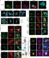
Fig 6. pUL71 vMIM2 is required for VPS4A recruitment to the cVAC during infection.
(A) MRC-5 cells infected with HCMV TB40/E were fixed at 5 dpi and stained using antibodies shown. The signal for pUL71 (green) overlaps with signals for tegument protein pp150 (red) and Golgi marker GM130 at DNA-containing (DAPI, cyan) perinuclear sites of virion assembly [28]. (B) MRC-5 cells transiently expressing VPS4A-FLAG from an inducible expression vector were infected (MOI 0.5) with the indicated strains of HCMV. Expression of VPS4A-FLAG was induced 1 dpi by addition of doxycycline and intracellular distribution VPS4A-FLAG was examined at 5 dpi via antibody detection of the FLAG epitope. The cVAC is denoted by additional staining for HCMV pUL71 (red) and nuclei are shown (DAPI, blue). (C, D) MRC-5 cells transiently expressing VPS4-FLAG were mock-infected or infected (MOI 0.5–1) with indicated viruses. Expression of VPS4A was induced 1 dpi and cells were fixed and immunostained with the antibodies shown at 5 dpi. Selected cells are outlined in single channel images and nuclei are shown (DAPI, cyan) in merge. (C) Cells were infected with WT HCMV, with a recombinant virus lacking pUL71 expression (TBstop71), or the revertant virus with restored pUL71 expression (TBrev71). Cells infected with TBstop71 were also stained for tegument protein pp28 to confirm successful cVAC formation (bottom). (D) Cells were infected with WT HCMV, with recombinant virus lacking the vMIM2 motif (TB71del315–326), with mutations P315A+P318A in the vMIM2 to disrupt VPS4A binding (TB71mutPPAA), or with the wild-type sequence subsequently restored (TB71revPPAA). (E) Bimolecular fluorescence complementation using split Citrine (residues 1–173, YN, and residues 156–239, YC) confirms a physical interaction between pUL71 and VPS4A during infection. MRC-5 cells conditionally expressing WT, MIM2-binding groove (V13D) or MIM1-binding surface (L64D) mutant human VPS4A that was N- and C-terminally tagged with YC and FLAG, respectively, were infected (MOI 1) with HCMV strain TB40/E where pUL71 was C-terminally tagged with YN (TB71-YN). Expression was induced 2 dpi by addition of doxycycline. Cells were fixed, immunostained (FLAG, magenta), and Citrine fluorescence from reconstitution of its constituent parts (yellow) was visualised at 5 dpi. Selected cells and their nuclei are outlined in single channel images are nuclei are shown (DAPI, blue) in merge. Scale bars = 10 μm.
Fig 7
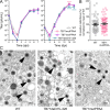
Fig 7. VPS4A recruitment to the cVAC is not required for efficient virus wrapping or spread in human fibroblasts.
(A) Mutations in the pUL71 vMIM2 motif do not affect virus release. For single-step kinetics (left), HFFs were infected (MOI 3) with HCMV WT (□), TB71mutPPAA (○), TB71revPPAA (Δ) or TB71del315–326 (♢). For multi-step kinetics (right) cells were infected at MOI 0.01. The supernatant of infected cells was harvested at the indicated times post infection and the virus yield was determined by titration on HFFs. Mean ± SD is shown (n = 2–3 independent experiments, each performed in technical duplicate). Virus yields of the inocula are given at time zero. A repeated measurement two-way ANOVA test shows no significant difference between viruses in the single-step growth curve (p = 0.5824) but a significant difference between viruses in the multi-step growth curve (p = 0.0005). Significance of differences between WT and TB71mutPPAA (pink) or TB71del315–326 (cyan) at specific time points are shown (Dunnett’s multiple comparison test; ****, p < 0.0001). (B) Relative size of plaques formed by HCMV WT and TB71mutPPAA after 9 days of infection in a focus expansion assay. Infected cells were detected by immunostaining for HCMV IE1/2 protein. Each data point represents the relative number of IE-positive nuclei per focus. Mean ± SEM (n = 56–61 foci) is shown for each virus (black line) normalised to the mean focus size for WT. A Mann-Whitney test shows no significant difference (p = 0.4888). (C) Electron micrographs showing representative areas of the cVAC at 5 dpi from cells infected with WT HCMV or with the indicated mutant viruses. Arrow heads mark fully enveloped virus particles. Scale bar = 200 nm.

References

    1. Rivera-Cuevas Y, Carruthers VB. The multifaceted interactions between pathogens and host ESCRT machinery. PLoS Pathog. 2023;19: e1011344. doi: 10.1371/journal.ppat.1011344 - DOI - PMC - PubMed
    1. Christ L, Raiborg C, Wenzel EM, Campsteijn C, Stenmark H. Cellular Functions and Molecular Mechanisms of the ESCRT Membrane-Scission Machinery. Trends Biochem Sci. 2017;42: 42–56. doi: 10.1016/j.tibs.2016.08.016 - DOI - PubMed
    1. McCullough J, Frost A, Sundquist WI. Structures, Functions, and Dynamics of ESCRT-III/Vps4 Membrane Remodeling and Fission Complexes. Annu Rev Cell Dev Biol. 2018;34: 85–109. doi: 10.1146/annurev-cellbio-100616-060600 - DOI - PMC - PubMed
    1. Maity S, Caillat C, Miguet N, Sulbaran G, Effantin G, Schoehn G, et al.. VPS4 triggers constriction and cleavage of ESCRT-III helical filaments. Sci Adv. 2019;5: eaau7198. doi: 10.1126/sciadv.aau7198 - DOI - PMC - PubMed
    1. Wenzel DM, Mackay DR, Skalicky JJ, Paine EL, Miller MS, Ullman KS, et al.. Comprehensive analysis of the human ESCRT-III-MIT domain interactome reveals new cofactors for cytokinetic abscission. Elife. 2022;11: e77779. doi: 10.7554/eLife.77779 - DOI - PMC - PubMed

MeSH terms

Substances

LinkOut - more resources