Review
. 2024 Jun 21;135(1):174-197.
doi: 10.1161/CIRCRESAHA.124.323067. Epub 2024 Jun 20.

G Protein-Coupled Receptors: A Century of Research and Discovery

Affiliations
Review

G Protein-Coupled Receptors: A Century of Research and Discovery

Samuel Liu et al. Circ Res. .

Abstract

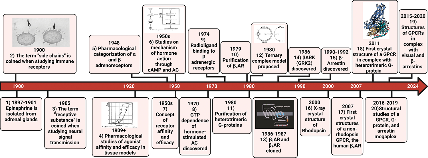
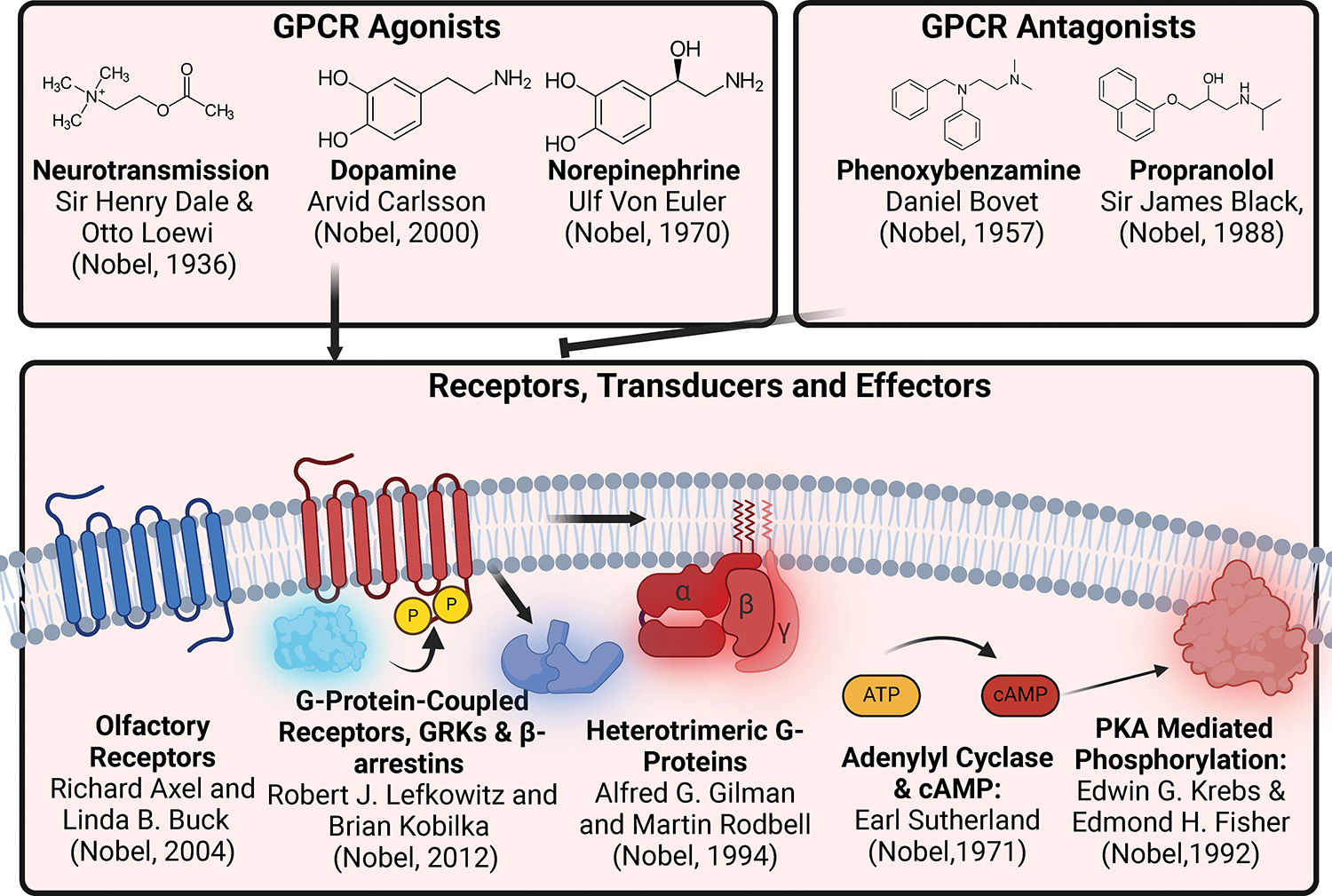
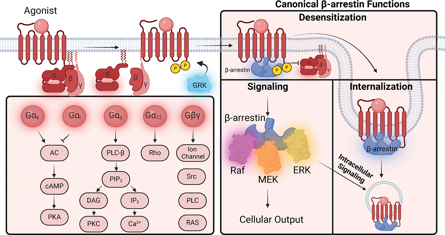
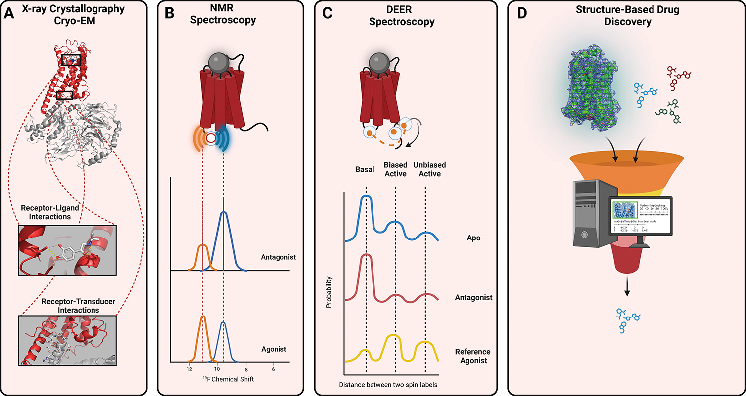
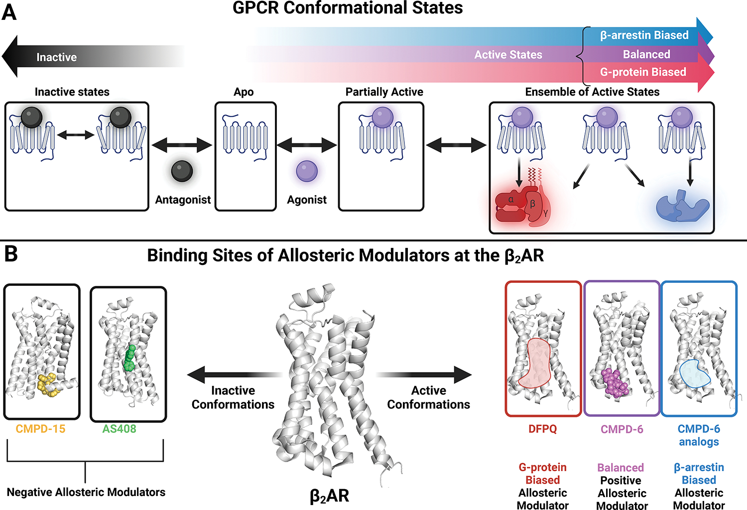
GPCRs (G protein-coupled receptors), also known as 7 transmembrane domain receptors, are the largest receptor family in the human genome, with ≈800 members. GPCRs regulate nearly every aspect of human physiology and disease, thus serving as important drug targets in cardiovascular disease. Sharing a conserved structure comprised of 7 transmembrane α-helices, GPCRs couple to heterotrimeric G-proteins, GPCR kinases, and β-arrestins, promoting downstream signaling through second messengers and other intracellular signaling pathways. GPCR drug development has led to important cardiovascular therapies, such as antagonists of β-adrenergic and angiotensin II receptors for heart failure and hypertension, and agonists of the glucagon-like peptide-1 receptor for reducing adverse cardiovascular events and other emerging indications. There continues to be a major interest in GPCR drug development in cardiovascular and cardiometabolic disease, driven by advances in GPCR mechanistic studies and structure-based drug design. This review recounts the rich history of GPCR research, including the current state of clinically used GPCR drugs, and highlights newly discovered aspects of GPCR biology and promising directions for future investigation. As additional mechanisms for regulating GPCR signaling are uncovered, new strategies for targeting these ubiquitous receptors hold tremendous promise for the field of cardiovascular medicine.

Keywords: adrenergic beta-antagonists; allosteric regulation; angiotensin II; beta-arrestins; g-protein-coupled receptor kinase 2; heart failure; receptors, g-protein-coupled.


Conflict of interest statement

Disclosures R.J. Lefkowitz is an investigator with the Howard Hughes Medical Institute. R.J. Lefkowitz and H.A. Rockman are scientific cofounders of Trevena Inc. R.J. Lefkowitz is a founder of Septerna Inc. Trevena and Septerna are companies that discover and develop novel GPCR-targeted therapeutics. R.J. Lefkowitz is on the board of Lexicon Pharmaceuticals. The other authors report no conflicts.

Figures

Figure 1:
Figure 1:. Notable Ligand, Receptor, and Transducer Discoveries.
Over the past century, there have been many discoveries from ligands to transducers. Here we have highlighted key studies that have contributed to our knowledge of GPCRs. Below are the corresponding publications for each discovery. Images were reproduced with permission from reference #’s 1. 1897–1901: Epinephrine is characterized and isolated from adrenal glands., 2. 1900: The term “side chains” is coined when studying immune receptors. 3. 1905: The term “receptive substance” is coined when studying neural signal transmission. 4. 1909+: Pharmacological studies of agonist affinity and efficacy in tissue models. 5. 1948: Pharmacological categorization of α and β adrenoreceptors. 6. 1950s: Studies on the mechanism of hormone action through cAMP and Adenylyl Cyclase (AC). ,, 7. 1950s: Concept of receptor affinity and efficacy., 8. 1971: GTP dependence of hormone-stimulated AC discovered. 9. 1974: Radioligand binding of β adrenergic receptors. 10. 1979: Purification of the β2AR. 11. 1980: Purification of heterotrimeric G-proteins., 12.1980: Ternary complex proposed. 13. 1986–8: β1AR and β2AR cloned.,, 14. 1986: βARK (GRK2) discovered. 15. 1990–1992: β-arrestins discovered., 16. 2000: X-ray crystal structure of Rhodopsin. 17. 2007: First crystal structures of a non-rhodopsin GPCR, the human β2AR., 18. 2011: First crystal structure of a GPCR in complex with heterotrimeric G-protein. 19. 2015–2020: Structures of GPCRs in complex with visual and β-arrestins. 20. 2016–2019: Structural studies of a GPCR, G-protein, and arrestin megaplex. ,
Figure 2:
Figure 2:. History of Nobel Prize winning GPCR Discoveries
The discoveries of components of the GPCR signaling system, ranging from ligands, receptors, and transducers have resulted in numerous Nobel Prizes being awarded. These include the development of clinically important agonists and antagonists, and basic discoveries related to the signaling mechanisms of GPCRs and their transducers. Names and Nobel Prizes were collected from the Nobel Foundation website (https://www.nobelprize.org/prizes/lists/all-nobel-prizes/)
Figure 3:
Figure 3:. GPCR signaling via G proteins, GRKs and β-arrestins.
Upon agonist stimulation, heterotrimeric G-proteins (Gα, Gβ, and G𝛾) are recruited to the receptor and there is guanine nucleotide exchange of GTP for GDP. The Gα-GTP subunit dissociates from the Gβ𝛾 transducer, and both signal to diverse downstream effectors. Gα has four distinctive families (Gαs, Gαi,q,12/13). GRKs phosphorylate the intracellular domains of the GPCR, which promote tight binding of β-arrestins. β-arrestins have three canonical functions: receptor desensitization, internalization, and signaling. In addition, there is intracellular signaling of β-arrestins from endosomes. GRK, G-protein receptor kinase; GDP, Guanosine diphosphate; GTP, Guanosine triphosphate; AC, adenyl cyclase; ATP, adenosine triphosphate; cAMP, cyclic adenosine monophosphate; PKA; Protein kinase A; PLC- β, phospholipase C- β; IP3, inositol trisphosphate; PIP2 phosphatidylinositol 4,5-bisphosphate; DAG, diacylglycerol; PKC, Protein kinase C; ERK, extracellular signaling-related kinases
Figure 4:
Figure 4:. Biased Signaling of GPCRs.
(A) Reference/endogenous agonists binding to receptors can signal through two different pathways, G-proteins and β-arrestin. (B) Ligand bias promotes the receptor:transducer complex to adopt certain conformations that bias the signaling through (B) G-proteins or (C) β-arrestin. (D) Biased receptors may have altered phosphorylation sites on their C-tail or other mutations that may bias signaling toward β-arrestin instead of G-proteins despite being stimulated with an endogenous agonist. (E) System bias occurs when there is a differential expression of signaling components. For example, some cells express different isoforms of GRK and β-arrestin and may bias signaling through β-arrestin. (F) Location bias refers to receptors promoting signaling from distinct intracellular locations, such as from endosomes, the nucleus, or the plasma membrane.
Figure 5:
Figure 5:. Structural studies of GPCRs.
(A) X-ray crystallography and CryoEM provide static structures of GPCRs. With improvements in cryoEM technology and workflow, cryoEM can now provide high-resolution data on both receptor-ligand and receptor-transducer interactions, making it particularly well-suited to the study of GPCRs. (B) NMR Spectroscopy with probes such as 19F can provide information on dynamics and conformational ensembles by measuring changes in the local environment of individually labeled probes. (C) EPR spectroscopy techniques such as DEER spectroscopy with two spin-label probes can provide information on distances, conformations, and their relative populations. (D) Structure-Based Drug Discovery techniques use receptor structures to allow molecular docking and other approaches to screen compounds that bind to a desired receptor conformation.
Figure 6:
Figure 6:. Allosteric Modulation of GPCRs.
(A) GPCR Conformational States. Receptors in the Apo (no ligands bound) state are largely in inactive conformations but can be stabilized by orthosteric agonists and antagonists in a variety of different active or inactive states. Active states are characterized by an opening of the transducer binding pocket. This pocket is closed to different degrees in inactive states. Orthosteric agonists can activate both G-protein and β-arrestin pathways or be biased towards one pathway. (B) Allosteric modulators bind to topographically distinct sites from the orthosteric site that can modulate receptor conformation. Allosteric modulators can either enhance agonist affinity and/or efficacy (positive allosteric modulators) or diminish the agonist affinity and/or efficacy (negative allosteric modulators). In addition, positive allosteric modulators can stabilize specific receptor conformations that promote bias towards G-protein or β-arrestin. Illustrated are the binding sites of allosteric modulators at the β2AR and the conformations they promote. The negative allosteric modulators of the β2AR, CMPD-15, and AS408, bind to distinct sites on the β2AR. CMPD-6, a balanced positive allosteric modulator, binds to another distinct site. Allosteric modulators can also display biased properties such as difluorophenyl quinazoline derivatives (DFPQ) which biases the β2AR towards G-protein signaling. The binding pose of DFPQ compounds has not been experimentally solved, but they are believed to bind close to the CMPD-6 binding site. β-arrestin biased analogs of CMPD-6 have also been proposed due to CMPD-6’s interactions with the β-arrestin biased orthosteric ligand carvedilol. Compound structures obtained from the Protein Data Bank (PDB) : CMPD-15: 5X7D, AS408: 60BA, CMPD-6: 6N48.
Figure 7:
Figure 7:. Location Bias in Cardiac Myocytes.
(A) In healthy cardiac tissue, βARs are found in T-tubules, which when stimulated with epinephrine/norepinephrine activate βAR-AC-PKA. PKA is tethered to intracellular compartments by AKAPs. PKA phosphorylates the RyR, inducing calcium release and resulting in increased cardiac contractility. (B) βARs at the plasma membrane enhance contractility through βAR-AC-PKA activation. (C) Recent work has evaluated cAMP nanodomains with different Gαs-mediated receptors. Nanodomains are membraneless compartments that enhance or sequester signaling molecules. For example, PDE is involved in regulating the size and shape of cAMP nanodomains, whereas AKAP (AKAP not pictured) serves as a PKA scaffolding protein to sequester cAMP signaling. (D) Contractility is promoted by Ca2+ binding to troponin C in addition to PKA mediated-phosphorylation of contractile proteins such as troponin I (E) OCT3 is found on the plasma membrane and the Golgi and facilitates the transportation of norepinephrine/epinephrine into the cell to promote Golgi-βARs-AC-PKA signaling. Unlike the pool of PKA found at the plasma membrane and T-tubules, Golgi PKA activates phospholamban and forms an inhibitory complex with SERCA. This reduces available Ca2+ and increases the rate of relaxation. βARs, β-adrenergic receptor; AC, adenyl cyclase; ATP, adenosine triphosphate; cAMP, cyclic adenosine monophosphate; PKA; Protein kinase A; AKAP; A-kinase anchoring protein; PDE, phosphodiesterase; PLB, phospholamban; SERCA, Sarcoendoplasmic Reticulum Calcium ATPase; OCT3, monoamine transporter; RyR, Ryanodine Receptor

Similar articles

Cited by

References

    1. Hauser AS, Attwood MM, Rask-Andersen M, Schioth HB, Gloriam DE. Trends in GPCR drug discovery: new agents, targets and indications. Nat Rev Drug Discov. 2017;16:829–842. doi: 10.1038/nrd.2017.178 - DOI - PMC - PubMed
    1. Insel PA, Sriram K, Gorr MW, Wiley SZ, Michkov A, Salmerón C, Chinn AM. GPCRomics: an approach to discover GPCR drug targets. Trends Pharmacol Sci. 2019;40:378–387. doi: 10.1016/j.tips.2019.04.001 - DOI - PMC - PubMed
    1. Chen KK, Schmidt CF. The action and clinical use of ephedrine: an alkaloid isolated from the Chinese drug Ma Huang. JAMA. 1926;87:836–842. doi: 10.1001/jama.1926.02680110036011 - DOI - PubMed
    1. Takamine J Adrenalin the active principle of the suprarenal glands and its mode of preparation. American Journal of Pharmacy (1835–1907). 1901:523.
    1. Abel Parascandola J., Takamine, and the isolation of epinephrine. J Allergy Clin Immunol. 2010;125:514–517. doi: 10.1016/j.jaci.2009.11.044 - DOI - PubMed

Substances