. 2024 Aug;23(8):100804.
doi: 10.1016/j.mcpro.2024.100804. Epub 2024 Jun 18.

DIA-Based Phosphoproteomics Identifies Early Phosphorylation Events in Response to EGTA and Mannitol in Arabidopsis

Affiliations

DIA-Based Phosphoproteomics Identifies Early Phosphorylation Events in Response to EGTA and Mannitol in Arabidopsis

Tian Sang et al. Mol Cell Proteomics. 2024 Aug.

Abstract

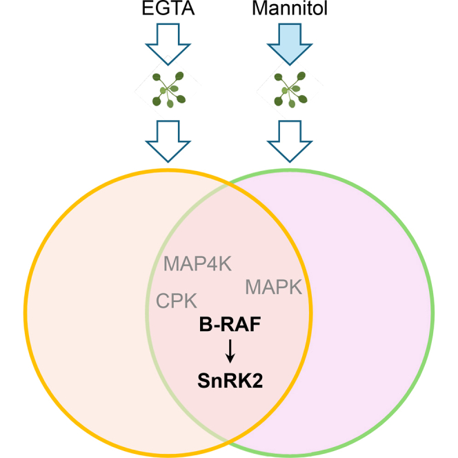
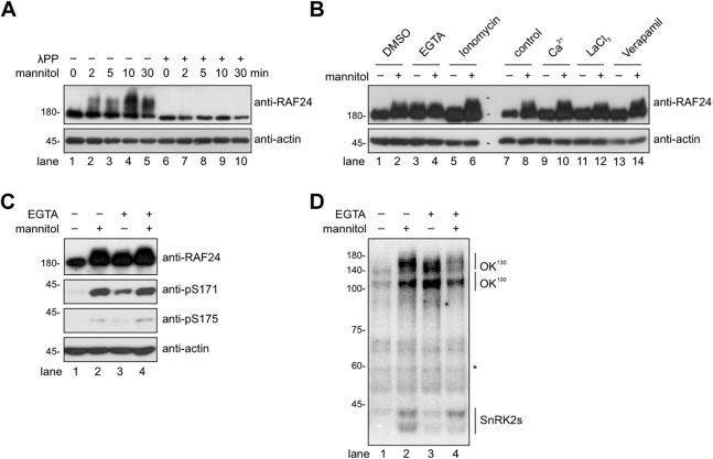
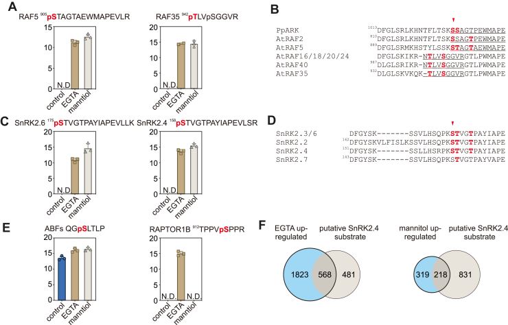
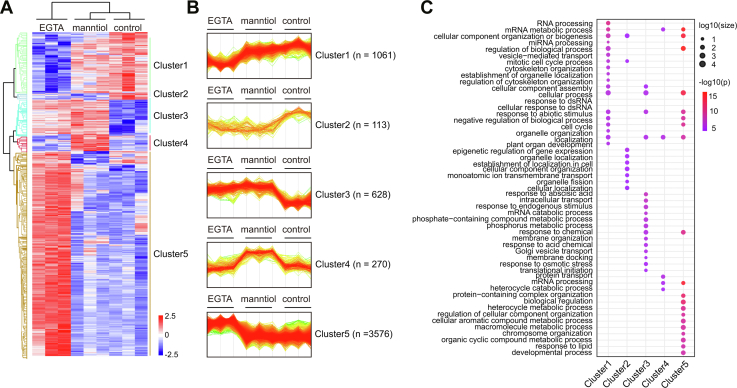
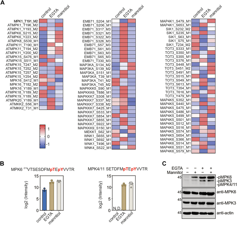
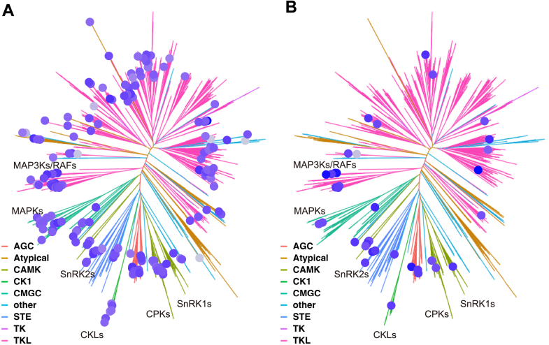
Osmotic stress significantly hampers plant growth and crop yields, emphasizing the need for a thorough comprehension of the underlying molecular responses. Previous research has demonstrated that osmotic stress rapidly induces calcium influx and signaling, along with the activation of a specific subset of protein kinases, notably the Raf-like protein (RAF)-sucrose nonfermenting-1-related protein kinase 2 (SnRK2) kinase cascades within minutes. However, the intricate interplay between calcium signaling and the activation of RAF-SnRK2 kinase cascades remains elusive. Here, in this study, we discovered that Raf-like protein (RAF) kinases undergo hyperphosphorylation in response to osmotic shocks. Intriguingly, treatment with the calcium chelator EGTA robustly activates RAF-SnRK2 cascades, mirroring the effects of osmotic treatment. Utilizing high-throughput data-independent acquisition-based phosphoproteomics, we unveiled the global impact of EGTA on protein phosphorylation. Beyond the activation of RAFs and SnRK2s, EGTA treatment also activates mitogen-activated protein kinase cascades, Calcium-dependent protein kinases, and receptor-like protein kinases, etc. Through overlapping assays, we identified potential roles of mitogen-activated protein kinase kinase kinase kinases and receptor-like protein kinases in the osmotic stress-induced activation of RAF-SnRK2 cascades. Our findings illuminate the regulation of phosphorylation and cellular events by Ca2+ signaling, offering insights into the (exocellular) Ca2+ deprivation during early hyperosmolality sensing and signaling.

Keywords: RAF; SnRK2; calcium signaling; osmotic stress; phosphorylation.


Conflict of interest statement

Conflict of interest The authors declare no competing interests.

Figures

None
Graphical abstract
Fig. 1
Fig. 1
EGTA triggers activation of RAF-SnRK2 cascades.A, the anti-RAF24 immunoblot showing RAF24 hyperphosphorylation after the indicated time of mannitol treatment. The λPP treatment was performed to remove all phosphorylation in the samples. B, the anti-RAF24 immunoblot showing the RAF24 phosphorylation after the treatment of indicated chemicals. C, the immunoblot showing the phosphorylation of RAF24 and conserved serines in SnRK2s corresponding to Ser175 in SnRK2.6. D, in-gel kinase assay showing the SnRK2, OK100 (B2/3 RAF), and OK130 (B4 RAF) activities after 5 min treatment of 800 mM mannitol or 120 min treatment of 20 mM EGTA. Anti-actin immunoblot or the nonspecific band indicated by an asterisk are used as loading controls. The images shown are representative of three independent experiments. λPP, lambda protein phosphatase; SnRK2, sucrose nonfermenting-1–related protein kinase 2.
Fig. 2
Fig. 2
EGTA induces phosphorylation of RAFs and SnRK2s.A, the relative intensity of the phosphopeptides from RAF5 (B3 subgroup) and RAF35 (B4 subgroup) in seedlings without or with EGTA or mannitol treatment (n = 3 biological replicates). N.D., not detected. B, sequence alignment showing the conserved phosphosites in the Arabidopsis RAFs and PpARK. The phosphosites in Arabidopsis RAFs identified by this study and in PpARK/PpCTR1 are highlighted in red, and an arrow indicates the conserved serine residue corresponding to Ser1029 in PpARK/PpCTR1. C, the relative intensity of the conserved phosphosite in SnRK2.6 and SnRK2.4 in seedlings without or with EGTA or mannitol treatment (n = 3 biological replicates). N.D., not detected. D, sequence alignment showing the conserved phosphosites in SnRK2s. The conserved phosphosites are highlighted in red, and the conserved serine residue corresponding to Ser175 in SnRK2.6 is indicated by an arrow. E, the phosphorylation of conserved phosphorylation site in ABFs (left) and Raptor1B in seedlings without or with EGTA or mannitol treatment (n = 3 biological replicates). N.D., not detected. F, venn diagrams showing the overlaps of putative SnRK2.4 substrates (Wang et al., 2020) and EGTA (left) or mannitol (right) upregulated phosphoproteins. ABF, ABRE-binding factor; PpARK, ABA and abiotic stress-responsive Raf-like kinase; SnRK2, sucrose nonfermenting-1–related protein kinase 2.
Fig. 3
Fig. 3
Comparison of EGTA- and mannitol-responsive phosphoproteomics.A, heat map showing the result of the hierarchy clustering analysis of z-score intensities of phosphorylation sites in control, EGTA, or mannitol treatment. B, the profile plot of the five different clusters. C, the bubble matrix plot showing the GO terms enriched in each cluster. GO, gene ontology.
Fig. 4
Fig. 4
MAPK cascade activated by EGTA and mannitol treatment.A, heat map showing the relative abundance of the phosphopeptides in MAPKs, MAPKKs, MAP3Ks, and MAP4Ks phosphoproteomics. The color intensity indicates the z-score intensities. B, the relative intensity of the phosphopeptides from MPK6 and MPK4/11 in seedlings without or with EGTA or mannitol treatment (n = 3 biological replicates). C, the immunoblot showing the EGTA- and mannitol-induced phosphorylation of MAPKs. The protein extract from 10-day-old seedlings with indicated treatments was subjected to immunoblots with anti-pERK1/2, anti-MPK3, and anti-MPK6 antibodies. The anti-actin immunoblot is shown as a protein loading control. The images shown are representative of two independent experiments. MAPK, mitogen-activated protein kinase; MAPKKK, mitogen-activated protein kinase kinase kinase.
Fig. 5
Fig. 5
KinMap showing the EGTA and mannitol upregulated protein kinase.A, the protein kinase induced by EGTA treatment in our phosphoproteomics as shown in the KinMap. B, the protein kinase induced by mannitol treatment in our phosphoproteomics as shown in the KinMap.

References

    1. Gorgues L., Li X., Maurel C., Martinière A., Nacry P. Root osmotic sensing from local perception to systemic responses. Stress Biol. 2022;2:36. - PMC - PubMed
    1. Waadt R., Seller C.A., Hsu P.K., Takahashi Y., Munemasa S., Schroeder J.I. Plant hormone regulation of abiotic stress responses. Nat. Rev. Mol. Cell Biol. 2022;23:680–694. - PMC - PubMed
    1. Wolf S. Cell wall signaling in plant development and defense. Annu. Rev. Plant Biol. 2022;73:323–353. - PubMed
    1. Dong Q., Wallrad L., Almutairi B.O., Kudla J. Ca2+ signaling in plant responses to abiotic stresses. J. Integr. Plant Biol. 2022;64:287–300. - PubMed
    1. Zhu J.K. Abiotic stress signaling and responses in plants. Cell. 2016;167:313–324. - PMC - PubMed

MeSH terms

LinkOut - more resources