. 2024 Aug;37(8):100540.
doi: 10.1016/j.modpat.2024.100540. Epub 2024 Jun 18.

Expression of L1 Cell Adhesion Molecule, a Nephronal Principal Cell Marker, in Nephrogenic Adenoma

Affiliations

Expression of L1 Cell Adhesion Molecule, a Nephronal Principal Cell Marker, in Nephrogenic Adenoma

Rahul Mannan et al. Mod Pathol. 2024 Aug.

Abstract

Nephrogenic adenoma (NA) is a benign, reactive lesion seen predominantly in the urinary bladder and often associated with antecedent inflammation, instrumentation, or an operative history. Its histopathologic diversity can create diagnostic dilemmas and pathologists use morphologic evaluation along with available immunohistochemical (IHC) markers to navigate these challenges. IHC assays currently do not designate or specify NA's potential putative cell of origin. Leveraging single-cell RNA-sequencing technology, we nominated a principal (P) cell-collecting duct marker, L1 cell adhesion molecule (L1CAM), as a potential biomarker for NA. IHC characterization revealed L1CAM to be positive in all 35 (100%) patient samples of NA; negative expression was seen in the benign urothelium, benign prostatic glands, urothelial carcinoma (UCA) in situ, prostatic adenocarcinoma, majority of high-grade UCA, and metastatic UCA. In the study, we also used single-cell RNA sequencing to nominate a novel compendium of biomarkers specific for the proximal tubule, loop of Henle, and distal tubule (DT) (including P and intercalated cells), which can be used to perform nephronal mapping using RNA in situ hybridization and IHC technology. Employing this technique on NA we found enrichment of both the P-cell marker L1CAM and, the proximal tubule type-A and -B cell markers, PDZKI1P1 and PIGR, respectively. The cell-type markers for the intercalated cell of DTs (LINC01187 and FOXI1), and the loop of Henle (UMOD and IRX5), were found to be uniformly absent in NA. Overall, our findings show that based on cell type-specific implications of L1CAM expression, the shared expression pattern of L1CAM between DT P cells and NA. L1CAM expression will be of potential value in assisting surgical pathologists toward a diagnosis of NA in challenging patient samples.

Keywords: L1CAM; cell type–specific marker; immunohistochemistry; kidney; nephrogenic adenoma; urothelial carcinoma.


Conflict of interest statement

Conflict of interest

The authors declare no conflict of interest.

Figures
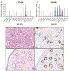
Figure 1.
Figure 1.
Nomination of L1CAM as a biomarker for NA. A&B) Violin plots comparing the expression of L1CAM and GATA3 genes among various epithelial cell types of the human kidney. L1CAM expressed only in PC cells, shows a highly restricted pattern while GATA3 shows broader expression among multiple distal nephron epithelial cell types; C&D) Cortico-medullary region of a kidney showing glomeruli and tubules (H&E) with membranous expression of L1CAM restricted in collecting duct revealed by IHC staining; E&F) Renal pelvis/ureter region of kidney shows L1CAM expression only in the collecting duct and not in the urothelium by IHC. Scale bar = 50 microns
Figure 2.
Figure 2.
Uniform and diffuse expression of L1CAM in NA by IHC. A&D) Two different representative samples of NA showing papillary and cystic features (H&E) with a corresponding high and low magnification of L1CAM IHC in NA specimens with papillary (B&C) and cystic (E&F) morphologies respectively. Scale bar = 50 and 20 microns.
Figure 3.
Figure 3.
Characterization of L1CAM in different NA morphological patterns. The morphological spectrum in the NA study cohort included (A) papillary (D) tubular, (G) flat, and (J) signet ring cell pattern (H&E). Corresponding uniform strong and moderate membranous positivity of L1CAM at low and high magnification B&C, E&F, H&I, and K&L. Scale bar = 100, 50, and 20 microns.
Figure 4.
Figure 4.
Negative expression of L1CAM in non-NA benign and malignant tissues. In corresponding H&E and IHC images, the absence of L1CAM expression in benign prostate and prostatic carcinoma (A-D), benign urothelium (E&F), urothelial carcinoma in situ (G&H), and high-grade urothelial carcinoma (I&J). Scale bar = 50 microns.
Figure 5.
Figure 5.
Focal, patchy, and variable to absent L1CAM IHC expression in clear cell adenocarcinoma (CCAC) and negative expression in adenocarcinoma NOS. H&E and corresponding L1CAM IHC images from clear cell adenocarcinoma (CCAC): urinary bladder CCAC (A-C), urethra CCAC (D-F), classic pattern of ovarian CCAC (G-I) and solid to oxyphilic pattern of ovarian CCAC (J-L), and adenocarcinoma NOS (M-O). Scale bar = 50 and 20 microns.
Figure 6.
Figure 6.
Leveraging the scRNA-Seq data to map human nephron epithelial cell types. A. Bubble plot showing the expression pattern of various genes of interest across 12 distinct epithelial cell types of the normal human kidney nephron- PDZK1IPl (labeling PT-A cell), PIGR (for PT-B cell), and WT1 (for PT-C cell) for PT lineage, IRX5 and UMOD for LOH lineage, FOXI1, and LINC01187 for IC-A and B cells of distal tubule lineage, respectively, and L1CAM as P-cell marker for distal tubule lineage. B. Schematic of normal human nephron mapping the location of epithelial cell types and their marker gene expression pattern.
Figure 7.
Figure 7.
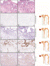
Summary of the expression pattern of biomarker panel used to map the normal human kidney nephron in a representative NA case. Corresponding high and low magnification in a NA case (A&B) showing positive mRNA transcripts of PDZK1IP1 (C&D), PIGR (E&F), and negative WT1 (G&H) by RNA-ISH technology. Scale bar = 100 and 50 microns.
Figure 8.
Figure 8.
Summary of the expression pattern of biomarker panel used to map the normal human kidney nephron in a representative NA case. Corresponding high and low magnification in a NA case (A&B) showing negative expression for loop of Henle marker UMOD (C&D) and IRX5 (E&F), positive P-cell distal tubule marker L1CAM (G&H) with negative IC-type A marker FOXI1 (I&J). Scale bar = 100 and 50 microns.

References

    1. Amin W, Parwani AV. Nephrogenic adenoma. Pathol Res Pract. 2010;206(10):659–662. - PubMed
    1. Li L, Williamson SR, Castillo RP, et al. Fibromyxoid Nephrogenic Adenoma: A Series of 43 Cases Reassessing Predisposing Conditions, Clinical Presentation, and Morphology. Am J Surg Pathol. 2023;47(1):37–46. - PubMed
    1. Santi R, Angulo JC, Nesi G, et al. Common and uncommon features of nephrogenic adenoma revisited. Pathol Res Pract. 2019;215(10):152561. - PubMed
    1. Turcan D, Acikalin MF, Yilmaz E, Canaz F, Arik D. Nephrogenic adenoma of the urinary tract: A 6-year single center experience. Pathol Res Pract. 2017;213(7):831–835. - PubMed
    1. Diolombi M, Ross HM, Mercalli F, Sharma R, Epstein JI. Nephrogenic Adenoma. Am J Surg Pathol. 2013;37(4):532–538. - PubMed

Substances

LinkOut - more resources