. 2024 Jun 20;15(1):5287.
doi: 10.1038/s41467-024-49603-4.

Guiding antibiotics towards their target using bacteriophage proteins

Affiliations

Guiding antibiotics towards their target using bacteriophage proteins

Xinghong Zhao et al. Nat Commun. .

Abstract

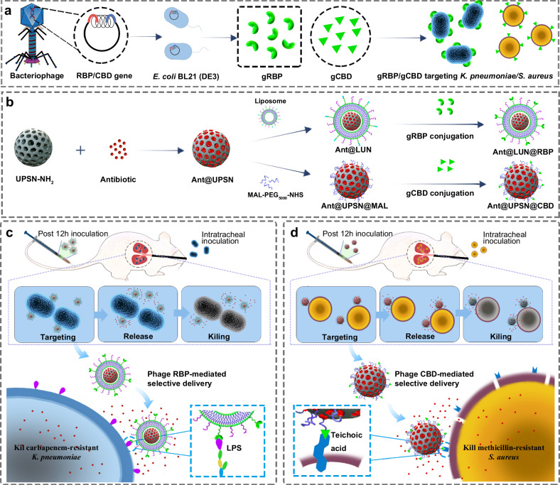
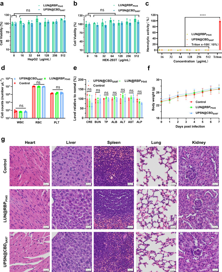
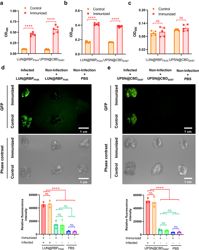
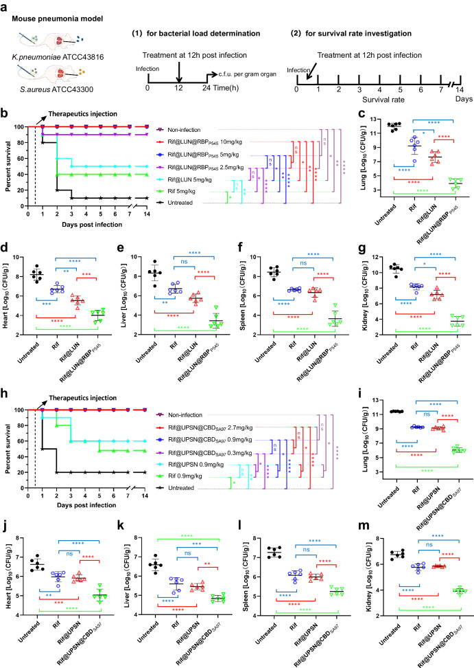
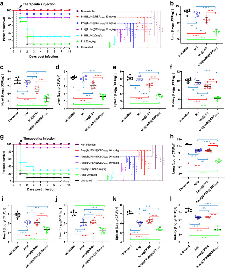
Novel therapeutic strategies against difficult-to-treat bacterial infections are desperately needed, and the faster and cheaper way to get them might be by repurposing existing antibiotics. Nanodelivery systems enhance the efficacy of antibiotics by guiding them to their targets, increasing the local concentration at the site of infection. While recently described nanodelivery systems are promising, they are generally not easy to adapt to different targets, and lack biocompatibility or specificity. Here, nanodelivery systems are created that source their targeting proteins from bacteriophages. Bacteriophage receptor-binding proteins and cell-wall binding domains are conjugated to nanoparticles, for the targeted delivery of rifampicin, imipenem, and ampicillin against bacterial pathogens. They show excellent specificity against their targets, and accumulate at the site of infection to deliver their antibiotic payload. Moreover, the nanodelivery systems suppress pathogen infections more effectively than 16 to 32-fold higher doses of free antibiotics. This study demonstrates that bacteriophage sourced targeting proteins are promising candidates to guide nanodelivery systems. Their specificity, availability, and biocompatibility make them great options to guide the antibiotic nanodelivery systems that are desperately needed to combat difficult-to-treat infections.


Conflict of interest statement

The authors declare no competing interests.

Figures

Fig. 1
Fig. 1. Schematic illustrations of the construction of bacterial targeted antibiotic nanodelivery systems.
a Heterologous expression of targeting proteins by employing bacteriophage origin RBPs and CBDs. b Development of two distinct nanodelivery systems by employing heterologous expressed RBP and CBD as targeting devices: lipid-coated UPSNs (LUN) bearing RBPs (LUN@RBP) and CBDs modified UPSNs (UPSN@CBD). c In a CRKP-induced mouse pneumonia model, antibiotic-loaded LUN@RBP (Ant@LUN@RBP) more effectively suppressed CRKP infections than untargeted antibiotic nanoparticles or of free antibiotics. d In an MRSA-induced mouse pneumonia model, antibiotic-loaded UPSN@CBD (Ant@UPSN@CBD) more effectively suppressed MRSA infections than untargeted antibiotic nanoparticles and free antibiotics.
Fig. 2
Fig. 2. Heterologous expressed gRBPP545 and gCBDSA97 show selective binding to cultured pathogenic bacteria in vitro and home to pathogenic bacteria-infected lungs in vivo.
a Schematic representation of the heterologous expression of gRBPP545 and gCBDSA97. SDS-PAGE Images (b) and anti-His6 western blot (c) of the heterologously expressed gRBPP545 and gCBDSA97. Three times the experiment was repeated with similar results. d Confocal laser scanning microscopy images of CRKP and MRSA after incubation with gRBPP545 and gCBDSA97 (green). Pathogenic bacteria are visualized under a phase contrast model, and bacterial nucleoid is stained with DAPI (blue). Three times the experiment was repeated with similar results. e Time-gated fluorescence images of gRBPP545 in lungs harvested from mice after 30 min of circulation. K. pneumoniae-induced lung infection was generated by intratracheal inoculation of CRKP. At 24 h post-infection, gRBPP545 was intravenously injected and allowed to circulate for 30 min. After that, lungs were harvested for time-gated fluorescence imaging using a FUSION FX7 EDGE Imaging System. Mice without CRKP infection were treated with the same dose of gRBPP545 or the same volume of PBS as controls. Data are presented as mean ± standard deviation (n = 3 biological replicates). The statistical significance of the data was assessed using one-way ANOVA followed by Tukey’s multiple comparisons test. ns, no significance; **p < 0.001. f Time-gated fluorescence image of gCBDSA97 in lungs harvested from mice after 30 min of circulation. S. aureus-induced lung infection was generated by intratracheal inoculation of MRSA. At 24 h post-infection, gCBDSA97 was intravenously injected and allowed to circulate for 30 min. After that, lungs were harvested for time-gated fluorescence imaging using a FUSION FX7 EDGE Imaging System. Mice without MRSA infection were treated with the same dose of gCBDSA97 or the same volume of PBS as controls. Data are presented as mean ± standard deviation (n = 3 biological replicates). The statistical significance of the data was assessed using one-way ANOVA followed by Tukey’s multiple comparisons test. ns, no significance; **p < 0.001. Source data are provided as a Source Data file.
Fig. 3
Fig. 3. Characterization of antibiotic-loaded nanodelivery systems.
a Preparation routes of Rif@LUN@RBPP545 and Rif@UPSN@CBDSA97. b Transmission electron microscope images of UPSN, UPSN-NH2, Rif@UPSN, Rif@UPSN@CBDSA97, Rif@LUN, and Rif@LUN@RBPP545. c Average hydrodynamic size of UPSN, UPSN-NH2, Rif@UPSN, Rif@UPSN@CBDSA97, Rif@LUN, and Rif@LUN@RBPP545 measured by dynamic light scattering. Data are presented as mean ± standard deviation (n = 2 independent experiments). d Surface zeta-potential of UPSN, UPSN-NH2, Rif@UPSN, Rif@UPSN@CBDSA97, Rif@LUN, and Rif@LUN@RBPP545 in ultrapure water. Data are presented as mean ± standard deviation (n = 2 independent experiments). e Confocal laser scanning microscopy images of Rif@LUN@RBPP545 and Rif@UPSN@CBDSA97 in which USPN was labeled with DyLight 633 (red) and the targeting devices were fused with GFP (green). f Release profiles of rifampicin payload from the nanoparticles in PBS at 37 °C. Data are presented as mean ± standard deviation (n = 3 independent experiments). Source data are provided as a Source Data file.
Fig. 4
Fig. 4. Antibiotic-loaded nanodelivery systems selectively bind to pathogenic bacteria and precisely target the sites of infection.
a Confocal laser scanning microscopy images of CRKP and MRSA after incubation with Rif@LUN@RBPP545 and Rif@UPSN@CBDSA97 in which USPN was labeled with DyLight 633 (red) and the targeting devices were fused with GFP (green). Pathogenic bacteria are visualized under a phase contrast model, and bacterial nucleoid is stained with DAPI (blue). Three times the experiment was repeated with similar results. b Time-gated fluorescence image of Rif@LUN@RBPP545, in which RBPP545 was fused with GFP (green), in lungs harvested from mice after 30 min of circulation. K. pneumoniae-induced lung infection was generated by intratracheal inoculation of CRKP. At 24 h post-infection, Rif@LUN@RBPP545 was intravenously injected and allowed to circulate for 30 min. After that, lungs were harvested for time-gated fluorescence imaging using a FUSION FX7 EDGE Imaging System. Mice without CRKP infection were treated with the same dose of Rif@LUN@RBPP545 or the same volume of PBS as controls. Data are presented as mean ± standard deviation (n = 3 biological replicates). The statistical significance of the data was assessed using one-way ANOVA followed by Tukey’s multiple comparisons test. ns, no significance; ****p < 0.001. c Time-gated fluorescence image of Rif@UPSN@CBDSA97, in which CBDSA97 was fused with GFP (green), in lungs harvested from mice after 30 min of circulation. S. aureus-induced lung infection was generated by intratracheal inoculation of MRSA. At 24 h post-infection, Rif@UPSN@CBDSA97 was intravenously injected and allowed to circulate for 30 min. After that, lungs were harvested for time-gated fluorescence imaging using a FUSION FX7 EDGE Imaging System. Mice without MRSA infection were treated with the same dose of Rif@UPSN@CBDSA97 or the same volume of PBS as controls. Data are presented as mean ± standard deviation (n = 3 biological replicates). The statistical significance of the data was assessed using one-way ANOVA followed by Tukey’s multiple comparisons test. ns, no significance; ***p < 0.001. Source data are provided as a Source Data file.
Fig. 5
Fig. 5. Biosafety evaluation of LUN@RBPP545 and UPSN@CBDSA97.
Viability of Hep G2 (a) and HEK-293T (b) after treatment with LUN@RBPP545 or UPSN@CBDSA97 at concentrations ranging from 16 to 512 μg/mL for 24 h. Data are presented as mean ± standard deviation (n = 3 biological replicates). The statistical significance of the data was assessed using one-way ANOVA followed by Tukey’s multiple comparisons test. ns, no significance, vs. untreated cells. c Rabbit erythrocytes were incubated with LUN@RBPP545 or UPSN@CBDSA97 at concentrations ranging from 16 to 512 μg/mL. Their hemolytic activity was assessed by the release of hemoglobin. Cells treated without a tested sample were used as no-lysis control. Cells treated with 10% Triton X-100 were used as complete lysis control. Data are presented as mean ± standard deviation (n = 3 biological replicates). The statistical significance of the data was assessed using one-way ANOVA followed by Tukey’s multiple comparisons test. ****p < 0.001 vs. 10% Triton X-100-treated cells. d Counts of various blood cells 7 days after that of LUN@RBPP545 and UPSN@CBDSA97 administration. WBC, white blood cells; RBC, red blood cells; PLT, platelets. Data are presented as mean ± standard deviation (n = 3 biological replicates). The statistical significance of the data was assessed using one-way ANOVA followed by Tukey’s multiple comparisons test. ns, no significance. e Comprehensive blood chemistry panel taken 7 days after that of LUN@RBPP545 and UPSN@CBDSA97 administration. CRE, creatinine; BUN Blood urea nitrogen; TP Total protein, ALB albumin; ALT alanine transaminase, AST Aspartate transaminase, ALP Alkaline phosphatase. Data are presented as mean ± standard deviation (n = 3 biological replicates). The statistical significance of the data was assessed using one-way ANOVA followed by Tukey’s multiple comparisons test. ns, no significance. f Body weight changes were recorded after treatment with LUN@RBPP545, UPSN@CBDSA97, or PBS over 7 d. Data are presented as mean ± standard deviation (n = 10 biological replicates). g Haematoxylin and eosin staining of histology sections from major organs 7 days after the intravenous administration of LUN@RBPP545 and UPSN@CBDSA97. Scale bars, 20 μm. Independent experiments (n = 3 biological replicates) were performed with similar results. Source data are provided as a Source Data file.
Fig. 6
Fig. 6. LUN@RBPP545 and UPSN@CBDSA97 are re-applicable.
Levels of RBPP545-specific and CBDSA97-specific antibodies, IgG (a), IgM (b), and IgA (c), after immunization of LUN@RBPP545 and UPSN@CBDSA97, respectively, for twice. Antibody titers for RBPP545 and CBDSA97 in mouse serum were determined by ELISA using 1000-fold diluted samples (IgG) or 100-fold diluted samples (IgM and IgA). Data are presented as mean ± standard deviation (n = 5 biological replicates). The statistical significance of the data was assessed using one-way ANOVA followed by Tukey’s multiple comparisons test. ns, no significance; ****p < 0.0001. d Time-gated fluorescence image of LUN@RBPP545, in which RBPP545 was fused with GFP (green), in lungs harvested from mice after 30 min of circulation. LUN@RBPP545 immunized and PBS-treated mice were intravenously inoculated with K. pneumoniae to generate the CRKP-induced lung infection mouse model. At 24 h post-infection, LUN@RBPP545 was intravenously injected and allowed to circulate for 30 min. After that, lungs were harvested for time-gated fluorescence imaging using a FUSION FX7 EDGE Imaging System. Mice without CRKP infection were treated with the same dose of LUN@RBPP545 or the same volume of PBS as controls. Data are presented as mean ± standard deviation (n = 3 biological replicates). The statistical significance of the data was assessed using one-way ANOVA followed by Tukey’s multiple comparisons test. ns, no significance; ****p < 0.0001. e Time-gated fluorescence image of UPSN@CBDSA97, in which CBDSA97 was fused with GFP (green), in lungs harvested from mice after 30 min of circulation. UPSN@CBDSA97 immunized and PBS-treated mice were intravenously inoculated with S. aureus to generate the MRSA-induced lung infection mouse model. At 24 h post-infection, UPSN@CBDSA97 was intravenously injected and allowed to circulate for 30 min. After that, lungs were harvested for time-gated fluorescence imaging using a FUSION FX7 EDGE Imaging System. Mice without MRSA infection were treated with the same dose of UPSN@CBDSA97 or the same volume of PBS as controls. Data are presented as mean ± standard deviation (n = 3 biological replicates). The statistical significance of the data was assessed using one-way ANOVA followed by Tukey’s multiple comparisons test. ns, no significance; ****p < 0.0001. Source data are provided as a Source Data file.
Fig. 7
Fig. 7. In vivo therapeutic efficacies of the two developed distinct bacterial targeted precise antibiotic nanodelivery systems, Rif@LUN@RBPP545 and Rif@UPSN@CBDSA97.
a Scheme of the experimental protocol for the mouse pneumonia models. b Survival rates of mice in the CRKP-induced mouse pneumonia model (n = 10 biological replicates). Survival was analyzed by the Log-rank (Mantel-Cox) test. ns, no significance; *p < 0.05; **p < 0.01; ****p < 0.0001. cg Treated with Rif@LUN@RBPP545 (5 mg/kg) significantly reduced the bacterial load of organs of the CRKP-induced pneumonia mouse relative to equivalent doses of untargeted rifampicin nanoparticles or of free rifampicin. At 24 h post-infection, the mice (n = 6) were euthanized by cervical dislocation. Bacterial loads (Log10 c.f.u. per gram of K. pneumoniae) of the lung (c), heart (d), liver (e), spleen (f), and kidney (g) were counted. Data are presented as mean ± standard deviation (n = 6 biological replicates). The statistical significance of the data was assessed using one-way ANOVA followed by Tukey’s multiple comparisons test. ns, no significance; *p < 0.05; **p < 0.01; ***p < 0.001; ****p < 0.0001. h Survival rates of mice in the MRSA-induced mouse pneumonia model (n = 10 biological replicates). Survival was analyzed by the Log-rank (Mantel-Cox) test. ns, no significance; *p < 0.05; **p < 0.01; ****p < 0.0001. im Treated with Rif@UPSN@CBDSA97 (0.9 mg/kg) significantly reduced the bacterial load of organs of the MRSA-induced pneumonia mouse relative to equivalent doses of untargeted rifampicin nanoparticles or of free rifampicin. At 24 h post-infection, the mice (n = 6) were euthanized by cervical dislocation. Bacterial loads (Log10 c.f.u. per gram of S. aureus) of the lung (i), heart (j), liver (k), spleen (l), and kidney (m) were counted. Data are presented as mean ± standard deviation (n = 6 biological replicates). The statistical significance of the data was assessed using one-way ANOVA followed by Tukey’s multiple comparisons test. ns, no significance; **p < 0.01; ***p < 0.001; ****p < 0.0001. Source data are provided as a Source Data file.
Fig. 8
Fig. 8. In vivo therapeutic efficacies of Imi@LUN@RBPP545 and Amp@UPSN@CBDSA97 against resistant pathogens.
a Survival rates of mice in the CRKP-induced mouse pneumonia model (n = 10 biological replicates). Survival was analyzed by the Log-rank (Mantel-Cox) test. ns, no significance; *p < 0.05; **p < 0.01; ***p < 0.001; ****p <  0.0001. bf Treated with Imi@LUN@RBPP545 (20 mg/kg) significantly reduced the bacterial load of organs of the CRKP-induced pneumonia mouse relative to equivalent doses of untargeted imipenem nanoparticles or of free imipenem. At 24 h post-infection, the mice (n = 6) were euthanized by cervical dislocation. Bacterial loads (Log10 c.f.u. per gram of K. pneumoniae) of the lung (b), heart (c), liver (d), spleen (e), and kidney (f) were counted. Data are presented as mean ± standard deviation (n = 6 biological replicates). The statistical significance of the data was assessed using one-way ANOVA followed by Tukey’s multiple comparisons test. ns, no significance; **p < 0.01; ***p < 0.001; ****p < 0.0001. g Survival rates of mice in the MRSA-induced mouse pneumonia model (n = 10 biological replicates). Survival was analyzed by the Log-rank (Mantel-Cox) test. ns, no significance; **p < 0.01; ***p < 0.001; ****p < 0.0001. hl Treated with Amp@UPSN@CBDSA97 (20 mg/kg) significantly reduced the bacterial load of organs of the MRSA-induced pneumonia mouse relative to equivalent doses of untargeted ampicillin nanoparticles or of free ampicillin. At 24 h post-infection, the mice (n = 6) were euthanized by cervical dislocation. Bacterial loads (Log10 c.f.u. per gram of S. aureus) of the lung (h), heart (i), liver (j), spleen (k), and kidney (l) were counted. Data are presented as mean ± standard deviation (n = 6 biological replicates). The statistical significance of the data was assessed using one-way ANOVA followed by Tukey’s multiple comparisons test. ns, no significance; ***p < 0.001; ****p < 0.0001. Source data are provided as a Source Data file.

References

    1. Andersson DI, et al. Antibiotic resistance: turning evolutionary principles into clinical reality. FEMS Microbiol. Rev. 2020;44:171–188. doi: 10.1093/femsre/fuaa001. - DOI - PubMed
    1. Huemer M, Mairpady Shambat S, Brugger SD, Zinkernagel AS. Antibiotic resistance and persistence—Implications for human health and treatment perspectives. EMBO Rep. 2020;21:e51034. doi: 10.15252/embr.202051034. - DOI - PMC - PubMed
    1. Murray CJL, et al. Global burden of bacterial antimicrobial resistance in 2019: a systematic analysis. Lancet. 2022;399:629–655. doi: 10.1016/S0140-6736(21)02724-0. - DOI - PMC - PubMed
    1. Ayobami O, Brinkwirth S, Eckmanns T, Markwart R. Antibiotic resistance in hospital-acquired ESKAPE-E infections in low-and lower-middle-income countries: a systematic review and meta-analysis. Emerg. Microbes Infect. 2022;11:443–451. doi: 10.1080/22221751.2022.2030196. - DOI - PMC - PubMed
    1. De Oliveira DMP, et al. Antimicrobial resistance in ESKAPE pathogens. Clin. Microbiol. Rev. 2020;33:10–1128. doi: 10.1128/CMR.00181-19. - DOI - PMC - PubMed

LinkOut - more resources