. 2024 Jun 20;15(1):5265.
doi: 10.1038/s41467-024-49611-4.

DYRK1A signalling synchronizes the mitochondrial import pathways for metabolic rewiring

Affiliations

DYRK1A signalling synchronizes the mitochondrial import pathways for metabolic rewiring

Adinarayana Marada et al. Nat Commun. .

Abstract

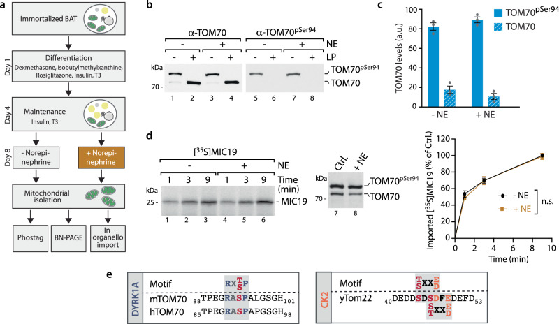
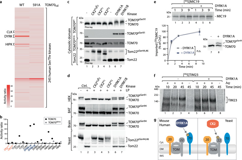
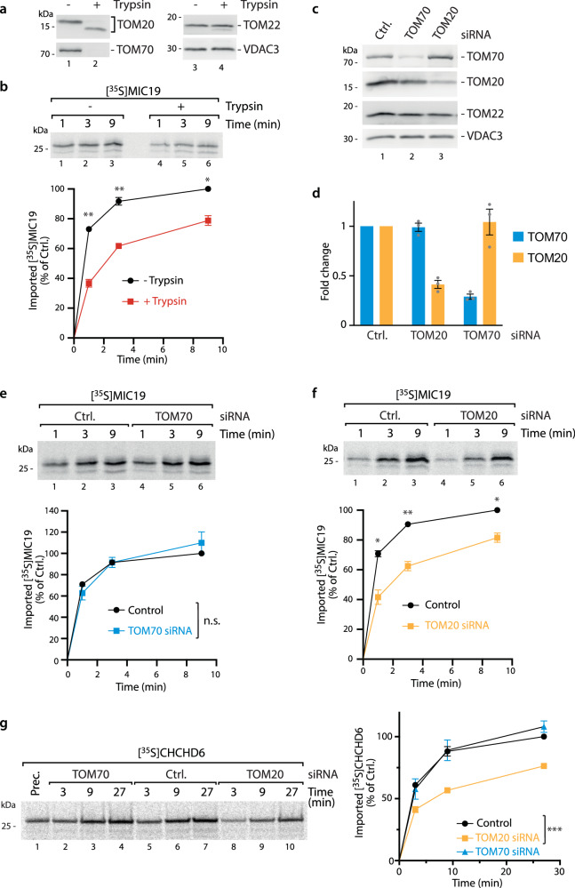
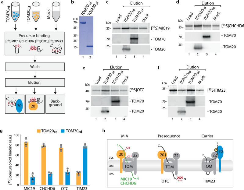
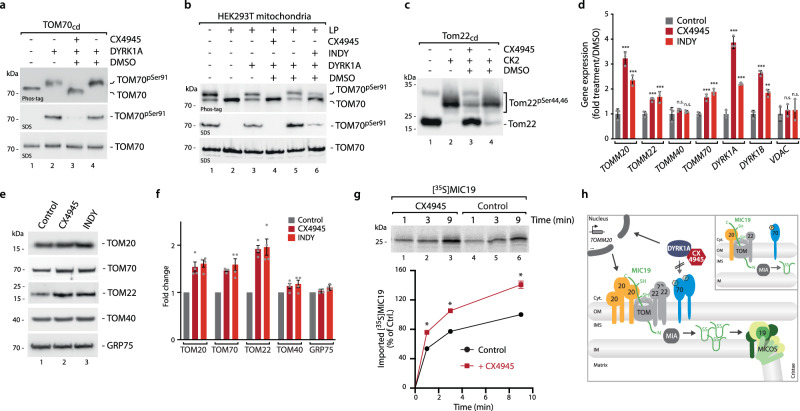
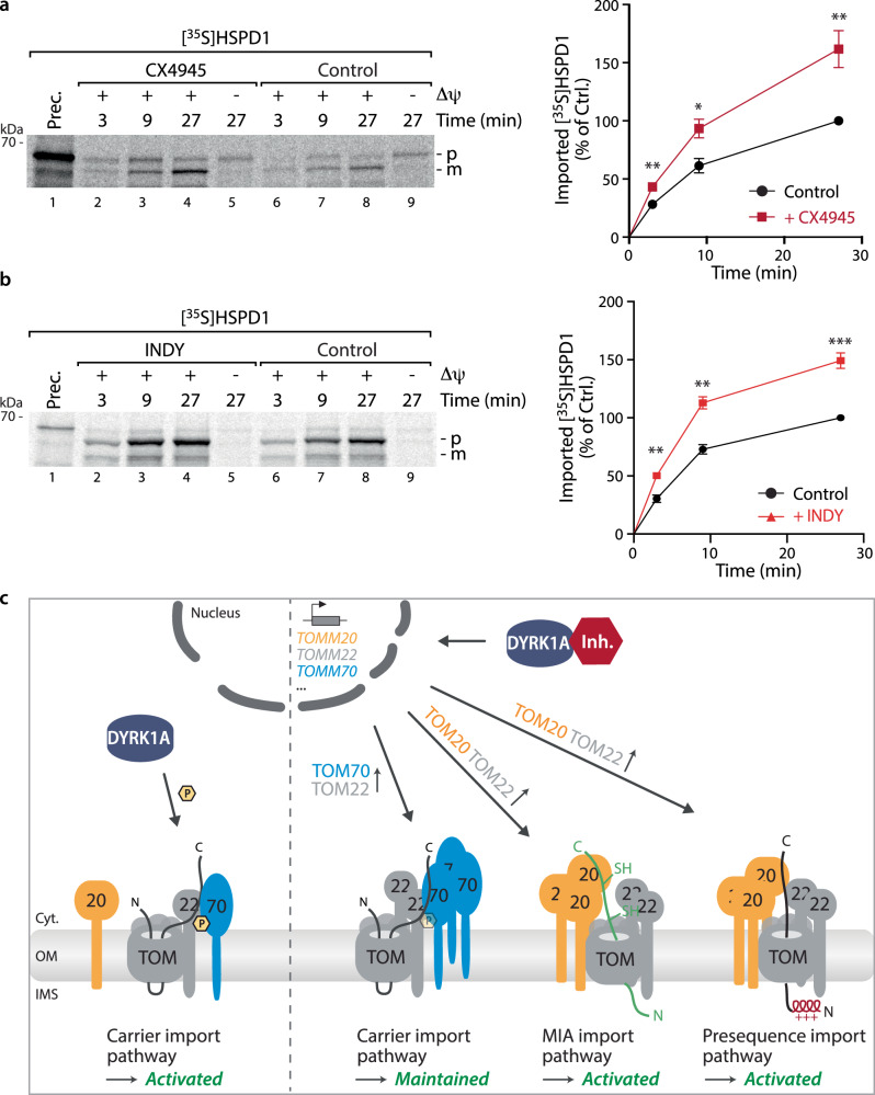
Mitochondria require an extensive proteome to maintain a variety of metabolic reactions, and changes in cellular demand depend on rapid adaptation of the mitochondrial protein composition. The TOM complex, the organellar entry gate for mitochondrial precursors in the outer membrane, is a target for cytosolic kinases to modulate protein influx. DYRK1A phosphorylation of the carrier import receptor TOM70 at Ser91 enables its efficient docking and thus transfer of precursor proteins to the TOM complex. Here, we probe TOM70 phosphorylation in molecular detail and find that TOM70 is not a CK2 target nor import receptor for MIC19 as previously suggested. Instead, we identify TOM20 as a MIC19 import receptor and show off-target inhibition of the DYRK1A-TOM70 axis with the clinically used CK2 inhibitor CX4945 which activates TOM20-dependent import pathways. Taken together, modulation of DYRK1A signalling adapts the central mitochondrial protein entry gate via synchronization of TOM70- and TOM20-dependent import pathways for metabolic rewiring. Thus, DYRK1A emerges as a cytosolic surveillance kinase to regulate and fine-tune mitochondrial protein biogenesis.


Conflict of interest statement

The authors declare no competing interests

Figures

Fig. 1
Fig. 1. β-adrenergic stimulation of differentiated BAT cells does not change TOM70 phosphorylation and MIC19 biogenesis.
a Schematic representation of the experimental strategy to generate differentiated brown adipocyte tissue (BAT) cells and analyze TOM70 phosphorylation and MIC19 biogenesis. Norepinephrine treatment was for 18 h,. b Mitochondria isolated from differentiated primary immortalized BAT cells incubated in the presence or absence of Norepinephrine (NE) as shown in a were analyzed via Phos-tag gels followed by immunodecoration with TOM70 antisera (lanes 1–4) and phospho-specific antibody recognizing TOM70pSer94 (lanes 5-8). Lambda phosphatase (LP) treatment allows distinction of phosphorylated and non-phosphorylated TOM70. c Quantification of phosphorylated and non-phosphorylated TOM70 of samples from b, via Phos-tag electrophoresis. Data represent mean ± SEM from three independent experiments. a.u. arbitrary units. d Import of [35S]MIC19 precursor protein into mitochondria from b. Non-imported precursor was removed by Proteinase K. Samples were analyzed by SDS-PAGE and imported MIC19 was detected by autoradiography. For quantification the control reaction (without NE) at 9 min import time point was set to 100%. Data represent mean ± SEM from three independent experiments. A multiple paired t test with a false discovery rate (FDR) of 1% and a two-stages step-up method of Benjamini, Krieger and Yekutieli was performed to compare between two groups. See the methods section for details on statistical analyses. The TOM70 phosphorylation status was monitored by Phos-tag gels (lanes 7 and 8). n.s. not significant. e Profiling of human TOM70Ser91 and mouse TOM70Ser94 for kinase motifs reveal consensus sites for DYRK1A. Yeast Tom22 has two consensus sites (Ser44/Ser46) for CK2. Source data are provided as a Source Data file.
Fig. 2
Fig. 2. TOM70 is phosphorylated by DYRK1A but not CK2.
a Heatmap of global profiling of TOM70WT and TOM70Ser91Ala receptor domains for phosphorylation activities of 245 Ser/Thr kinases of the human kinome (KinaseFinder assay,). HIPK, CLK and DYRK indicate the members of the DYRK-family. b Detailed comparison of phosphorylation activities for CK2 isoforms and the DYRK family members for TOM70WT and TOM70Ser91Ala receptor domains as shown in a. c In vitro phosphorylation assay of purified receptor domains of human TOM70WT, TOM70Ser91Ala and yeast Tom22 in the presence of indicated kinases. Three different CK2 variants were tested (lanes 2–4). Samples were analyzed via Phos-tag gels and probed with TOM70 or Tom22 antisera or via standard SDS-PAGE and probed with phospho-specific TOM70pSer91 antibody. d Isolated mitochondria from HEK293T cells, yeast, mice brain or brown adipocyte tissue (BAT) were analyzed via Phos-tag gels. For testing phosphorylation with indicated kinases endogenous phosphorylation sites were removed by lambda phosphatase (LP) treatment prior to kinase incubation. Three different CK2 variants were tested (lanes 3–5). e Import of [35S]MIC19 precursor protein into LP treated mitochondria after re-phosphorylation of TOM70Ser91 by DYRK1A (+). (−) mock treated control sample. n.s. not significant. Data were obtained from three biological replicates and represent mean ± SEM. A multiple paired t test with a false discovery rate (FDR) of 1% and a two-stages step-up method of Benjamini, Krieger and Yekutieli was performed to compare between two groups. See the methods section for details on statistical analyses. f Import of [35S]TIM23 precursor protein into mitochondria obtained from e. Import was monitored via autoradiography after BN-PAGE. g Cartoon illustrating targeting of the human, mouse and yeast TOM complex by cytosolic kinases. DYRK1A phosphorylates the human and murine TOM70 receptor at position Ser91 and Ser94, respectively. CK2 targets the yeast Tom22 protein at position Ser44 and Ser46. Source data are provided as a Source Data file.
Fig. 3
Fig. 3. In organello import of human MIA substrates MIC19 and CHCHD6 depend on TOM20 but not TOM70.
a Analysis of mitochondria that were incubated with (+) or without (−) Trypsin after SDS-PAGE and Immunoblotting. b Import of [35S]MIC19 precursor protein into mitochondria obtained from a. For quantification the control reaction (without Trypsin) at 9 min import time point was set to 100%. Data represent mean ± SEM from three independent experiments. A multiple paired t test with a false discovery rate (FDR) of 1% and a two-stages step-up method of Benjamini, Krieger and Yekutieli was performed to compare between two groups. See the methods section for details on statistical analyses. **p < 0.01, and *p < 0.05, n.s. not significant, p > 0.05. c Immunoblots of isolated mitochondria after depletion of indicated TOM receptors via siRNA. Ctrl., non-targeting siRNA. d Quantification of TOM20 and TOM70 knockdown efficiency analyzed in c. Data represent mean ± SEM from three independent experiments. e Import of [35S]MIC19 precursor protein into TOM70 depleted and control (Ctrl.) mitochondria. Quantification was performed as in b. Data represent mean ± SEM from three independent experiments. For statistical analyses see b and methods section. f Import of [35S]MIC19 precursor protein into TOM20 depleted and control (Ctrl.) mitochondria. Quantification was performed as in b. Data represent mean ± SEM from three independent experiments. For statistical analyses see b and methods section. g Import of [35S]CHCHD6 precursor protein into TOM20 and TOM70 depleted and control (Ctrl.) mitochondria. Quantification was performed as in b. Data represent mean ± SEM from four independent experiments. For statistical analyses see b and methods section. Source data are provided as a Source Data file.
Fig. 4
Fig. 4. Human MIC19 and CHCHD6 precursor bind to TOM20 receptor domain.
a Schematic overview of the precursor-receptor binding assay. b Coomassie staining of SDS-PAGE showing the purified receptor domains of human import receptors TOM20 and TOM70. cd, cytosolic domain. c Binding assay for [35S]MIC19 precursor (upper panel) to TOM70cd and TOM20cd (immobilized via decaHis-tag on Ni-NTA beads). Load, loaded radiolabelled precursor; Elution, elution fraction containing released TOM70cd or TOM20cd and radiolabelled precursor in case of specific binding. Mock, same treatment without TOM receptor domains detects non-specific binding. d Binding assay for [35S]CHCHD6 precursor performed as in a. e Binding assay for [35S]OTC precursor performed as in a. f Binding assay for [35S]TIM23 precursor performed as in a. g Quantification of specific precursor binding to TOM20cd or TOM70cd, respectively. Quantification from three independent experiments ± SEM. a.u., arbitrary units. h Cartoon illustrating MIC19 precursor import via TOM20- and TIM23 carrier import via TOM70-associated TOM core complexes. Source data are provided as a Source Data file.
Fig. 5
Fig. 5. CK2 inhibitor CX4945 inhibits DYRK1A leading to a transcriptional response upregulating the mitochondrial import receptors.
a Inhibition of DYRK1A dependent phosphorylation of TOM70 receptor domain in the presence of the CK2 inhibitor CX4945. Upper panel, Phos-tag gel; Middle and lower panel, SDS-PAGE. b Inhibition of DYRK1A-dependent phosphorylation of mitochondrial TOM70 by CX4945 and INDY. Endogenous phosphorylation was removed by lambda phosphatase (LP) before DYRK1A incubation. c Phos-tag gel showing inhibition of CK2-dependent phosphorylation of yeast Tom22 by CX4945. d Analysis of changes of indicated transcript levels upon INDY and CX4945 treatment (12 h) by qRT-PCR. n = 3, data represent mean ± SEM. Statistical analysis was performed using a two-sided Student’s t test to compare between two groups (***p < 0.001; **p < 0.01; n.s. not significant.) Data are representative of three independent experiments. e Analysis of indicated protein levels after in vivo treatment with CX4945 or INDY (12 h) by SDS-PAGE and immunoblotting. f Quantification of indicated proteins from e. Statistical analysis was performed using a two-sided Student’s t test to compare between two groups (Control vs. CX4945 or Control vs. INDY). Data represent mean ± SEM from four independent experiments. *p < 0.05. g Import of [35S]MIC19 precursor protein into mitochondria from cells incubated in the presence or absence of CX4945 for 12 h. For quantification the control reaction at 9 min import time point was set to 100%. A multiple paired t test with a false discovery rate (FDR) of 1% and a two-stages step-up method of Benjamini, Krieger and Yekutieli was performed to compare between two groups. See the methods section for details on statistical analyses. Data represent mean ± SEM from three independent experiments. h Model for stimulation of MIC19 import via CX4945: Import of human MIC19 requires the import receptor TOM20 but is independent of TOM70 (inset). Inhibition of DYRK1A which normally phosphorylates TOM70 for activation of the carrier import pathway triggers a transcriptional response stimulating expression of TOM receptors including the MIC19 receptor TOM20. This stimulates the TOM20-dependent import pathway. Imported MIC19 precursor become trapped in the intermembrane space by oxidation at the MIA machinery. This may then stimulate MICOS biogenesis and ultimately control of cristae structure and respiration. Source data are provided as a Source Data file.
Fig. 6
Fig. 6. DYRK1A signaling links the main mitochondrial import pathways.
a Import of [35S]HSPD1 precursor protein into mitochondria from cells incubated in the presence or absence of the CK2 inhibitor CX4945 for 12 h. p precursor, m mature. For quantification the control reaction at 9 min import time point was set to 100%. Data represent mean ± SEM from three independent experiments. A multiple paired t test with a false discovery rate (FDR) of 1% and a two-stages step-up method of Benjamini, Krieger and Yekutieli was performed to compare between two groups. See the methods section for details on statistical analyses. *p < 0.05; **p < 0.01. b Import of [35S]HSPD1 precursor protein into mitochondria from cells incubated in the presence or absence of the DYRK1A inhibitor INDY for 12 h. Quantification performed as in a. ***p < 0.001. c Schematic showing the role of DYRK1A as central regulatory hub linking the main mitochondrial import routes. While DYRK1A is essential to activate the carrier import pathway via TOM70pSer91 phosphorylation (left panel), its impairment leads to a transcriptional activation of all three TOM import receptors followed by a remodeling of the TOM complex that allows an activation of the MIA- and the presequence import pathways (right panel). Source data are provided as a Source Data file.

Similar articles

Cited by

References

    1. Pagliarini DJ, et al. A mitochondrial protein compendium elucidates complex I disease biology. Cell. 2008;134:112–123. doi: 10.1016/j.cell.2008.06.016. - DOI - PMC - PubMed
    1. Meisinger C, Sickmann A, Pfanner N. The mitochondrial proteome: from inventory to function. Cell. 2008;134:22–24. doi: 10.1016/j.cell.2008.06.043. - DOI - PubMed
    1. Chacinska A, Koehler CM, Milenkovic D, Lithgow T, Pfanner N. Importing mitochondrial proteins: machineries and mechanisms. Cell. 2009;138:628–644. doi: 10.1016/j.cell.2009.08.005. - DOI - PMC - PubMed
    1. Calvo SE, Mootha VK. The mitochondrial proteome and human disease. Annu. Rev. Genomics Hum. Genet. 2010;11:25–44. doi: 10.1146/annurev-genom-082509-141720. - DOI - PMC - PubMed
    1. Nunnari J, Suomalainen A. Mitochondria: in sickness and in health. Cell. 2012;148:1145–1159. doi: 10.1016/j.cell.2012.02.035. - DOI - PMC - PubMed

MeSH terms

Substances