. 2024 Jun 20;15(1):5282.
doi: 10.1038/s41467-024-49637-8.

Multiple roles for hypoxia inducible factor 1-alpha in airway epithelial cells during mucormycosis

Affiliations

Multiple roles for hypoxia inducible factor 1-alpha in airway epithelial cells during mucormycosis

Povilas Kavaliauskas et al. Nat Commun. .

Abstract

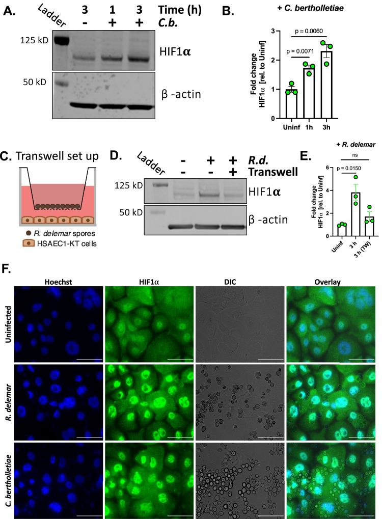
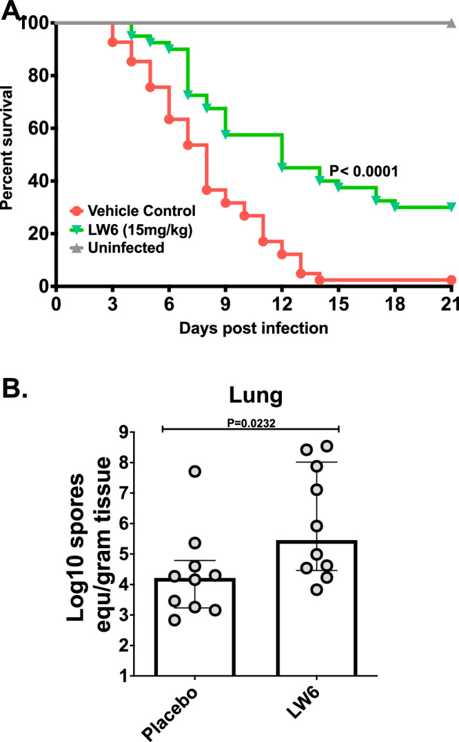
During pulmonary mucormycosis, inhaled sporangiospores adhere to, germinate, and invade airway epithelial cells to establish infection. We provide evidence that HIF1α plays dual roles in airway epithelial cells during Mucorales infection. We observed an increase in HIF1α protein accumulation and increased expression of many known HIF1α-responsive genes during in vitro infection, indicating that HIF1α signaling is activated by Mucorales infection. Inhibition of HIF1α signaling led to a substantial decrease in the ability of R. delemar to invade cultured airway epithelial cells. Transcriptome analysis revealed that R. delemar infection induces the expression of many pro-inflammatory genes whose expression was significantly reduced by HIF1α inhibition. Importantly, pharmacological inhibition of HIF1α increased survival in a mouse model of pulmonary mucormycosis without reducing fungal burden. These results suggest that HIF1α plays two opposing roles during mucormycosis: one that facilitates the ability of Mucorales to invade the host cells and one that facilitates the ability of the host to mount an innate immune response.


Conflict of interest statement

A.S.I. owns shares in Vitalex Biosciences, a start-up company that is developing immunotherapies and diagnostics for mucormycosis. T.J.W. has received grants for experimental and clinical antimicrobial pharmacology, therapeutics, and diagnostics to his institution from Amplyx, Astellas, Gilead, Lediant, Merck, Scynexis, Shionogi, T2 Biosystems, Viosera; and has served as a consultant to Abbott, Astellas, Karyopharm, Leadiant, Partner Therapeutics, Scynexis, Shionogi, Statera, and T2 Biosystems. The remaining authors declare no competing interests.

Figures

Fig. 1
Fig. 1. Interaction of R. delemar with small airway epithelial cells.
A R. delemar invasion of HSAEC1-KT cells was determined with a differential fluorescence assay using 1% Uvitex. Data represents the mean ± SEM of two independent experiments performed in triplicate. n = 6. Two-tailed Student’s t test was used for statistical analysis. B R. delemar-induced damage of HSAEC1-KT cell was determined with a 51Cr release assay. Data represents the mean ± SEM of two independent experiments with several biological replicates per experiment. n = 8 for 6 h, n = 13 for 12 h, n = 12 for 24 h, n = 14 for 36 h, n = 14 for 48 h. Two-tailed Student’s t test was used for statistical analysis to compare the 6 h and 48 h timepoints. C Expression of select genes that have been annotated as a “Cytokine” or other pro-inflammatory genes by Ingenuity Pathway analysis (IPA). Plotted are log transformed TPM values that have been normalized across all 12 samples. Yellow indicates high gene expression; blue indicates low expression. Each columns represents an individual sample. D HSAEC1-KT cells were infected with R. delemar spores for 16 h, after which the levels of IL1α, IL1β, IL8, and GM-CSF protein levels were determined in the culture supernatants by ELISA. Values represent mean ± SEM of 2 experiments, each performed in biological triplicate. n = 6. Two-tailed Student’s t test was used for statistical analysis.
Fig. 2
Fig. 2. Upstream regulator analysis and validation.
A Each regulator was predicted by Ingenuity Pathway Analysis (IPA) to be activated (red, Z-score >2) or repressed (blue, Z-score <2) during infection of HSAEC1-KT cells with R. delemar strain 99-880. Each of the depicted pathways have achieved a Z-score ≥ |3.0| in at least one of the timepoints. The complete analysis is displayed in Supplementary Data 5. White indicates no predicted activation or repression. Red arrows indicate pathways that were previously tied to R. delemar infection. The green arrow indicates a regulator of biological interest that was selected for functional follow-up experiments. Blue arrows indicate signaling pathways that are known to govern innate immune responses. B Representative immunoblot examining phosphorylation of ERK1/2 in response to infection ofHSAEC1-KT cells with R. delemar. C Densitometric analysis of the immunoblot in (B). Data represents the mean ± SEM of three independent experiments performed in singlicate. Two-tailed Student’s t test was used for statistical analysis. D Immunoblot representing HIF1α accumulation from whole cell lysates collected 1 or 3 h post-infection of HSAEC1-KT cells with R. delemar in the presence or absence of HIF1α inhibitor, LW6. E Densitometric analysis of the immunoblot in (D). Data represents the mean ± SEM of three independent experiments performed in singlicate. Two-tailed Student’s t test was used for statistical analysis. ns, not significant.
Fig. 3
Fig. 3. Mucorales infection activates the HIF1α pathway in human airway epithelial cells.
A Immunoblot representing HIF1α accumulation from whole cell lysates collected 1 or 3 h post-infection of HSAEC1-KT cells with C. bertholletiae. B Densitometric analysis of the immunoblot in (A). Data represents the mean ± SEM of three independent experiments performed in singlicate. Two-tailed Student’s t test was used for statistical analysis. C Schematic of transwell experiment. D Immunoblot representing HIF1α accumulation from whole cell lysates collected 3 h post-infection of HSAEC1-KT cells with R. delemar under standard conditions or in a transwell. E Densitometric analysis of the immunoblot in panel D. Data represents the mean ± SEM of three independent experiments performed in singlicate. Two-tailed Student t test was used for statistical analysis. ns, not significant. F HSAEC1-KT cells were infected with R. delemar for 3 h and localization of HIF1α was assessed by indirect immunofluorescence with an anti-HIF1α antibody (green). Hoechst stain was used to visualize nuclei (blue). DIC, differential inference contrast. Scale bars = 100 μm. Microscopy experiments were performed three times independently with similar results.
Fig. 4
Fig. 4. Inhibition of HIF1α signaling in airway epithelial cells.
A Invasion of human small airway epithelial cells 3 h post-infection with R. delemar in the presence or absence of LW6 demonstrated by fluorescent microscopy. Values represent mean ± SEM of two independent experiments performed with 5 biological replicates each time. n = 10. Two-tailed Student’s t test was used for statistical analysis. B Immunoblot representing depletion of HIF1α by siRNA knockdown. HSAEC1-KT cells were exposed to siRNAs for 48 h prior to infection, which was allowed to progress for 3 h. Experiment was performed two times independently with similar results. C Invasion of human small airway epithelial cells lacking HIF1α 3hrs post-infection with R. delemar demonstrated by fluorescent microscopy. Values represent mean ± SEM of two independent experiments performed with 8 biological replicates each time. n = 16. Two-tailed Student t-test was used for statistical analysis. NS, not significant. D Expression of select LW6-sensitive genes following 3 h of HSAEC1-KT cells infection with R. delemar in the presence or absence of HIF1α inhibitor, LW6. Plotted are log transformed TPM values that have been normalized across all 9 samples. Yellow indicates high gene expression; blue indicates low expression. Each column represents an individual sample. E mRNA levels for select genes determined by qRT-PCR performed on total RNA isolates. Data represents the mean ± SEM of three independent experiments performed in singlicate and independent of those depicted in panel (D). R.d., R. delemar. Two-tailed Student t-test was used for statistical analysis.
Fig. 5
Fig. 5. R. delemar infection induces HIF1α accumulation in infected lungs.
Indirect immunofluorescence staining was performed on lung sections from uninfected mice (A), mice infected with R. delemar (B), and mice infected with R. delemar and treated with LW6 (C). Staining was performed with antibodies raised against HIF1α (red). The nuclei were stained with DAPI (blue). Scale bars = 50 μm; 10 μm in magnified images). Magnification = 8X. Experiment was performed two times independently with similar results.
Fig. 6
Fig. 6. LW6 protects mice from pulmonary mucormycosis.
(A Survival of neutropenic mice (40 per group from three experiments) infected intratracheally with R. delemar (average inoculum of 1.1×10^4 spores per mouse) and treated with vehicle control (placebo) or 15 mg/kg LW6 24 h post infection for 4 consecutive days. (B) Inhibition of the HIF1α by LW6 did not affect the fungal burden in the lungs of mice harvested on Day +4 post infection. Data from one experiment (10 mice per group) and presented as median + interquartile range. Statistical analysis was performed by using Mann-Whitney non-parametric (two-tailed) test comparing treated groups vs. placebo group.

References

    1. Prakash, H. & Chakrabarti, A. Global Epidemiology of MucormycosisJ. Fungi (Basel)5 (2019). - PMC - PubMed
    1. Ribes JA, Vanover-Sams CL, Baker DJ. Zygomycetes in human disease. Clin. Microbiol. Rev. 2000;13:236–301. doi: 10.1128/CMR.13.2.236. - DOI - PMC - PubMed
    1. Roden MM, et al. Epidemiology and outcome of zygomycosis: a review of 929 reported cases. Clin. Infect. Dis. 2005;41:634–653. doi: 10.1086/432579. - DOI - PubMed
    1. Spellberg B, Edwards J, Jr., Ibrahim A. Novel perspectives on mucormycosis: pathophysiology, presentation, and management. Clin. Microbiol. Rev. 2005;18:556–569. doi: 10.1128/CMR.18.3.556-569.2005. - DOI - PMC - PubMed
    1. Skiada A, et al. Zygomycosis in Europe: analysis of 230 cases accrued by the registry of the European Confederation of Medical Mycology (ECMM) Working Group on Zygomycosis between 2005 and 2007. Clin. Microbiol. Infect. 2011;17:1859–1867. doi: 10.1111/j.1469-0691.2010.03456.x. - DOI - PubMed

MeSH terms

Substances