. 2024 Jun 20;14(1):12680.
doi: 10.1038/s41598-024-62820-7.

In vivo brain estrogen receptor density by neuroendocrine aging and relationships with cognition and symptomatology

Affiliations

In vivo brain estrogen receptor density by neuroendocrine aging and relationships with cognition and symptomatology

Lisa Mosconi et al. Sci Rep. .

Abstract

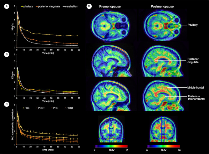
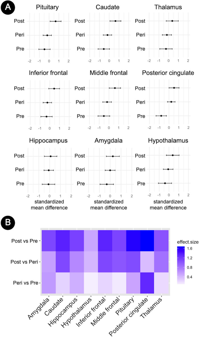
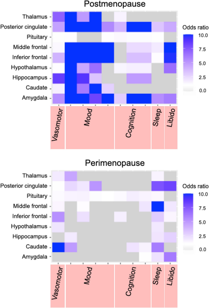
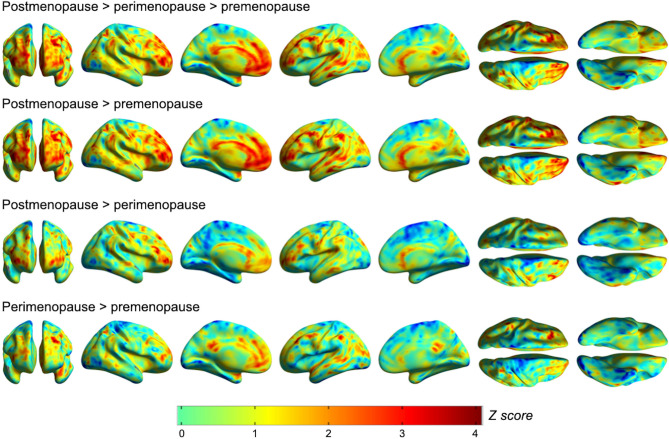
17β-estradiol, the most biologically active estrogen, exerts wide-ranging effects in brain through its action on estrogen receptors (ERs), influencing higher-order cognitive function and neurobiological aging. However, our knowledge of ER expression and regulation by neuroendocrine aging in the living human brain is limited. This in vivo brain 18F-fluoroestradiol (18F-FES) Positron Emission Tomography (PET) study of healthy midlife women reveals progressively higher ER density over the menopause transition in estrogen-regulated networks. Effects were independent of age, plasma estradiol and sex hormone binding globulin, and were highly consistent, correctly classifying all women as being postmenopausal or premenopausal. Higher ER density in target regions was associated with poorer memory performance for both postmenopausal and perimenopausal groups, and predicted presence of self-reported mood and cognitive symptoms after menopause. These findings provide novel insights on brain ER density modulation by female neuroendocrine aging, with clinical implications for women's health.


Conflict of interest statement

The authors declare no competing interests.

Figures

Figure 1
Figure 1
Brain [18F]Fluoroestradiol time activity curves and distribution. Left panel: Time-activity curves (TACs) of 18F-Fluoroestradiol (18F-FES) in target estrogen receptor (ER)-rich brain regions. Representative TACs are shown in pituitary, posterior cingulate and cerebellum from representative (A) postmenopausal and (B) premenopausal participants. (C) Mean TACs (standard errors) in pituitary and posterior cingulate normalized by cerebellar measures are displayed for the entire postmenopausal (squares) and the premenopausal (circles) groups. Right panel: 18F-FES PET images (summed frames over 30–90 min, pseudo-colored using a rainbow spectrum scale) overlaid on the coregistered structural MRI of the two representative premenopausal and postmenopausal participants in (A) and (B). PET images are scaled identically, with a range of 0–16 standardized uptake values (SUV) for each participant (see color-coded scales), depicting higher tracer uptake in ER-rich regions in the postmenopausal participant. From top to bottom, 18F-FES PET images are displayed in the axial, sagittal and coronal views at the level of pituitary, posterior cingulate, frontal cortex and thalamus. Comparatively low to negligible uptake is evident in the lateral inferior cerebellar gray matter, which was used as the reference region for quantification. Note that tracer uptake is also present in portions of the cortical and cerebellar white matter, as well as corpus callosum and brainstem, which was generally higher in postmenopausal than in premenopausal women across the entire dataset. However, since 18F-FES white matter uptake is predominantly non-specific,, white matter regions were excluded from our statistical analysis.
Figure 2
Figure 2
Regional brain estrogen receptor density by menopause stage. (A) Forest plots showing standardized brain 18F-FES distribution volume ratios (DVR) in pre-specified regions of interest by menopausal status, adjusted by age, plasma estradiol (E2) and sex hormone binding globulin (SHBG). Error bars are 95% confidence intervals. (B) Heatmaps showing standardized pairwise mean regional 18F-FES DVR differences between menopause statuses expressed as Cohen’s d coefficients, where d ≥ 0.8 reflects a large effect size. Abbreviations: Peri, perimenopause; Post, postmenopause; Pre, premenopause.
Figure 3
Figure 3
Associations between brain estrogen receptor density and menopause symptoms. Heatmaps show associations between 18F-FES distribution volume ratios (DVR), reflecting estrogen receptor (ER) density, and presence of self-reported menopause symptoms by domain. From left to right, vasomotor symptoms (hot flashes, night sweats), mood symptoms (low mood, mood fluctuations, tearfulness, irritability), cognition (reduced focus, memory complaints, brain fog), disturbed sleep, and low libido. Multivariable-adjusted odds ratios (OR) are displayed on a purple-to-grey color-coded scale, with purple indicating positive associations and grey indicating neutral associations. Estimates are presented separately for the postmenopausal and perimenopausal groups.
Figure 4
Figure 4
Voxel-wise analysis of brain estrogen receptor density by menopause stage. Surface statistical parametric maps (SPMs) of voxel-wise differences in parametric 18F-FES binding potential images, reflecting estrogen receptor (ER) density, between premenopausal, perimenopausal and postmenopausal groups, at P < 0.05 cluster-level corrected for family-type wise error (FWE), adjusted by age, plasma estradiol (E2) and sex hormone binding globulin (SHBG). SPMs are represented on a spectrum color-coded scale with corresponding Z scores. Corresponding statistics are reported in Supplementary Table 1.

References

    1. McEwen B. Estrogen actions throughout the brain. Recent Prog. Horm. Res. 2002;57:357–384. doi: 10.1210/rp.57.1.357. - DOI - PubMed
    1. McEwen BS, Alves SE, Bulloch K, Weiland NG. Ovarian steroids and the brain: Implications for cognition and aging. Neurology. 1997;48:8S–15S. doi: 10.1212/WNL.48.5_Suppl_7.8S. - DOI - PubMed
    1. Brinton RD, Yao J, Yin F, Mack WJ, Cadenas E. Perimenopause as a neurological transition state. Nat. Rev. Endocrinol. 2015;11:393–405. doi: 10.1038/nrendo.2015.82. - DOI - PMC - PubMed
    1. Arevalo MA, Azcoitia I, Garcia-Segura LM. The neuroprotective actions of oestradiol and oestrogen receptors. Nat. Rev. Neurosci. 2015;16:17–29. doi: 10.1038/nrn3856. - DOI - PubMed
    1. Heldring N, et al. Estrogen receptors: How do they signal and what are their targets. Physiol. Rev. 2007;87:905–931. doi: 10.1152/physrev.00026.2006. - DOI - PubMed

LinkOut - more resources