. 2024 Aug;20(8):991-999.
doi: 10.1038/s41589-024-01642-0. Epub 2024 Jun 20.

Rationally seeded computational protein design of ɑ-helical barrels

Affiliations

Rationally seeded computational protein design of ɑ-helical barrels

Katherine I Albanese et al. Nat Chem Biol. 2024 Aug.

Abstract

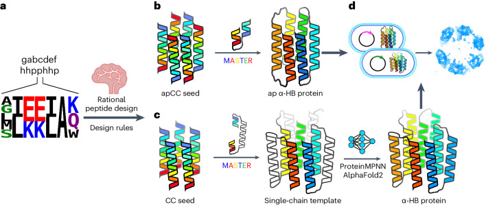
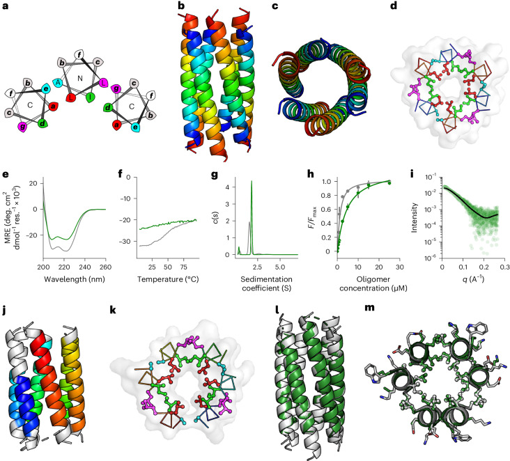
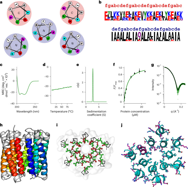
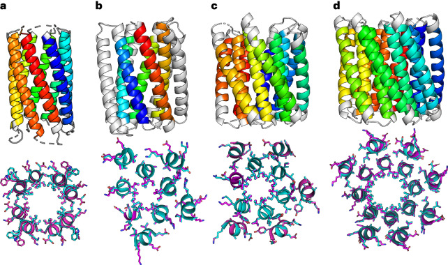
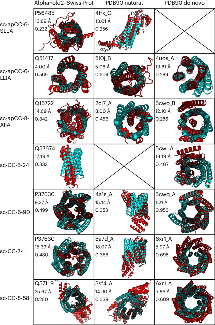
Computational protein design is advancing rapidly. Here we describe efficient routes starting from validated parallel and antiparallel peptide assemblies to design two families of α-helical barrel proteins with central channels that bind small molecules. Computational designs are seeded by the sequences and structures of defined de novo oligomeric barrel-forming peptides, and adjacent helices are connected by loop building. For targets with antiparallel helices, short loops are sufficient. However, targets with parallel helices require longer connectors; namely, an outer layer of helix-turn-helix-turn-helix motifs that are packed onto the barrels. Throughout these computational pipelines, residues that define open states of the barrels are maintained. This minimizes sequence sampling, accelerating the design process. For each of six targets, just two to six synthetic genes are made for expression in Escherichia coli. On average, 70% of these genes express to give soluble monomeric proteins that are fully characterized, including high-resolution structures for most targets that match the design models with high accuracy.


Conflict of interest statement

R.P. is a South West Biosciences Collaborative Awards in Science and Engineering student supported by Rosa Biotech. D.A.S. is a cofounder and was an employee at Rosa Biotech from 2019 to 2024. D.N.W. is a cofounder and director of Rosa Biotech. All other authors have no competing interests.

Figures

Fig. 1
Fig. 1. Pipeline for rationally seeded computational design of de novo protein folds.
a, Robust sequence-to-structure relationships for coiled-coil oligomers were used as rules to seed the design of new protein scaffolds. b,c, Antiparallel (b) and parallel (c) α-helical barrel protein design targets. For both targets, MASTER, was used to search known experimental protein structures for segments with the potential to connect adjacent helices and generate single-chain models. For the antiparallel designs (b), the sequences and structures of identified short connectors were used directly. However, the parallel targets required longer structured loops (c), for which we targeted helix–turn–helix–turn–helix motifs. ProteinMPNN and AlphaFold2 (refs. ,) were then used iteratively to optimize the sequences and models of these three-helix bundle motifs. d, For each design, a small number of synthetic genes were made and expressed in E. coli for biophysical and structural characterization. Peptide and protein chains are shown in chainbows from the N termini to the C termini (blue to red), except for the initially placed central helices of the helix–turn–helix–turn–helix motifs in the parallel designs, which are shown in white. α-HB, α-helical barrel.
Fig. 2
Fig. 2. Biophysical and structural characterization of the apCC-Hex peptide and the sc-apCC-6-LLIA protein.
a, Helical-wheel representation of part of an antiparallel α-helical barrel highlighting the ag heptad repeats: red, a sites; green, d sites; magenta, g sites; and cyan, e sites; N and C labels refer to the termini of the helices closest to the viewer. bd, X-ray crystal structure (1.4-Å resolution) of apCC-Hex (PDB ID, 8QAB). Coiled-coil regions identified by Socket2 (ref. ) (packing cutoff, 7.0 Å) are colored as chainbows from N termini to C termini (blue to red) (b,c). d, A slice through the structure of a heptad repeat with KIH packing colored the same as in the helical wheel in a. eh, Comparison of the biophysical data for the apCC-Hex α-helical barrel peptide (gray) and the sc-apCC-6-LLIA α-helical barrel protein (green). Circular dichroism spectra were recorded at 5 °C (e). f, Thermal responses of the α-helical circular dichroism signal at 222 nm. g, AUC sedimentation velocity data at 20 °C are fitted to a single-species model; fits returned a peptide assembly of 18.7 kDa (hexamer) and a protein of 24.0 kDa (monomer). h, Fitted data for DPH binding to the peptide and protein; fits returned dissociation constant (Kd) values of 0.8 ± 0.3 µM and 4.0 ± 0.4 µM, respectively. Fitted data are the mean and s.d. of three independent repeats. i, SEC-SAXS data for sc-apCC-6-LLIA fitted using FoXS, to an AlphaFold2 model of the design (χ2 = 1.50). j, X-ray crystal structure (2.25 Å) of sc-apCC-6-LLIA (PDB ID, 8QAD) with coiled-coil regions identified by Socket2 (ref. ) (packing cutoff, 7.0 Å) colored as chainbows. k, A slice through the structure of a heptad repeat showing KIH packing, colored as in a. l,m, Overlays of the experimental apCC-Hex (gray) and sc-apCC-6-LLIA protein (green) structures (RMSD for backbone atoms (RMSDbb) = 1.177 Å). The conditions were as follows: circular dichroism spectroscopy, 50 µM peptide, 10 µM protein in PBS, pH 7.4; AUC, 100 µM peptide, 15 µM protein in PBS, pH 7.4; DPH binding, oligomer concentration was 0–30 µM peptide, 0–30 µM protein in PBS, pH 7.4, 20 °C, final concentration was 1 µM DPH (5% v/v DMSO); SEC-SAXS, 10 mg ml−1 protein in PBS, pH 7.4. deg., degrees; MRE, mean residue ellipticity; res., residue. Source data
Fig. 3
Fig. 3. Biophysical and structural characterisation of sc-CC-7 de novo proteins.
a, Helical-wheel representation for part of a parallel single-chain α-helical barrel showing KIH packing for the buttressing helices (shaded red) and the inner barrel (shaded blue): red, a sites; green, d sites; magenta, g sites; and cyan, e sites; N and C labels refer to the termini of the helices closest to the viewer. b, Sequence pileups and registers for the inner (blue register) and buttressing (red register) helices of sc-CC-7-LI. c,d, Circular dichroism spectrum recorded at 5 °C (c) and thermal-response curve (d) for sc-CC-7-LI. e, AUC sedimentation velocity data for sc-CC-7-LI fitted to a single-species model, which returned MW = 37.4 kDa (monomer). f, Fitted binding data of DPH to sc-CC-7-LI, which returned Kd = 3.8 ± 0.8 µM. Fitted data are the mean and s.d. of three independent repeats. g, SEC-SAXS data fitted using the final AlphaFold2 model and FoXS (χ2 = 1.43),. h, X-ray crystal structure of sc-CC-7-LI at a 2.5-Å resolution (PDB ID, 8QAI). Coiled-coil regions identified by Socket2 (ref. ) (packing cutoff, 7 Å) are colored as chainbows from N termini to C termini (blue to red). i, A slice through the structure of a heptad repeat showing KIH packing with a-type (red) and d-type (green) knobs. j, Overlay of the middle helical turns from the sc-CC-7-LI structure (cyan) and the final AlphaFold2 model (magenta) (RMSDbb = 0.433 Å). The conditions were as follows: circular dichroism spectroscopy, 5 µM protein in PBS, pH 7.4; AUC, 25 µM protein in PBS, pH 7.4; DPH binding, 0–24 µM protein in PBS, pH 7.4, final concentration was 0.5 µM DPH (5% v/v DMSO); SEC-SAXS, 10 mg ml−1 protein in PBS, pH 7.4. Source data
Fig. 4
Fig. 4. Structural characterization of five-helix, six-helix and eight-helix targets.
ad, Top, X-ray crystal structures of sc-apCC-8 at a 2.0-Å resolution (PDB ID, 8QAF) (a), sc-CC-5 at a 1.9-Å resolution (PDB ID, 8QKD) (b), sc-CC-6-95 at a 2.8-Å resolution (PDB ID, 8QAG) (c) and sc-CC-8-58 at a 2.35-Å resolution (PDB ID, 8QAH) (d). Coiled-coil regions identified by Socket2 (ref. ) (packing cutoff, 7.5 Å for sc-apCC-8, sc-CC-5-24, sc-CC-6-95 and sc-CC-8-58 at 7.0 Å) are colored as chainbows from N termini (blue) to C termini (red). Bottom, overlays for the middle helical turns of each crystal structure (cyan) and the corresponding AlphaFold2 (refs. ,) model (magenta); RMSDbb = 0.413 Å (a), RMSDbb = 0.371 Å (b), RMSDbb = 0.300 Å (c) and RMSDbb = 0.530 Å (d). Source data
Fig. 5
Fig. 5. Comparison of de novo α-helical barrel proteins against existing and predicted protein folds.
Foldseek was used for this comparison. Each de novo α-helical barrel protein structure determined in this study (cyan) is overlaid with the top match from the AlphaFold2–Swiss-Prot database,, and natural and de novo sequences from the PDB, (red). Within each box, the top value is the ID of the matched structure, the middle value is the backbone RMSD between the query and match, and the bottom value is the template modeling score between the two structures. Source data

References

    1. Korendovych, I. V. & DeGrado, W. F. De novo protein design, a retrospective. Q. Rev. Biophys.53, e3 (2020). - PMC - PubMed
    1. Pan, X. & Kortemme, T. Recent advances in de novo protein design: principles, methods, and applications. J. Biol. Chem.296, 100558 (2021). - PMC - PubMed
    1. Woolfson, D. N. A brief history of de novo protein design: minimal, rational, and computational. J. Mol. Biol.433, 167160 (2021). - PubMed
    1. Dawson, W. M., Rhys, G. G. & Woolfson, D. N. Towards functional de novo designed proteins. Curr. Opin. Chem. Biol.52, 102–111 (2019). - PubMed
    1. Ovchinnikov, S. & Huang, P.-S. Structure-based protein design with deep learning. Curr. Opin. Chem. Biol.65, 136–144 (2021). - PMC - PubMed

Publication types