. 2024 Jun 21;19(1):48.
doi: 10.1186/s13062-024-00491-0.

PNSC928, a plant-derived compound, specifically disrupts CtBP2-p300 interaction and reduces inflammation in mice with acute respiratory distress syndrome

Affiliations

PNSC928, a plant-derived compound, specifically disrupts CtBP2-p300 interaction and reduces inflammation in mice with acute respiratory distress syndrome

Fan Li et al. Biol Direct. .

Abstract

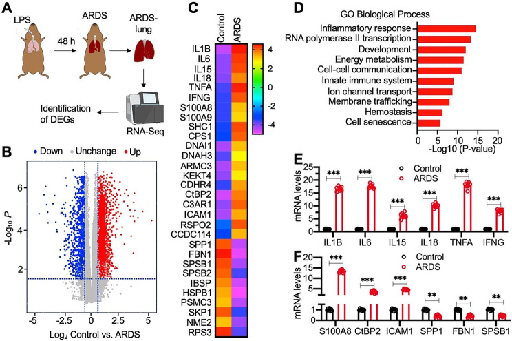
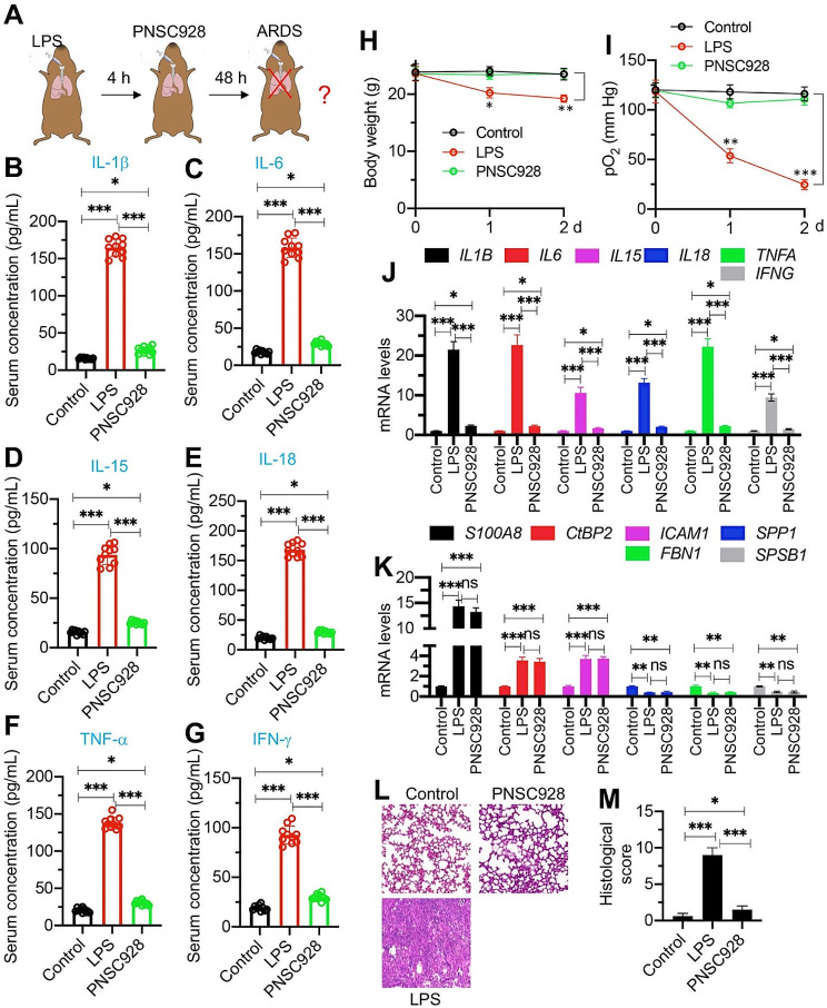
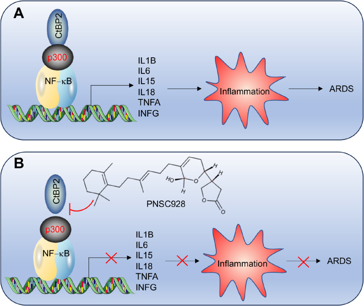
Background: Prior research has highlighted the involvement of a transcriptional complex comprising C-terminal binding protein 2 (CtBP2), histone acetyltransferase p300, and nuclear factor kappa B (NF-κB) in the transactivation of proinflammatory cytokine genes, contributing to inflammation in mice with acute respiratory distress syndrome (ARDS). Nonetheless, it remains uncertain whether the therapeutic targeting of the CtBP2-p300-NF-κB complex holds potential for ARDS suppression.

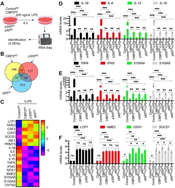
Methods: An ARDS mouse model was established using lipopolysaccharide (LPS) exposure. RNA-Sequencing (RNA-Seq) was performed on ARDS mice and LPS-treated cells with CtBP2, p300, and p65 knockdown. Small molecules inhibiting the CtBP2-p300 interaction were identified through AlphaScreen. Gene and protein expression levels were quantified using RT-qPCR and immunoblots. Tissue damage was assessed via histological staining.

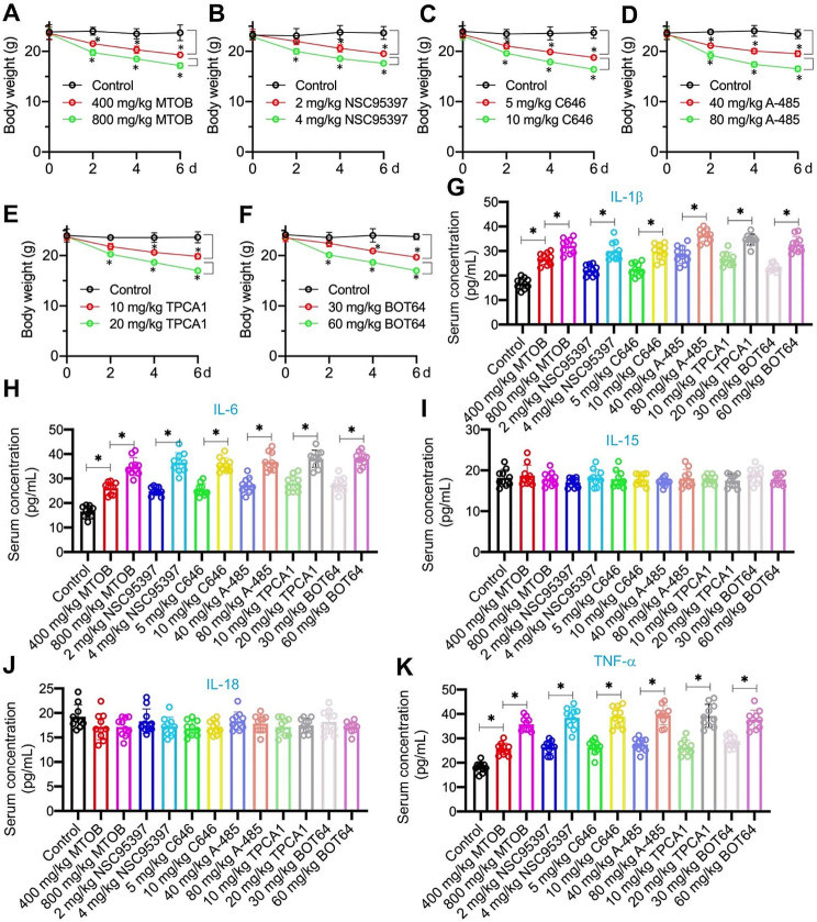
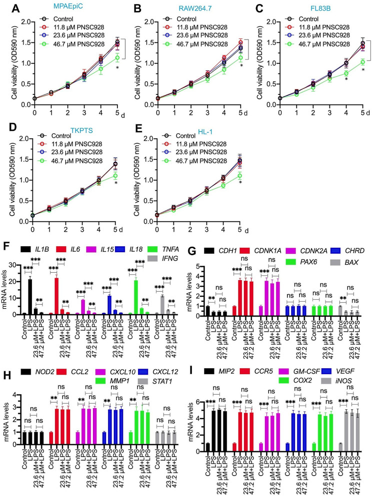
Key findings: We elucidated the specific role of the CtBP2-p300-NF-κB complex in proinflammatory gene regulation. RNA-seq analysis in LPS-challenged ARDS mice and LPS-treated CtBP2-knockdown (CtBP2KD), p300KD, and p65KD cells revealed its significant impact on proinflammatory genes with minimal effects on other NF-κB targets. Commercial inhibitors for CtBP2, p300, or NF-κB exhibited moderate cytotoxicity in vitro and in vivo, affecting both proinflammatory genes and other targets. We identified a potent inhibitor, PNSC928, for the CtBP2-p300 interaction using AlphaScreen. PNSC928 treatment hindered the assembly of the CtBP2-p300-NF-κB complex, substantially downregulating proinflammatory cytokine gene expression without observable cytotoxicity in normal cells. In vivo administration of PNSC928 significantly reduced CtBP2-driven proinflammatory gene expression in ARDS mice, alleviating inflammation and lung injury, ultimately improving ARDS prognosis.

Conclusion: Our results position PNSC928 as a promising therapeutic candidate to specifically target the CtBP2-p300 interaction and mitigate inflammation in ARDS management.

Keywords: ARDS; CtBP2; PNSC928; Proinflammatory cytokine genes; p300.


Conflict of interest statement

The authors declare no competing interests.

Figures

Fig. 1
Fig. 1
Proinflammatory cytokine genes were predominately upregulated in ARDS mice. (A) Experimental design: RNA-Seq analysis to identify differentially expressed genes (DGEs) in ARDS mice. (B) Volcano plot displaying RNA-Seq results. (C) Heatmap of top 20 upregulated genes and top 10 downregulated genes in ARDS lung tissues. (D) Biological processes of DGEs revealed by gene ontology (GO) analysis. (E and F) Verification of 12 DGEs by RT-qPCR analysis (n = 3). (E) IL-1B, IL-6, IL-15, IL-18, TNFA, and IFNG. (F) S100A8, CtBP2, ICAM1, SPP1, FBN1, and SPSB1. **P < 0.01; ***P < 0.001
Fig. 2
Fig. 2
Proinflammatory cytokine genes were downregulated in knockdown cell lines of CtBP2-p300-NF-κB complex members. (A) Experimental design: RNA-Seq analysis to DGEs in LPS-challenged ControlKD, CtBP2KD, p300KD, and p65KD cells. (B) Venn diagram illustrating the altered gene profiles in LPS-challenged ControlKD, CtBP2KD, p300KD, and p65KD cells. (C) Heatmap displaying the top 7 upregulated genes and top 11 downregulated genes in LPS-challenged ControlKD, CtBP2KD, p300KD, and p65KD cells. (D-F) Verification of 12 DGEs by RT-qPCR analysis (n = 3). (D) IL-1B, IL-6, IL-15, and IL-18; (E) TNFA, IFNG, S100A8, and S100A9; (F) LCP1, NME2, CDH1, and SOCS1. *P < 0.05; **P < 0.01; ***P < 0.001
Fig. 3
Fig. 3
Inhibitors of CtBP2, p300, and NF-κB exhibited cytotoxicity in vivo. C57BL/6 mice were administrated with MTOB (400 and 800 mg/kg), NSC95397 (2 and 4 mg/kg), C646 (5 and 10 mg/kg), A-485 (40 and 80 mg/kg), TPCA1 (10 and 20 mg/kg), BOT64 (30 and 60 mg/kg) for a duration of 6 days (n = 10 for each group). (A-F) Body weights were measured every two days. (G-K) Serum concentrations of different groups of mice. (G) IL-1β, (H) IL-6, (I) IL-15, (J) IL-18, and (K) TNF-α. *P < 0.05
Fig. 4
Fig. 4
Identification of PNSC928 and determination of its in vitro and in vivo effects on disruption of CtBP2-p300 interaction. (A) A schematic representation illustrating four regions of the p300 amino acid sequence. (B) Co-IP results. Different combinations of plasmids were cotransfected into MPAEpiC cells. After a 48-hour incubation, cells were lysed and used for immunoprecipitation with anti-Flag agarose. The input and output proteins were probed with anti-Flag and anti-Myc antibodies. n = 3. (C) A schematic representation illustrating the in vitro AlphaScreen setup. (D) AlphaScreen signals for different concentrations of His-CtBP2 and GST-p300BRD. (E) Chemical structure of PNSC928. (F) IC50 of PNSC928 disrupting CtBP2-p300BRD interaction. (G and H) In vitro pulldown assay in a reaction consisting of His-CtBP2, GST-p300BRD, and incremental concentrations of PNSC928 (0, 11.8, 23.6, and 47.2 µM). (G) Pulldown with Ni-NTA beads; (H) Pulldown with GST beads. (I) In vivo immunoprecipitation with anti-CtBP2-coated protein G beads in MPAEpiC cells exposed to varying doses of PNSC928 (0, 11.8, 23.6, and 47.2 µM) over a 6-hour period. n = 3
Fig. 5
Fig. 5
Determination of PNSC928 cytotoxicity and evaluation of PNSC928 effects on the expression levels of CtBP2/p300/NF-κB target genes. (A-E) Cell viability assessment. Five cell lines, including MPAEpiC (A), RAW264.7 (B), FL83B (C), TKPTS (D), and HL-1 (E) were treated with varying concentrations of PNSC928 (0, 11.8, 23.6, and 46.7 µM). Cell viability was assessed every 24 h for 5 days. (F-I) Effects of PNSC928 on gene expression levels. The RAW264.7 cells were co-treated with LPS and PNSC928 (23.6 and 47.2 µM) for a duration of 6 h. Subsequently, RNA isolation and RT-qPCR analysis were performed to quantify mRNA levels of various genes. (F) The mRNA levels of IL-1B, IL-6, IL-15, IL-18, TNFA, and IFNG. (G) The mRNA levels of CDH1, CDKN1A, CDKN2A, CHRD, PAX6, and BAX. (H) The mRNA levels of NOD2, CCL2, CXCL10, CXCL12, MMP1, and STAT1 mRNA levels, (I) The mRNA levels of MIP2, CCR5, GM-CSF, COX2, and iNOS. n = 3 for each experiment. ns: no significant difference. **P < 0.01; ***P < 0.001
Fig. 6
Fig. 6
Administration of PNSC928 significantly improve the inflammatory outcomes of ARDS mice. (A) A schematic representation illustrating PNSC928 administration. (B-G) Serum concentrations of proinflammatory cytokines by ELISA assays. (B) IL-1β, (C) IL-6, (D) IL-15, (E) IL-18, (F) TNF-α, and (G) IFN-γ. (H) Body weights of mice measured at 0, 1, and 2 days. (I) pO2 levels in mice measured at 0, 1, and 2 days. (J) Effects of PNSC928 on the expression levels of IL-1B, IL-6, IL-15, IL-18, TNFA, and IFNG. (K) Effects of PNSC928 on the expression levels of S100A8, CtBP2, ICAM1, SPP1, FBN1, and SPSB1. (L) Representative H&E staining images of lung from control, ARDS, PNSC928 groups of mice. Bars = 100 μm. (M) Quantification of histological scores. Images in (L) were quantified. n = 3 for each experiment. ns: no significant difference. *P < 0.05; **P < 0.01; ***P < 0.001
Fig. 7
Fig. 7
A schematic model of targeting CtBP2-p300 by PNSC928 to suppress the expression of proinflammatory cytokine genes and improve ARDS outcomes. (A) This schematic model illustrates the role of the CtBP2-p300-NF-κB complex in the activation of proinflammatory cytokine genes. CtBP2 forms a transcriptional complex with p300 and NF-κB subunits, leading to the activation of proinflammatory cytokine genes (IL-1B, IL-6, IL-15, IL-18, TNFA, and IFNG). The induction of these proinflammatory cytokines promotes the inflammatory response, contributing to the pathogenesis of ARDS. (B) This schematic model demonstrates the mechanism of action of PNSC928 in targeting the CtBP2-p300 complex. PNSC928 specifically disrupts the interaction between CtBP2 and p300, effectively suppressing the expression of proinflammatory cytokine genes. This intervention ultimately leads to improved outcomes in ARDS by mitigating the inflammatory response

References

    1. Matthay MA, Zemans RL, Zimmerman GA, et al. Acute respiratory distress syndrome. Nat Rev Dis Primers. 2019;5:18. doi: 10.1038/s41572-019-0069-0. - DOI - PMC - PubMed
    1. Meyer NJ, Gattinoni L, Calfee CS. Acute respiratory distress syndrome. Lancet. 2021;398:622–37. doi: 10.1016/S0140-6736(21)00439-6. - DOI - PMC - PubMed
    1. Gonzales JN, Lucas R, Verin AD. The Acute Respiratory Distress Syndrome: mechanisms and perspective therapeutic approaches. Austin J Vasc Med. 2015;2:1009. - PMC - PubMed
    1. Han S, Mallampalli RK. The acute respiratory distress syndrome: from mechanism to translation. J Immunol. 2015;194:855–60. doi: 10.4049/jimmunol.1402513. - DOI - PMC - PubMed
    1. Griffiths MJD, McAuley DF, Perkins GD, et al. Guidelines on the management of acute respiratory distress syndrome. BMJ Open Respir Res. 2019;6:e000420. doi: 10.1136/bmjresp-2019-000420. - DOI - PMC - PubMed

Publication types

MeSH terms

LinkOut - more resources