Review
. 2024 Jun 21;11(1):40.
doi: 10.1186/s40779-024-00544-5.

Role of signaling pathways in age-related orthopedic diseases: focus on the fibroblast growth factor family

Affiliations
Review

Role of signaling pathways in age-related orthopedic diseases: focus on the fibroblast growth factor family

Heng-Zhen Li et al. Mil Med Res. .

Abstract

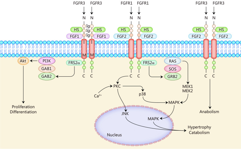
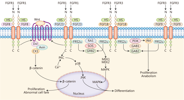
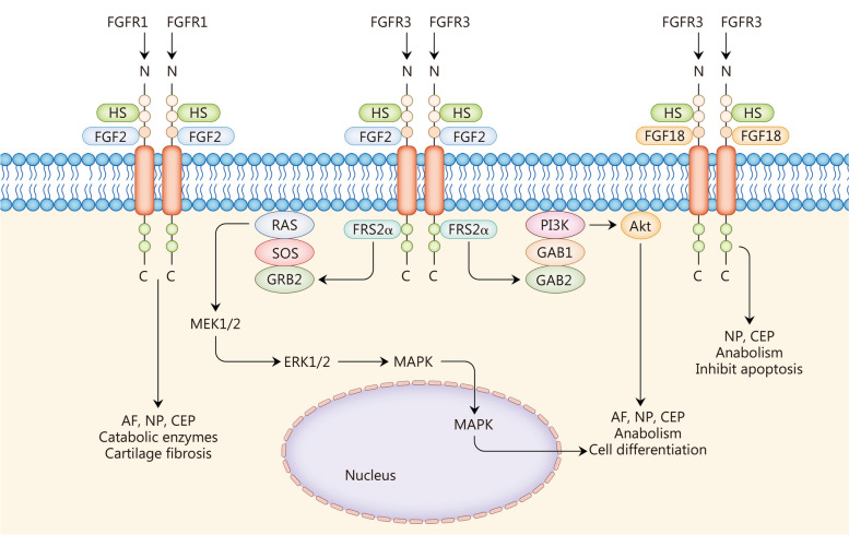
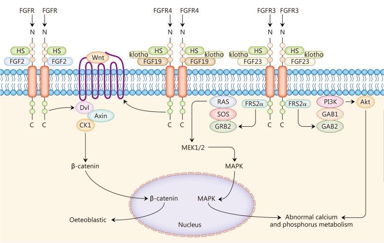
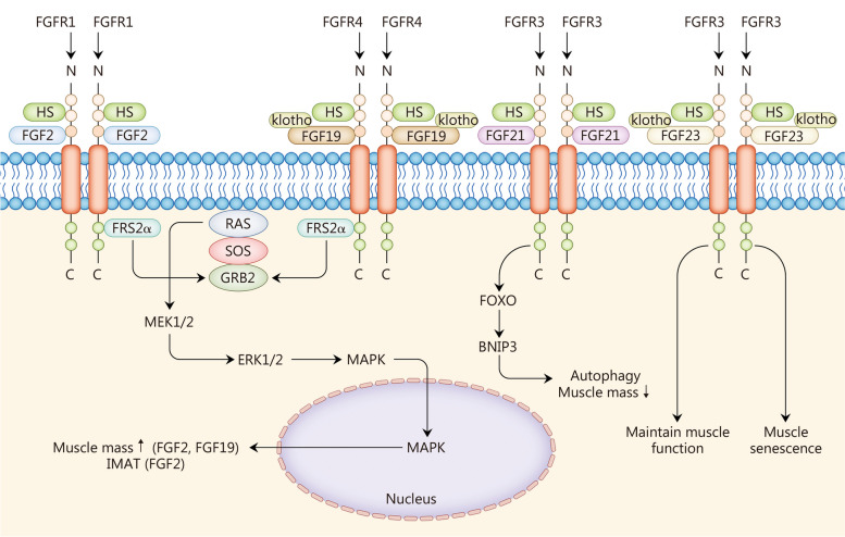
Fibroblast growth factor (FGF) signaling encompasses a multitude of functions, including regulation of cell proliferation, differentiation, morphogenesis, and patterning. FGFs and their receptors (FGFR) are crucial for adult tissue repair processes. Aberrant FGF signal transduction is associated with various pathological conditions such as cartilage damage, bone loss, muscle reduction, and other core pathological changes observed in orthopedic degenerative diseases like osteoarthritis (OA), intervertebral disc degeneration (IVDD), osteoporosis (OP), and sarcopenia. In OA and IVDD pathologies specifically, FGF1, FGF2, FGF8, FGF9, FGF18, FGF21, and FGF23 regulate the synthesis, catabolism, and ossification of cartilage tissue. Additionally, the dysregulation of FGFR expression (FGFR1 and FGFR3) promotes the pathological process of cartilage degradation. In OP and sarcopenia, endocrine-derived FGFs (FGF19, FGF21, and FGF23) modulate bone mineral synthesis and decomposition as well as muscle tissues. FGF2 and other FGFs also exert regulatory roles. A growing body of research has focused on understanding the implications of FGF signaling in orthopedic degeneration. Moreover, an increasing number of potential targets within the FGF signaling have been identified, such as FGF9, FGF18, and FGF23. However, it should be noted that most of these discoveries are still in the experimental stage, and further studies are needed before clinical application can be considered. Presently, this review aims to document the association between the FGF signaling pathway and the development and progression of orthopedic diseases. Besides, current therapeutic strategies targeting the FGF signaling pathway to prevent and treat orthopedic degeneration will be evaluated.

Keywords: Fibroblast growth factor (FGF); Fibroblast growth factor receptor (FGFR); Intervertebral disc degeneration (IVDD); Orthopedic degeneration; Osteoarthritis (OA); Osteoporosis (OP); Sarcopenia.


Conflict of interest statement

The authors declare that they have no competing interests.

Figures

Fig. 1
Fig. 1
Fibroblast growth factors (FGFs) 1 and 2 in osteoarthritis (OA). FGF1 activates the PI3K-Akt pathway; FGF2 activates the Ras-MAPK pathway and PKC. The effects of each FGF-activating downstream signaling pathway on articular chondrocytes are shown. PI3K phosphoinositide 3-kinase, Akt protein kinase B, MAPK mitogen-activated protein kinase, PKC protein kinase C, GRB guanine nucleotide exchange factor, GAB GRB2-associated binding protein, HS heparan sulfates, FRS2α FGFR substrates 2α, SOS son of sevenless, MEK mitogen-activated extracellular signal-regulated kinase, FGFR3 fibroblast growth factor receptor 3, JNK c-Jun N-terminal kinase
Fig. 2
Fig. 2
Fibroblast growth factors (FGFs) 8, 9, 18, and 23 in osteoarthritis (OA). FGF8 and FGF23 activate the Wnt/β-catenin pathway; FGF9 and FGF18 activate the PI3K-Akt pathway; FGF18 and FGF23 activate the Ras-MAPK pathway. The effects of each FGF-activating downstream signaling pathway on articular chondrocytes are shown. Dvl dishevelled, Axin axis inhibition, CK1 casein kinases 1, HS heparan sulfates, FRS2α FGFR substrates 2α, SOS son of sevenless, GRB guanine nucleotide exchange factor, GAB GRB2-associated binding protein, PKC protein kinase C, JNK c-Jun N-terminal kinase, MEK mitogen-activated extracellular signal-regulated kinase, MAPK mitogen-activated protein kinase, PI3K phosphoinositide 3-kinase, Akt protein kinase B
Fig. 3
Fig. 3
Fibroblast growth factors (FGFs) in intervertebral disc degeneration (IVDD). FGF2 promotes the catabolism of AF, NP, and CEP through FGFR1, and fibrosis of IVD cartilage. FGF2 works through FGFR3 to activate downstream Ras-MAPK and PI3K-Akt signaling pathways to promote anabolism and cell differentiation in various tissues of IVDs. FGF18 promotes tissue synthesis and apoptosis via FGFR3. AF annulus fibrosus, NP nucleus pulposus, CEP cartilaginous endplate, ERK extracellular signal-regulated kinase, MAPK mitogen-activated protein kinase, HS heparan sulfates, FRS2α FGFR substrates 2α, SOS son of sevenless, GRB guanine nucleotide exchange factor, GAB GRB2-associated binding protein, PI3K phosphoinositide 3-kinase, Akt protein kinase B
Fig. 4
Fig. 4
Fibroblast growth factors (FGFs) in osteoporosis (OP). FGF2 and FGF19 promote osteogenesis through the Wnt-β-catenin signaling pathway. FGF23 binds to FGFR3 to affect osteoblast metabolism through the RAS-MAPK and PI3K-Akt signaling pathways, as well as calcium and phosphate metabolism. It also affects calcium and phosphate metabolism by influencing hormone secretion and cell metabolism. Dvl dishevelled, Axin axis inhibition, CK1 casein kinases 1, MEK mitogen-activated extracellular signal-regulated kinase, MAPK mitogen-activated protein kinase, PI3K phosphoinositide 3-kinase, Akt protein kinase B, GRB guanine nucleotide exchange factor, GAB GRB2-associated binding protein, HS heparan sulfates, FRS2α FGFR substrates 2α, SOS son of sevenless
Fig. 5
Fig. 5
Fibroblast growth factors (FGFs) in sarcopenia. FGF2 and FGF19 increase muscle mass through the RAS-MAPK signaling pathway, and FGF2 promotes IMAT. FGF21 promotes autophagy of muscle tissue and decreases muscle mass. FGF23 accelerates the senescence of muscle tissue and maintains muscle function. IMAT intramuscular adipose tissue, FOXO forkhead box protein O, BNIP3 Bcl-2/E1B-19 kD interacting protein 3, HS heparan sulfates, FRS2α FGFR substrates 2α, SOS son of sevenless, MEK mitogen-activated extracellular, MAPK mitogen-activated protein kinase, ERK extracellular signal-regulated kinase

References

    1. Yun YR, Won JE, Jeon E, Lee S, Kang W, Jo H, et al. Fibroblast growth factors: biology, function, and application for tissue regeneration. J Tissue Eng. 2010;2010:218142. doi: 10.4061/2010/218142. - DOI - PMC - PubMed
    1. Potthoff MJ, Kliewer SA, Mangelsdorf DJ. Endocrine fibroblast growth factors 15/19 and 21: from feast to famine. Genes Dev. 2012;26(4):312–24. doi: 10.1101/gad.184788.111. - DOI - PMC - PubMed
    1. Bailey KN, Alliston T. At the crux of joint crosstalk: TGF-β signaling in the synovial joint. Curr Rheumatol Rep. 2022;24(6):184–97. doi: 10.1007/s11926-022-01074-6. - DOI - PMC - PubMed
    1. Xie Y, Zinkle A, Chen L, Mohammadi M. Fibroblast growth factor signalling in osteoarthritis and cartilage repair. Nat Rev Rheumatol. 2020;16(10):547–64. doi: 10.1038/s41584-020-0469-2. - DOI - PubMed
    1. Oh WT, Yang YS, Xie J, Ma H, Kim JM, Park KH, et al. WNT-modulating gene silencers as a gene therapy for osteoporosis, bone fracture, and critical-sized bone defects. Mol Ther. 2023;31(2):435–53. doi: 10.1016/j.ymthe.2022.09.018. - DOI - PMC - PubMed

MeSH terms

Substances